
A pathology report is a detailed medical document outlining the findings from an expert examination of tissues, organs, or fluids. For families navigating loss, this report often contains crucial information regarding a loved one's health. Understanding its contents can provide clarity during a difficult time.
This guide is designed to serve as a compassionate translator for the complex medical terminology found within a pathology report. Dealing with such a document while grieving can be overwhelming. Our aim is to walk through each section, demystifying the jargon and structure, to help you read and understand these findings with confidence.
The importance of clear pathological analysis is growing. As the global population ages, with projections indicating the number of people over 60 will reach 1.5 billion by 2030, the demand for detailed pathology services is increasing, particularly for understanding chronic conditions. For more information on the standards behind these documents, you can review essential medical documentation guidelines that pathologists follow.
Disclaimer: This article is for informational purposes only and does not constitute medical or legal advice. Please consult with a qualified physician or legal professional to discuss your specific situation and the findings of any medical report.
A pathology report serves several vital roles:
- It provides medical information. The report offers a clear explanation of diseases or conditions present.
- It can support legal processes. Findings may be important for insurance claims or other legal matters.
- It informs family health. The report can identify hereditary conditions, providing vital information to surviving family members about potential health risks.
For families seeking definitive answers about a cause of death, a https://www.texasautopsyservices.com/private-autopsies/ can be a valuable option.
Navigating the Key Sections of the Report
A pathology report is structured like a detailed story, with each section serving as a distinct chapter. While the exact layout may vary between laboratories, most follow a similar, logical progression. Understanding this structure is the first step toward interpreting the report's content.
Knowing the purpose of each part—from basic patient details to the final diagnosis—will help you navigate the document with greater confidence. This standardized format ensures that every critical piece of information is included.
This image provides an overview of how a typical pathology report is organized.
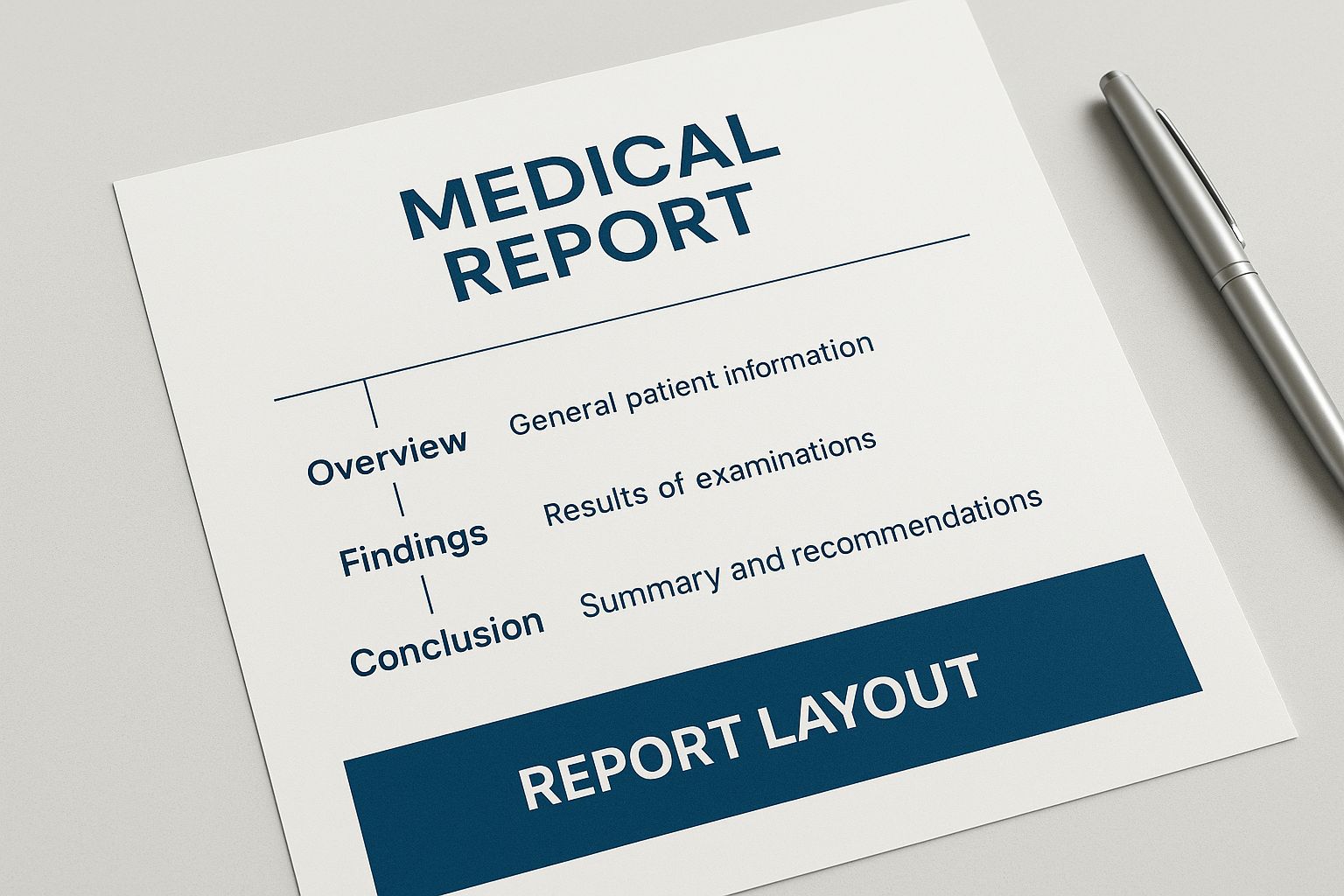
The report is designed to flow from general information to specific findings, creating a complete medical narrative.
Let's break down the main sections you will encounter. Here is a summary of what to expect in a standard report.
Anatomy of a Pathology Report
| Section Title | Purpose and Content |
|---|---|
| Identifying Information | Contains patient name, date of birth, medical record number, and case identifiers to ensure the report is accurately linked to the correct individual. |
| Clinical History | A brief summary provided by the treating physician explaining the patient's medical background and the reason for the pathology examination. |
| Gross Description | The pathologist's notes on the physical characteristics of the tissue or organ as seen by the naked eye, including size, weight, color, and texture. |
| Microscopic Description | Details of what the pathologist observed under a microscope, describing cellular appearance and arrangement. This is often the most technical section. |
| Final Pathologic Diagnosis | The pathologist's definitive conclusion and diagnosis based on all the evidence gathered from the examination. |
| Comments/Notes | Additional context or recommendations. The pathologist may add notes to clarify findings, suggest further tests, or provide other relevant insights. |
This table provides a roadmap to the document. Now, let's explore these sections in more detail.
Identifying Information and Clinical History
Every report begins with foundational information to ensure the findings are matched to the right person and clinical situation. This section serves as the administrative core of the document.
You will find details such as:
- Patient Demographics: The individual's full name, date of birth, and unique medical record numbers.
- Case Identifiers: Unique numbers assigned by the laboratory to track tissue samples throughout the analysis process.
- Clinical History: A brief summary from the ordering physician that gives the pathologist context about the patient’s health and the reason for the examination. This background is critical for an accurate interpretation.
Ensuring the accuracy of these details is vital for medical and legal integrity, preventing potential errors and confirming the validity of the entire report.
Gross and Microscopic Descriptions
This is where the pathologist's direct observations are recorded. These two sections describe what was seen, first with the naked eye and then through the powerful lens of a microscope. This is the raw data that informs the final conclusion.
The Gross Description (sometimes called macroscopic) details the physical traits of the tissue or organs. The pathologist notes their size, weight, color, and texture. For instance, a report might note that an organ appeared unusually large or had an abnormal discoloration.
The Microscopic Description provides a much deeper view. For this part, the pathologist examines thinly sliced, stained pieces of tissue under a microscope. This is often the most technical section, containing precise medical terms describing the cells' health, shape, and arrangement. It is here that the cellular-level evidence is gathered to support the final diagnosis.
According to research in the Journal of Pathology Informatics, a pathology report contains valuable information for prognosis that is not always present in other secondary data sources. Understanding its structure unlocks this value for both medical and legal review.
From here, the report moves toward its conclusion. Next, we will look at the most critical parts: the Final Pathologic Diagnosis and any special tests that were performed, which together deliver the ultimate answers from the examination.
Making Sense of Medical Observations
This part of the report is where the pathologist lays out direct observations. It is often the most technical section, but it contains the core evidence for the final diagnosis. It is broken down into two main parts: what the pathologist could see with the naked eye (gross description) and what was discovered under the microscope (microscopic description).

Gross Description: What the Pathologist Saw
The gross description is a description of the large-scale, visible characteristics of the tissues and organs. The pathologist records details like size, weight, color, and texture. Think of it as a detailed physical inventory of the body's internal state.
For example, the report might mention "cardiac hypertrophy." This term refers to an enlarged heart. Just as a muscle grows larger with exercise, the heart muscle can thicken and enlarge when it is forced to work too hard over a prolonged period. To a pathologist, cardiac hypertrophy is a significant physical clue.
Microscopic Description: A Cellular-Level View
Next, the pathologist examines tissues at a microscopic level. After preparing very thin slices of tissue and staining them with special dyes, they are examined under a microscope. This is where signs of disease that are completely invisible to the naked eye can be identified.
This section is dense with specific medical terms that describe what is happening at a cellular level. To make sense of an example pathology report, it is helpful to become familiar with a few common findings.
- Inflammation: This is the body’s natural response to injury or infection. Under the microscope, a pathologist sees an accumulation of immune cells in a specific area.
- Necrosis: This is the medical term for cell or tissue death. It can occur when blood supply is cut off or from a severe infection or injury. Microscopically, necrotic cells appear disorganized and broken down.
- Atrophy: This term describes the shrinking of cells or an entire organ. It often results from disuse, poor nutrition, or lack of nerve signals.
These microscopic findings are not just isolated details; they are crucial pieces of a larger puzzle. By identifying specific changes in the cells, the pathologist can connect them to broader disease processes and build a clearer picture of what was happening in the body. For instance, finding both inflammation and necrosis in the heart muscle is powerful microscopic evidence of a recent heart attack.
By understanding both the gross and microscopic descriptions, you can begin to follow the pathologist's line of reasoning. Each observation is a clue that builds the foundation for the report's final conclusions.
Understanding the Final Pathologic Diagnosis

This is the section of the report where all detailed observations come together to form a final medical opinion. It is typically found near the end of the document, following the descriptive sections, and represents the pathologist's expert interpretation of all the evidence.
Key Terms in the Diagnosis
Diagnoses are usually separated into two main categories: primary and secondary. This helps create a clear hierarchy of the findings.
- Primary Diagnosis: This is the main condition that was the direct cause of death or the primary disease process. It carries the most weight in the pathologist's medical opinion.
- Contributing Diagnosis: These are other conditions that were present and likely played a role, either by worsening the primary condition or contributing to the individual's overall decline in health.
The Final Pathologic Diagnosis is the culmination of rigorous analysis, blending gross and microscopic findings into a definitive conclusion. This distinction is important because it helps families, physicians, and legal professionals focus on the most critical findings, especially when multiple health issues were present.
Analyzing a Sample Diagnosis
Let's look at an example. In a sample report, the top line might be Atherosclerotic Cardiovascular Disease. This would be the primary diagnosis, indicating that hardened and narrowed arteries were the main issue.
Below that, we might see Hepatic Steatosis (fatty liver disease). While a significant health issue, it would be listed as a secondary or contributing finding.
| Diagnosis Type | Example Entry |
|---|---|
| Primary Diagnosis | Atherosclerotic Cardiovascular Disease |
| Contributing Diagnosis | Hepatic Steatosis (Fatty Liver) |
This table illustrates how the report separates the main finding from supporting details.
Practical Tips for Interpretation
When reading this section, pay close attention to the order in which diagnoses are listed.
- The first diagnosis is almost always the most significant one.
- View any secondary entries as factors that may have complicated the main issue.
- When discussing the report with family, it can be helpful to explain complex medical terms in plain language.
Why This Matters: A Growing Field
The need for accurate, clear pathology reports is growing. The global market for pathology laboratories is projected to expand significantly, with one report from market projections on Statista showing a valuation of around USD 380.88 billion and an expected rise to USD 818.4 billion by 2034. This growth is driven by the rise of chronic diseases and an increasing emphasis on preventive care.
What is behind this growth?
- A higher prevalence of chronic conditions like heart disease and diabetes.
- A greater reliance on diagnostic testing in preventive medicine.
- New technologies making analysis faster and more precise.
These numbers highlight how vital an accurate final diagnosis is for everything from patient care to medical research. For families and legal teams, focusing on the primary diagnosis helps clarify the probable cause of death. The secondary findings can open important conversations about contributing factors or hereditary risks.
It is essential to discuss these findings with a qualified physician to understand the full context. Next, we will explore ancillary studies and special tests that often provide deeper evidence for these definitive conclusions.
What Are Ancillary Studies and Special Tests?
Sometimes, the gross and microscopic examinations do not provide the complete story. To investigate further, pathologists may perform a series of specialized procedures called ancillary studies. This section of the report details the results of any additional tests that were ordered.
These extra steps help uncover clues that are not immediately visible, which can confirm a diagnosis, rule out other possibilities, and add a higher degree of certainty to the final conclusions.
Common Types of Ancillary Studies
The specific tests a pathologist orders depend on the questions presented by each case. However, some common studies you might see include:
- Toxicology Screening: This is used to check for the presence and quantity of medications, alcohol, illegal drugs, or other chemicals. It is crucial for determining if substance use or poisoning played a role in the cause of death.
- Microbiology Cultures: If an infection is suspected, tissue or fluid samples may be cultured to grow microorganisms. This can identify the specific bacteria, virus, or fungus responsible for conditions like pneumonia, sepsis, or meningitis.
- Immunohistochemistry (IHC): This is an advanced technique where special antibodies are used to "stain" and identify specific proteins within cells. It is a powerful tool for differentiating between types of cancer cells, which can appear similar under a standard microscope.
In cases involving cancer, a genetic cancer test may also be performed. This type of ancillary study can provide detailed information that refines the diagnosis.
Knowing about these additional tests helps you appreciate the thoroughness of a modern pathological investigation. Each study is a deliberate step taken to ensure the final diagnosis is as accurate and complete as possible.
The technology behind these tests is constantly evolving. According to digital pathology's growth on Future Market Insights, the field of digital pathology is expected to grow at a compound annual rate of 13.1%, reaching an estimated worth of USD 31.3 billion by 2035. The integration of AI is also helping pathologists spot subtle patterns that might be missed by the human eye.
Ultimately, these specialized tests are a vital part of a complete investigation, particularly in complex situations. For families wanting the most definitive answers, understanding the full range of tests performed during private autopsies can bring needed clarity.
Your Next Steps After Reviewing the Report
After reviewing a pathology report, it is normal to feel overwhelmed and have more questions. The report provides medical facts, but it may not offer the complete picture or human context that a conversation with a professional can provide.
Your most critical next step is to schedule a follow-up discussion with a physician. A doctor can translate complex medical jargon into a coherent narrative, connecting the report's findings with your loved one’s health history. This is an opportunity to ask questions and gain a clearer understanding of what the conclusions mean for you and your family.
Looking Beyond the Medical Chart
A pathology report is a foundational document that can be important in handling practical matters that follow a loss.
What else is it used for?
Legal and Insurance Claims: This report is often a key piece of evidence needed to settle life insurance policies, address workers' compensation issues, or answer other legal questions. It provides the objective, scientific information required by these institutions.
Insights for Family Health: The findings can sometimes point to hereditary or genetic conditions. This can be difficult information to process, but it is also valuable for surviving family members. Knowing about potential risks empowers relatives to be proactive with their own health screenings and care.
It can also be helpful to understand how this information is used in broader contexts, like legal cases or insurance reviews. Learn more about how AI can assist in compiling medical chronologies, which often include pathology report data.
Answering Your Questions About the Pathology Report
Receiving a pathology report often brings up many questions. The document is filled with technical medical terminology, and you are often reviewing it during an emotionally challenging time. Let's address some of the most common concerns.
How Long Until We Get the Final Report?
The timeline for a final report can vary, ranging from a few days to several weeks. A straightforward examination may be completed quickly. However, if the pathologist needs to conduct specialized ancillary tests—such as toxicology screenings or microbiology cultures—the process will take longer. These additional steps are crucial for a complete and accurate analysis.
Who Is Allowed to See the Report?
Pathology reports contain protected health information, and access is tightly controlled by privacy laws. Generally, only the legal next of kin or a designated representative of the deceased's estate can request and receive a copy. Proper identification and legal authorization are required before the report can be released.
It is important not to feel overwhelmed if you do not understand every word in the report. The best course of action is to schedule time with a physician who can explain the findings and connect them to your loved one's medical history.
What if I Disagree With the Findings?
If something in the report does not seem correct or if you have lingering questions, seeking a second opinion is a valid option. This typically involves having an independent pathologist review the original tissue slides and all case materials. A second review can provide peace of mind by either confirming the initial diagnosis or offering a different interpretation. Families seeking this level of clarity can learn more about how private autopsies are conducted.
We understand that navigating this process is challenging, and you do not have to do it alone. If you have questions about private autopsy services, our compassionate team is here to assist you. Please feel free to contact us with your questions. You may reach our team by phone at (214) 920-5630 or by email at support@texasautopsyservices.com.