May 17, 2026 · Texas Autopsy Services
Mole Biopsy Results: Understanding Your Mole Biopsy
Anxious about mole biopsy results? A pathologist explains what benign, atypical, and melanoma mean on your report, and when a second opinion is wise.
On this page
When people wait for mole biopsy results, the hardest part is often the silence between the procedure and the phone call. I know that from the other side of the process. I'm our forensic pathology team, a board-certified pathologist, and the answer you're waiting for often begins with someone like me looking at thin sections of tissue on a glass slide.
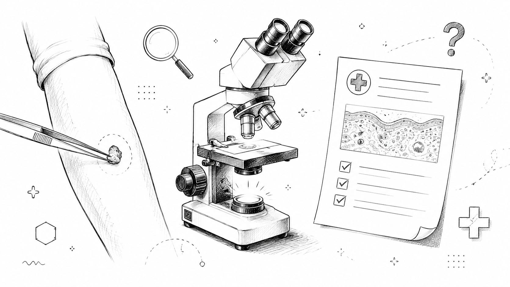
Most patients meet their dermatologist, not their pathologist. That makes sense. The dermatologist sees the mole on your skin. I see the cells after the tissue reaches the lab, after it's processed, stained, and placed under the microscope. My job is to turn patterns in tissue into a diagnosis that is accurate, clear, and useful.
A pathology report can look cold at first glance. It uses exact words because small wording changes can affect what happens next. “Benign,” “atypical,” “melanoma in situ,” “invasive,” “clear margins,” and “positive margins” are not interchangeable phrases. Each one points your doctor toward reassurance, follow-up, or more treatment.
That precision can feel intimidating when you or a family member is already anxious. It shouldn't stay mysterious. A good report should guide care, and a good explanation should let you understand what the report is saying.
Receiving the News About Your Mole Biopsy
A common scene plays out the same way every week. A patient has a spot removed in a dermatology office, goes home with a bandage, and then starts replaying every possibility while waiting for the result. By the time the report is ready, the words can feel heavier than they are.
From my side of the microscope, that report didn't appear instantly. The tissue was fixed, processed, embedded, cut into very thin sections, stained, and reviewed. If the lesion is straightforward, the diagnosis may be direct. If the lesion sits near the border between categories, the review becomes slower and more careful, because a rushed label helps no one.
What I'm looking for under the microscope
For a mole biopsy, I'm not only asking whether cells are present. I'm asking how they are arranged, whether they mature normally as they descend into the skin, whether the architecture is orderly or irregular, and whether there are features that support benign change, atypia, or melanoma.
That's why two reports that both sound “abnormal” can mean very different things in practice. One may describe a harmless mole with mild irregular features. Another may describe melanoma that requires additional surgery.
Practical rule: A pathology report is not written to alarm you. It is written to be exact enough that your treating physician can act on it.
Why the wording can seem technical
Pathology reports are medical documents. They are meant to communicate with dermatologists, surgeons, and other physicians who need specific findings, not general impressions. Patients then receive that same report, often without much translation.
That gap creates worry. People often assume technical language means bad news. It often doesn't. In daily practice, I see many reports where the final meaning is reassuring even though the wording is dense.
A useful way to read the report is this:
- Look first at the final diagnosis. That's the main answer.
- Then look at any comment section. This often explains uncertainty, margins, or recommended clinical correlation.
- Treat microscopic detail as context. Important, yes. But it usually supports the diagnosis rather than replacing it.
If you understand those layers, the report becomes easier to read and easier to discuss with your dermatologist.
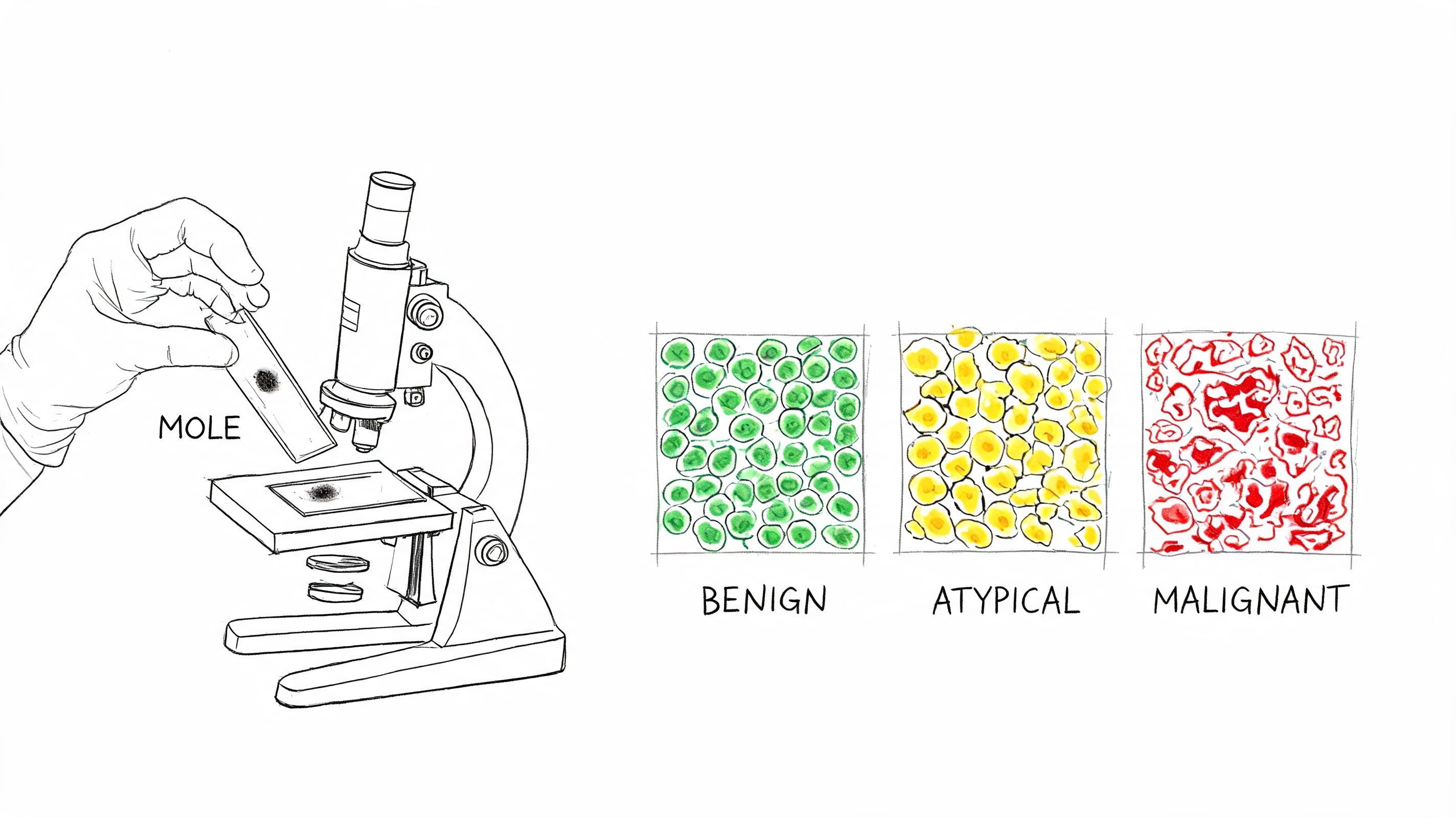
The Three Main Outcomes Benign Atypical and Malignant
Most patients want the same plain answer first. Is it harmless, concerning, or cancer? Most mole biopsy results fall into one of three broad groups.
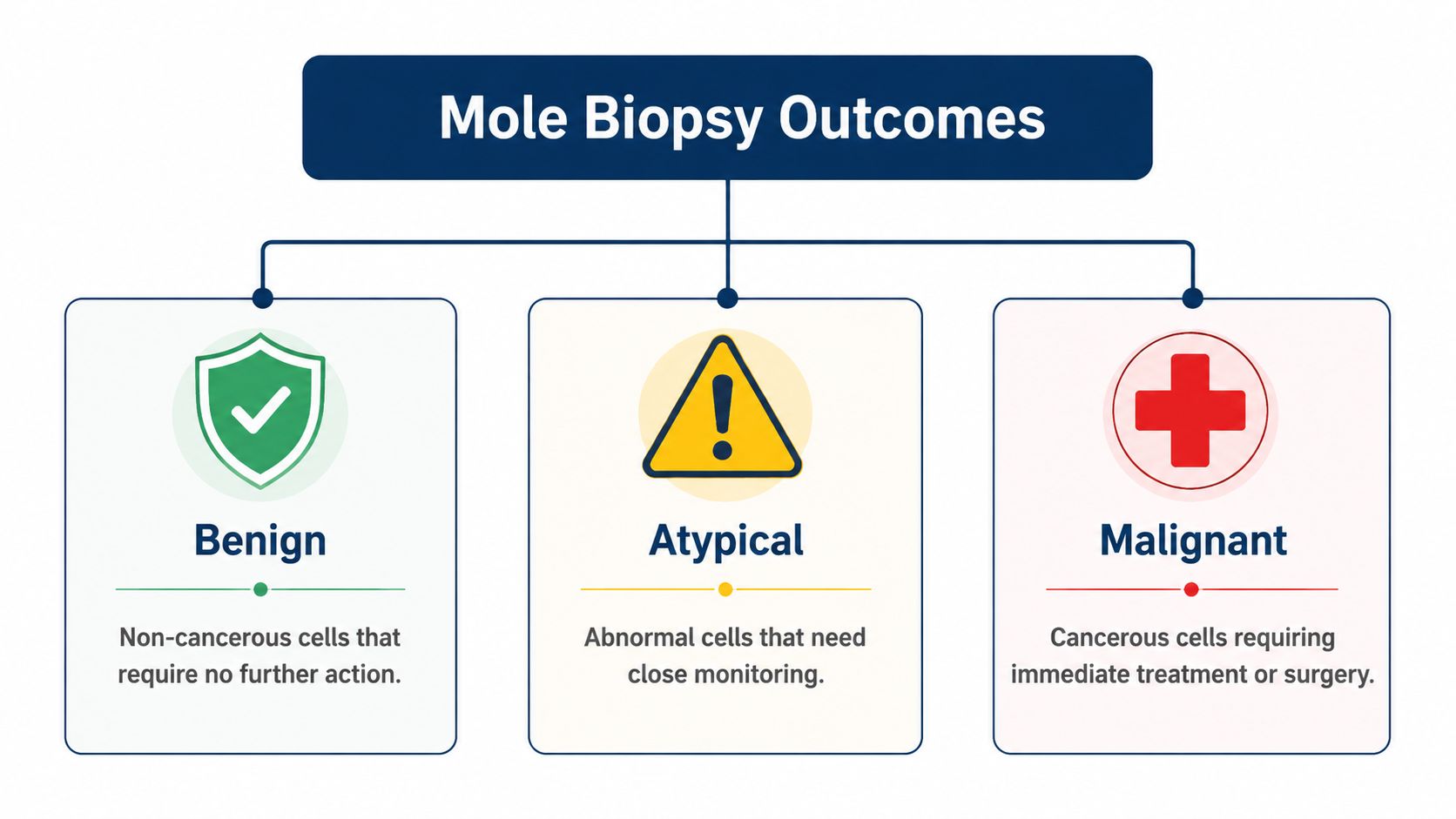
A large biopsy series helps put that into perspective. In an analysis of about 80,000 skin biopsies, melanocytic lesions made up 23% of all diagnoses. Within those melanocytic cases, 83% were benign nevi, 8.3% moderately dysplastic, 4.5% melanoma in situ, and 4.1% invasive melanoma, according to the University of Washington summary of biopsy text analysis. That's why biopsy is often a rule-out tool rather than proof of cancer.
Benign
A benign result means the lesion is non-cancerous. Under the microscope, the cells may still have recognizable mole features, but they do not show the pattern of melanoma.
This is the most reassuring outcome. In many cases, no further treatment is needed if the lesion has been adequately removed and the clinical picture matches the pathology.
Benign doesn't always mean “perfectly ordinary.” Some benign moles are irritated, inflamed, traumatized by shaving, or altered by sun exposure. Those changes can make them look worrisome to the eye, which is exactly why the biopsy was useful.
Atypical
An atypical or dysplastic mole is the category that causes the most confusion. These lesions are not the same as melanoma, but they aren't entirely routine either.
I often explain atypia this way: the cells are acting unusually, not necessarily dangerously. They may show irregular architecture or cytologic features that make a pathologist pause, describe the lesion carefully, and recommend attention to margins or follow-up.
Not every atypical mole leads to more surgery. The decision often depends on the degree of atypia, whether the lesion extends to the edge of the specimen, and how the lesion looked clinically.
Atypia exists on a spectrum. That's one reason expert review matters. Borderline lesions can be difficult, and slight interpretive differences can change management.
When a report says “atypical,” the next question is not “Is it cancer?” The next question is “How atypical, and were the abnormal cells fully removed?”
Malignant
A malignant result means melanoma is present. At that point, the report shifts from describing a suspicious mole to defining a cancer that needs treatment planning.
There are two broad forms patients commonly hear about:
- Melanoma in situ means the malignant cells are confined to the top layer of the skin.
- Invasive melanoma means those cells have moved deeper into the skin.
That distinction matters because depth of invasion affects prognosis and next steps. A lesion limited to the surface is very different from one that has entered deeper tissue.
In situ versus invasive
Think of in situ melanoma as a cancer that has not yet broken through its original boundary. Invasive melanoma has crossed that line. Under the microscope, that boundary is not theoretical. It is something we examine directly.
Once invasion is present, the report becomes more than a yes-or-no diagnosis. It becomes a map for treatment.
Decoding Your Pathology Report Key Terms Explained
Even an accurate report can be hard to live with if the language feels opaque. A few terms carry most of the practical meaning.
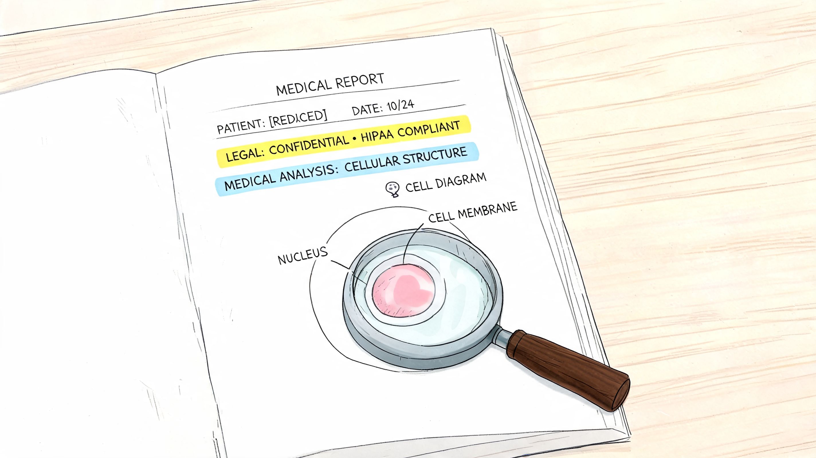
For melanoma, the report typically records lesion type, anatomic site, ulceration, and especially Breslow thickness, which is the measured depth of invasion in millimetres and one of the most important prognostic variables. It also comments on margins. Negative or clear margins indicate the abnormal cells were fully removed, while positive margins suggest residual disease may remain and usually lead to wider excision, as outlined in this overview of melanoma pathology report terms.
Margins
A margin is the edge of the tissue the dermatologist removed. When I examine the specimen, I'm looking to see whether the abnormal cells stop short of that edge or run right up to it.
Here is the practical meaning:
| Term | Plain meaning | Why it matters |
|---|---|---|
| Negative margins | The abnormal cells do not reach the edge | The lesion appears fully removed in that specimen |
| Positive margins | Abnormal cells extend to the edge | Some of the lesion may still remain in the skin |
Positive margins do not automatically mean something has spread widely. They usually mean the lesion may not be completely removed, so your doctor may recommend taking a wider piece of tissue.
Breslow thickness
Breslow thickness is the measured depth of a melanoma in millimetres. If you remember one technical term from a melanoma report, this is often the one that matters most.
Why? Because melanoma behaves differently depending on how deep it goes. A very superficial lesion and a deeper invasive lesion do not carry the same implications.
A shave biopsy can sometimes make this harder to judge if the base of the lesion is not fully sampled.
Ulceration and related features
Ulceration means the surface over the melanoma is no longer intact. It is one of the standard features included in melanoma reporting because it contributes to risk assessment.
You may also see comments on lesion type and anatomic site. Those details are not filler. They help place the microscopic findings into the right clinical setting.
If you want a broader plain-language primer on pathology wording, our article on what a pathology report means gives the same kind of translation for patients and families who are new to pathology reports.
Why reports sometimes include comments
A comment section often appears when a lesion is technically difficult, partially sampled, or best interpreted with the dermatologist's clinical impression. That's not a sign that something went wrong. It's often the pathologist doing exactly what careful practice requires, which is naming both the diagnosis and its limits.
What Happens Next From Surveillance to Surgery
The biopsy result matters because it drives the next decision. That decision is rarely random. It follows the pathology.
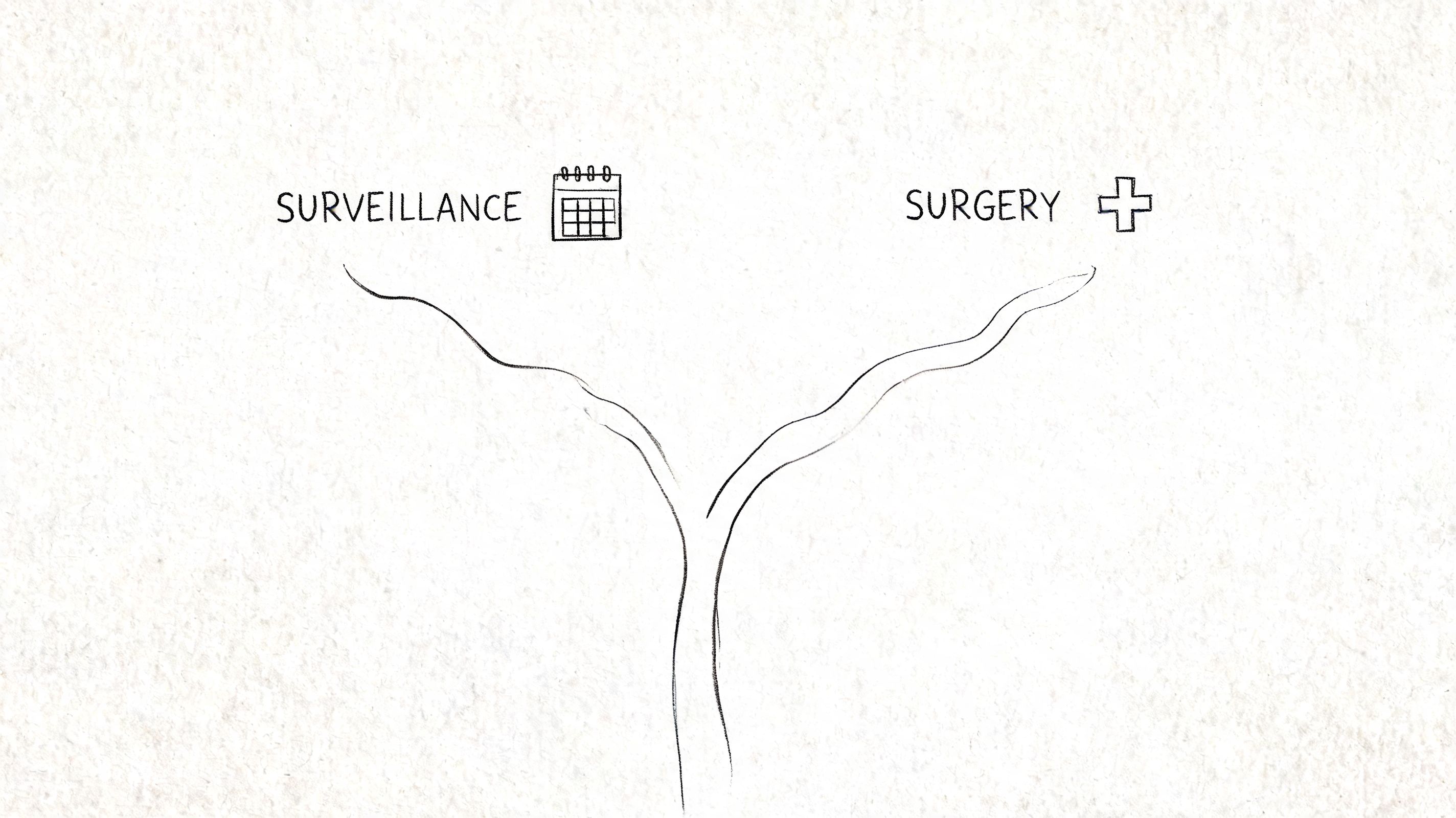
If the lesion is benign
For a benign mole, the next step is often simple reassurance. Your dermatologist may just recommend routine skin checks and watching for new or changing lesions elsewhere on the body.
That is one reason biopsies are valuable even when they do not show cancer. They can end uncertainty and prevent overtreatment.
If the lesion is atypical
For an atypical lesion, the next step depends on the report details. A mildly irregular mole that appears fully removed may lead to observation. A more concerning atypical lesion, especially one involving a margin, may lead to re-excision.
That conversation is usually about balancing caution with proportional treatment. Not every abnormality needs aggressive surgery, but some do merit a cleaner and wider removal.
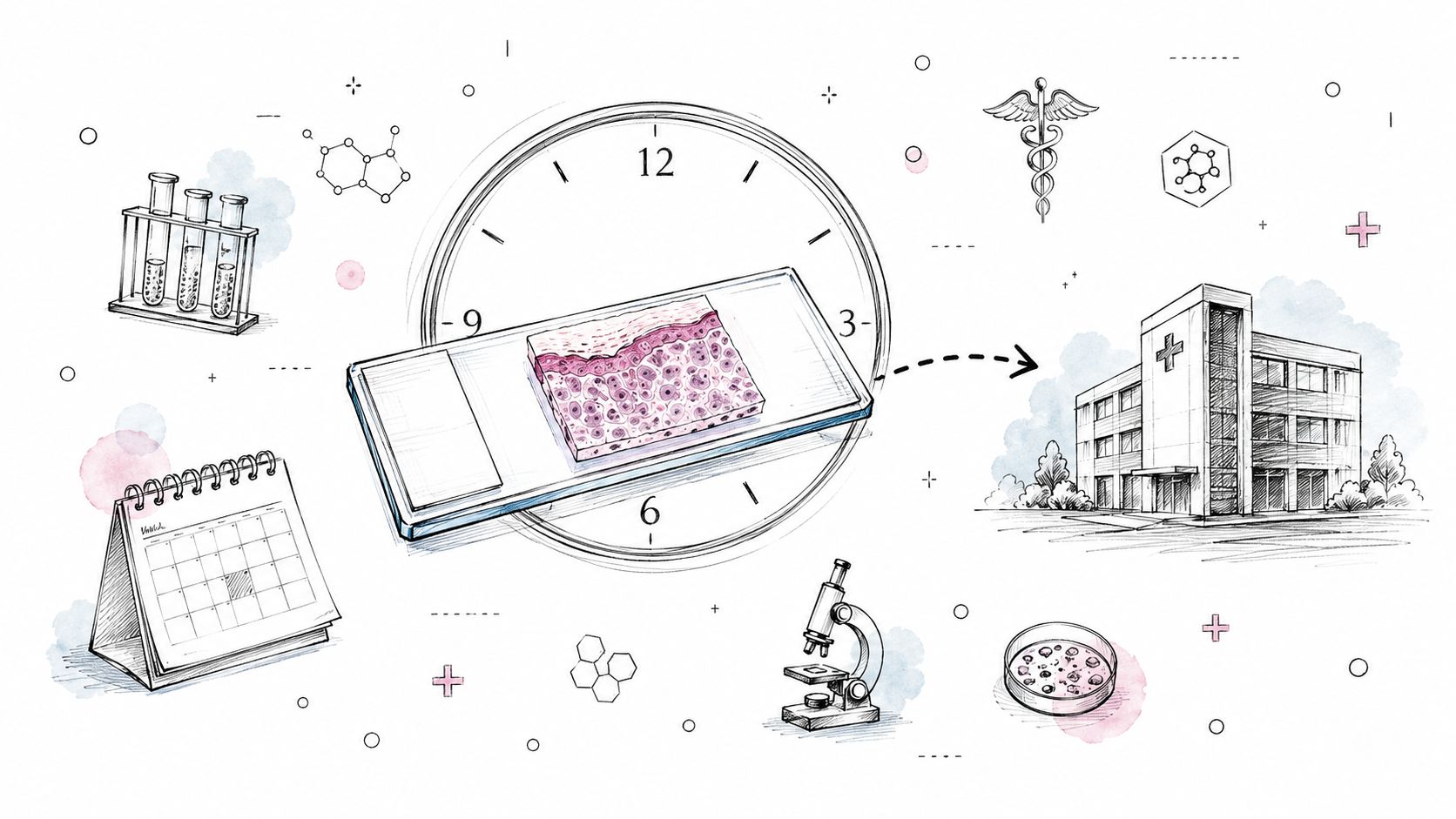
Biopsy technique also affects this decision. Shave biopsies are efficient but can under-sample lesion depth, potentially affecting Breslow thickness assessment. Excisional biopsy is often preferred for clinically suspicious lesions because it captures the entire lesion, improving margin assessment and reducing sampling error. Pathology turnaround is typically 5 to 10 working days, though complex testing may take longer, as described in this practical review of mole biopsy technique and timing.
If the lesion is melanoma
If melanoma is diagnosed, the next step commonly includes a wider excision to remove additional normal-appearing skin around the site. That is done to reduce the chance that residual melanoma cells remain.
In deeper or otherwise significant cases, the treating team may also discuss whether a sentinel lymph node biopsy should be considered. That decision depends on the pathology details and the clinical context.
For readers who want a separate plain-language guide to skin pathology follow-up, this overview of skin biopsy results may help frame the usual sequence after the report is issued.
A short visual explanation can also make the treatment pathway easier to follow:
What does not work well
What doesn't help is trying to guess the treatment plan from one isolated word in the report. “Atypical” without the margin status is incomplete information. “Melanoma” without depth and other report features is also incomplete information.
The right way to read mole biopsy results is to connect three things:
- The diagnosis itself
- Whether the lesion was fully sampled or fully removed
- What the treating dermatologist or surgeon sees clinically
That combination, not a single phrase, tells you what comes next.
When to Consider a Second Opinion on Your Biopsy
In pathology, a second opinion is not an insult. It is a quality step. I say that as someone who has spent a career making decisions from tissue evidence. Some cases are straightforward. Some are not.
A population-level study found that from 1986 to 2001, the biopsy rate among people 65 and older increased 2.5-fold, from 2,847 to 7,222 biopsies per 100,000, while melanoma incidence increased 2.4-fold, from 45 to 108 per 100,000. The authors estimated that every 1,000 additional biopsies were associated with 12.6 extra melanoma diagnoses, and even after adjustment, 6.9 extra melanoma cases, according to the BMJ study on biopsy rates and melanoma diagnosis. The practical lesson is simple. As more lesions are sampled, more borderline and early lesions enter the diagnostic gray zone.
When I think another review is reasonable
A second opinion often makes sense in a few situations:
- Borderline pathology. Severe atypia, melanoma in situ, or wording that sounds qualified rather than definitive.
- A mismatch between clinic and microscope. The report and the dermatologist's impression do not fit well together.
- A major treatment decision is about to follow. Before a larger excision or another procedure, some patients want confirmation.
- Peace of mind. That is a legitimate reason. Patients live with the consequences of these labels.
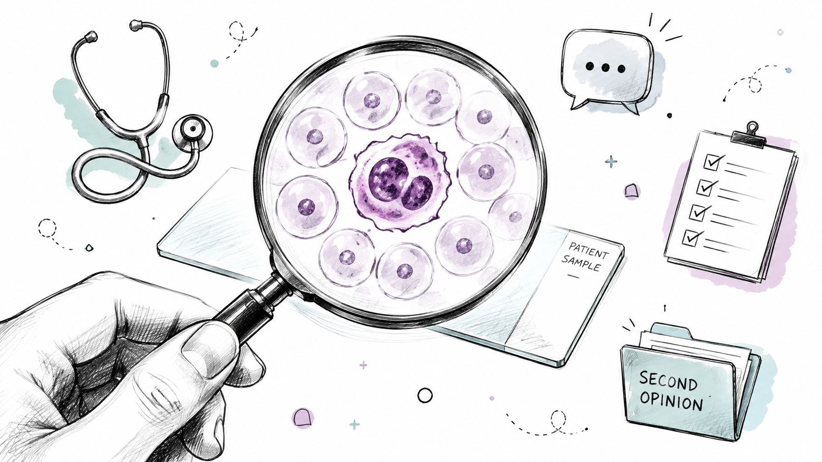
Careful medicine sometimes means asking another pathologist to look at the same slide and see whether the interpretation holds.
What a second opinion actually does
A second review does not guarantee a different diagnosis. Often, it confirms the first report and gives the patient more confidence to proceed. In other cases, it refines the category, clarifies the margins, or better explains the uncertainty.
For families or patients seeking an independent pathology-style review process, Texas Autopsy Services second opinion services provide that kind of outside evaluation in a separate context. The principle is the same in surgical pathology. Independent review can help when clarity matters.
What works is targeted review by a qualified pathologist who understands why the distinction matters. What does not work is treating every difficult lesion as if the answer is obvious when the slide says otherwise.
Frequently Asked Questions About Biopsy Reports
| Question | Answer |
|---|---|
| How long do mole biopsy results usually take? | Many practices report turnaround in 5 to 10 working days, though more technical testing can take longer. |
| What's the difference between a dermatologist and a pathologist? | The dermatologist examines the lesion on your skin and performs the biopsy. The pathologist examines the tissue under the microscope and issues the diagnosis. |
| Can a shave biopsy miss part of a melanoma? | It can under-sample depth in some cases. That matters because incomplete depth sampling can affect assessment of invasion and later treatment planning. |
| Do positive margins mean the cancer has spread? | Not necessarily. Positive margins usually mean abnormal cells extend to the edge of the sampled tissue, so more tissue may need to be removed locally. |
| Should I ask for a second opinion? | It's reasonable when the diagnosis is borderline, when major surgery is being considered, or when the report feels unclear in light of the clinical picture. |
If you're trying to make sense of a pathology finding and need a calm, independent explanation, contact Texas Autopsy Services. I approach every pathology question the same way I approach forensic work. With precision, restraint, and respect for the person behind the report.