June 14, 2026 · Texas Autopsy Services
Understanding Your Skin Cancer Biopsy Results
Get a clear explanation of your skin cancer biopsy results. Experts explain margins, depth, and tumor type for your diagnosis & what to ask your doctor.
On this page
- A skin biopsy report is a structured medical document, not just a one-line answer.
- Histopathology is the gold standard for diagnosing skin disease, which means the tissue under the microscope is the main evidence used to confirm what a lesion is.
- Results can range from benign to precancerous to malignant, and some reports fall into a gray zone that needs follow-up or a second opinion.
- If cancer is found, details such as tumor type, depth, and margins help guide treatment, not just the word “cancer” alone.
- Turnaround varies, with some results returning in a few days and others taking longer when additional testing is needed.
Waiting for skin cancer biopsy results can make time feel unusually slow. Many people have already replayed the appointment in their mind, searched unfamiliar terms online, and tried to guess what the report will say before anyone has called.
We understand that kind of waiting from the pathology side. Our daily work at Texas Autopsy Services centers on tissue analysis, medical evidence, and careful written conclusions. In private autopsy work, every examination is performed by a forensic pathologist certified by the American Board of Pathology. Although a skin biopsy is very different from a postmortem examination, the underlying principle is the same. A pathologist studies real tissue, documents what is present, and turns microscopic findings into a clear medical report.
That's why a skin biopsy report is such a useful example for explaining how pathology works. It shows, in a very human and familiar setting, how tissue becomes evidence and how that evidence becomes a diagnosis.
The Wait for Answers After a Skin Biopsy
A familiar scene plays out every day. A dermatologist removes a spot, places the tissue in a small jar, and says the office will call with the results. The procedure is brief. The silence afterward can feel much longer.
Part of that anxiety comes from expecting a yes-or-no answer. Patients often picture the result as a simple label: cancer or not cancer. A pathology report is built differently. It is a structured medical document written to record what the pathologist observed, how those findings fit together, and what diagnosis the tissue supports.
That structure can feel intimidating at first.
From the pathologist's side of the microscope, each line has a job. The report is not written to sound dramatic or reassuring. It is written to be accurate, traceable, and useful to the clinician deciding what happens next. In that sense, a skin biopsy report works like any other pathology report we create, including in postmortem practice. The conclusion must follow the findings, and the findings must be documented clearly enough that another physician can understand exactly how that conclusion was reached.
That is why a report may include terms such as atypia, dysplastic, inflamed, involved margins, or suspicious for. Those phrases are not filler. They mark specific observations or limits of certainty. A pathologist is trained to describe what the tissue shows on the slide, even when the answer is reassuring, borderline, or still incomplete.
Many biopsied skin lesions do turn out to be noncancerous, but the value of the biopsy is not only in ruling out melanoma or another skin cancer. It also creates a written record of what was sampled and how the diagnosis was made. For readers who want background on when tissue is taken to check for melanoma, our overview of a skin biopsy for suspected melanoma explains the clinical side of that decision.
One more point often surprises patients. The pathology report is also a communication tool between the lab, the dermatologist, and the health system. Clear documentation supports treatment planning, follow-up, and accurate billing workflows that can reduce biopsy claim denials.
The helpful shift is to stop viewing the report as a form and start viewing it as evidence organized on purpose. Once you know that the document is built section by section by the pathologist, the wording becomes less mysterious and much easier to read.
The Journey From Your Skin to the Microscope
You leave the dermatology office with a small bandage and one big question. What happens to that tiny piece of skin before a diagnosis appears in your chart?
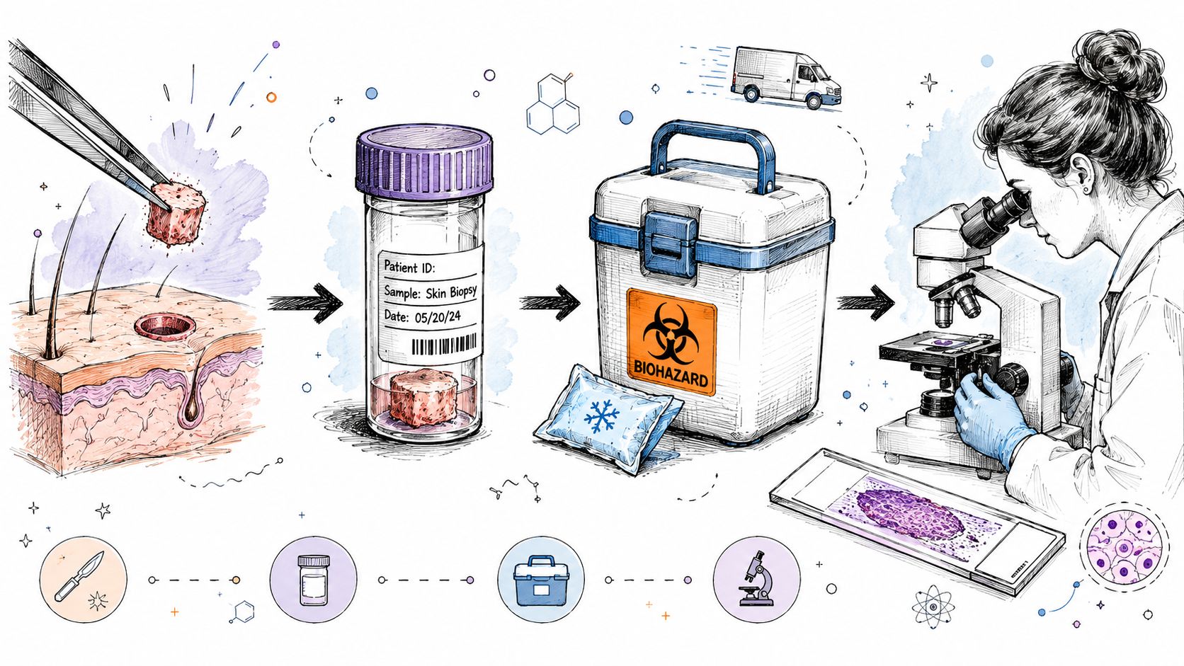
A biopsy report starts long before the pathologist writes the final diagnosis. The specimen has to be preserved, identified, prepared, examined, and documented in a way that another physician can follow from start to finish. In pathology, that chain of steps matters because the report is not just an answer. It is the written record of how that answer was reached.
Why pathology is the deciding step
For skin lesions, the diagnosis is made from tissue review under the microscope. Clinical appearance helps the dermatologist decide what to sample, but the pathologist studies the actual architecture of the lesion, how the cells are arranged, how they mature, and whether they show features of inflammation, atypia, or invasion. This tissue-based approach is why histopathology remains the standard method for confirming many skin diseases, as explained in this review of histopathology in skin disease diagnosis.
A useful comparison is a map and the ground itself. A photo, memory, or office note can suggest where to look. The slide shows the terrain.
That perspective shapes the report. A pathologist is not only naming a condition. The pathologist is building a structured medical document that ties the diagnosis to a specific specimen from a specific site, processed in a specific way, with findings that support the conclusion.
A typical workflow includes these steps:
Specimen receipt
The lab verifies patient identifiers, the body site, and the biopsy type so the case is connected to the right person and the right lesion.Gross examination
The tissue is inspected without a microscope, measured, and described. This becomes the gross description in the report and documents what physically arrived in the lab.Processing and slide preparation
The sample is fixed, embedded, sliced into very thin sections, placed on glass slides, and stained so cell patterns can be seen clearly.Microscopic review
The pathologist examines the slides and determines which findings are diagnostic, which are supportive, and which details need to be mentioned because they affect treatment.Final report
The diagnosis is issued with the details needed for clinical follow-up, such as lesion type, margin status, depth, or uncertainty when the findings are limited.
If you want the clinical side of why a dermatologist chooses to sample a lesion in the first place, our overview of a skin biopsy for suspected melanoma and how clinicians use the result gives that context.
Documentation also has a practical role outside diagnosis. Precise labeling, specimen handling, and complete records protect patient safety and support billing accuracy. In clinic workflows, better documentation can also help reduce biopsy claim denials.
Why timing can vary
Biopsy results do not all move at the same speed through the lab. Some cases are straightforward and can be signed out quickly. Others need deeper tissue levels, special stains, or a second review by a dermatopathologist before the pathologist is comfortable finalizing the report.
The wait usually reflects the work required to make the report accurate and complete.
As noted earlier, skin biopsy turnaround can range from a few days to a few weeks depending on case complexity and lab workflow. A longer wait does not automatically point to bad news. It often means the lab is taking the extra steps needed to support a diagnosis that can guide treatment with confidence.
How to Read Your Pathology Report
Many people open their report and go straight to the last line. That instinct is understandable, but the rest of the document explains how the pathologist reached that conclusion.
The parts of the report
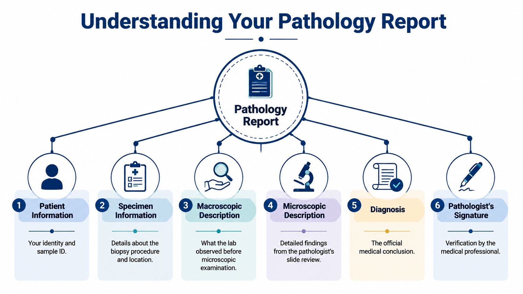
Most skin biopsy reports contain several standard sections.
| Section | What it usually tells you | Why it matters |
|---|---|---|
| Patient information | Your name or identifier and case number | Confirms the report belongs to the correct person |
| Specimen information | Where the tissue came from and what type of biopsy was done | Connects the diagnosis to the exact body site |
| Gross description | What the tissue looked like before slides were made | Documents specimen size and appearance |
| Microscopic description | What the pathologist saw under the microscope | Shows the evidence behind the diagnosis |
| Diagnosis | The final medical conclusion | This is the key answer your clinician uses |
| Signature or authentication | Verification by the pathologist | Confirms professional review and responsibility |
The gross description can sound dry, but it serves an important purpose. It records what arrived in the lab. A line describing a small tan skin fragment or an excised pigmented lesion may seem routine, yet it confirms the specimen's size and character before microscopic processing.
The microscopic description is where the specialized language often appears. This section may describe how cells are arranged, whether there is invasion into deeper tissue, whether atypia is present, or whether inflammation is part of the picture.
For a companion explanation focused on the report document itself, our resource on how to read a biopsy pathology report gives a related pathology-centered walkthrough.
A short visual explanation can also help orient first-time readers.
Why these sections matter
The report is structured this way because pathology is both interpretive and documentary. The pathologist is not only giving an opinion. The pathologist is also creating a formal medical record that other physicians can rely on.
That structure should feel familiar to anyone who has reviewed forensic documentation. In our field, the report must show how the conclusion was reached. The same principle applies to skin pathology. A diagnosis has more value when the supporting observations are clearly recorded.
The diagnosis line matters most, but the language above it often explains what the pathologist felt certain about, what was borderline, and what details influenced that final call.
If any term is unclear, ask your treating clinician to review the report line by line. That is a reasonable request, not an inconvenience.
Understanding Common Biopsy Diagnoses
A skin biopsy report often answers the first big question, but it also does something more practical. It places the finding into a category that other clinicians can act on. From the pathologist's side of the microscope, that is why the diagnosis line is written in a precise, standardized way. It is not only naming what was seen. It is creating a shared medical record that tells your dermatologist whether the lesion is harmless, abnormal but not cancer, or cancerous.
In skin pathology, many reports fall into a few broad diagnostic groups. Some describe a benign lesion. Some describe abnormal changes that are not cancer but may need follow-up or complete removal. Others diagnose skin cancer, most often basal cell carcinoma, squamous cell carcinoma, or melanoma.
What malignant findings often include
A large analysis published on PubMed examining biopsy outcomes by specialty found that malignant diagnoses were common in dermatology practice, although the frequency differed by specialty. The same study reported that basal cell carcinoma, squamous cell carcinoma, squamous cell carcinoma in situ, melanoma in situ, and melanoma were among the most frequent malignant results.
Those names can sound blunt on the page. In the report, each one serves as a label with a specific meaning.
Basal cell carcinoma
This diagnosis identifies a cancer arising from basal cells in the skin. In many reports, it is one of the more common malignant findings and is often described with subtype language that helps classify its growth pattern.Squamous cell carcinoma
This means malignant squamous cells are present. If the report says in situ, the abnormal cells are limited to the upper layer. If it says invasive, the tumor has grown beyond that surface boundary.Melanoma
This diagnosis usually comes with more qualifiers because the tumor name alone is not enough to guide care. Pathologists often include carefully defined features that help the treating team judge risk and choose treatment.
For readers dealing specifically with a melanoma report, our article on what a melanoma pathology report means explains how pathology terms shape the next clinical steps.
When families face a serious diagnosis, they often need support beyond the pathology language itself. Resources on navigating illness stages can help people organize questions and communication during treatment discussions.
What benign and borderline language can mean
Benign findings are common, and a benign result still answers an important medical question. If a report identifies a mole, seborrheic keratosis, or another noncancerous lesion, the biopsy has done its job. It has tested a suspicious spot and shown that cancer is not present.
Some reports land in a middle zone that causes understandable confusion. Terms such as atypical, dysplastic, or severe atypia do not automatically mean cancer. They tell you the pathologist saw cells or tissue architecture that depart from the usual pattern, and the report is grading how strong that departure is.
A useful comparison is a traffic light. Benign is green. Malignant is red. Atypical or dysplastic is yellow, meaning the finding needs context from the full report, the appearance of the lesion, and the treating clinician's plan.
In practical terms:
- Benign means no cancer is identified.
- Precancerous means the lesion shows changes linked to early transformation or increased risk.
- Malignant means cancer is present.
- Atypical or dysplastic means abnormal features are present, and management depends on the degree of abnormality and the rest of the report.
That last point matters because pathology is pattern-based, not keyword-based. A single term rarely tells the whole story. The diagnosis line gives the headline. The exact wording around it explains how definite the finding is and why the report is written that way.
Critical Details That Guide Your Treatment
A skin cancer diagnosis is only the first line of the story. The rest of the report tells the treating clinician what kind of problem is present, how far it extends in the tissue that was sampled, and whether the biopsy appears to have captured all of it.
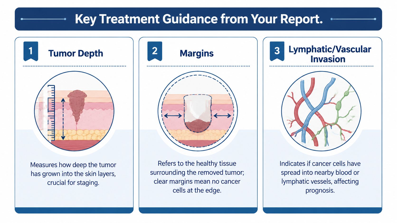
From the pathologist's side of the microscope, this part of the report functions like a map legend. The diagnosis names the lesion. The supporting details explain size, location within the skin, and boundaries. Those details are included because treatment decisions depend on them.
Depth and extent
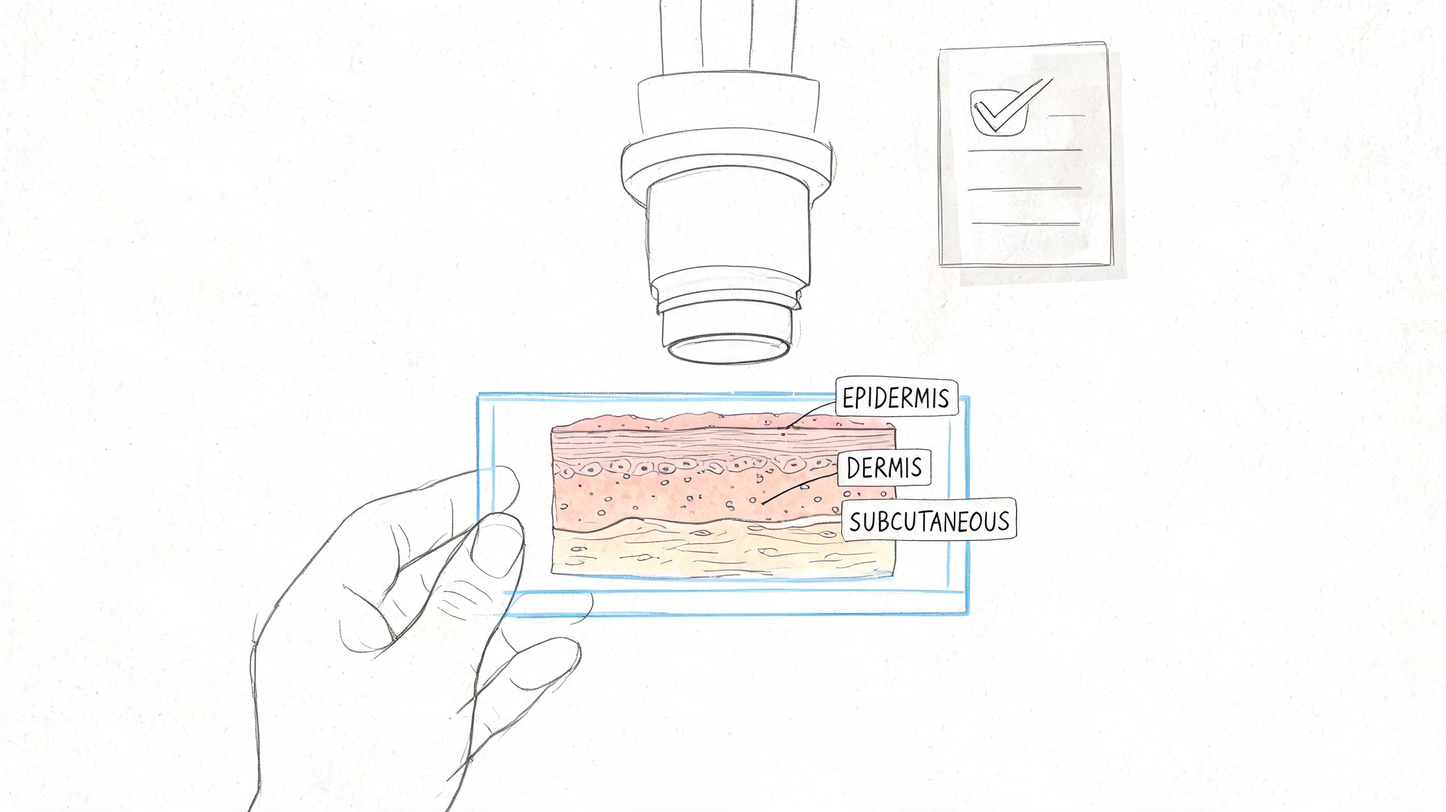
Skin is built in layers, and pathology reports describe disease in relation to those layers. A lesion limited to the surface is a different finding from one that has grown downward into deeper tissue. Two reports can use the same cancer name but point to different next steps because the extent of growth is different.
That is why reports often include wording about depth, invasion, or whether the lesion is confined to upper portions of the skin. If deeper structures are involved, the report may say so directly. That information helps the dermatologist or surgeon judge how much additional treatment may be needed.
A short diagnosis line can look simple. The measured details around it often carry the treatment implications.
Margins and next steps
Margins are the outer edges of the tissue piece the pathologist examines. If no tumor is seen touching those edges, the report may describe the margins as clear or negative in the sampled specimen. If tumor reaches an edge, the report signals that abnormal tissue may still remain in the skin.
Patients often find margin language confusing because it sounds final. It is more accurate to read it as a statement about the tissue submitted to the lab. In other words, the pathologist is reporting what is visible at the borders of that specimen, not making a broad promise about the entire area beyond it.
For melanoma and many other malignant lesions, margin status helps shape the plan for excision, re-excision, or follow-up. The report is structured this way on purpose. It gives the clinician a set of facts that can be turned into action.
Clinicians often focus on four parts of the report after a malignant result:
Tumor type
Basal cell carcinoma, squamous cell carcinoma, melanoma, and other lesions follow different treatment pathways.Depth or level of invasion
This describes how far the tumor extends into the skin or underlying tissue.Margin status
This shows whether tumor is present at the edges of the specimen examined.Additional microscopic features
Some reports include other findings that help estimate behavior and guide management.
Patients do not need to decode every line on their own. They should know why these lines exist. A pathology report is a structured medical document, built to translate what the pathologist sees under the microscope into decisions about surgery, follow-up, and risk.
When Results Are Negative or Uncertain
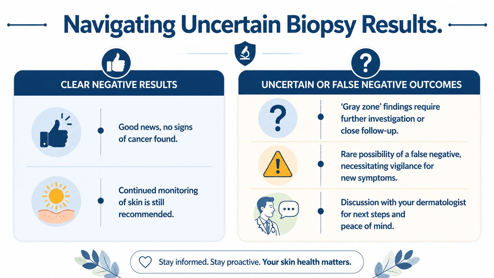
A negative biopsy can bring relief, but it does not always end the conversation. Pathology is strongest when the tissue sample, the lesion on the skin, and the clinical impression all line up.
The gray zone in pathology
Research discussed by UW Medicine notes that pathologists agree most often on clearly benign or clearly malignant biopsies, but they frequently disagree on lesions in the middle gray zone, which is why some patients receive ambiguous reports and may be advised to seek a second opinion, as explained in this UW Medicine discussion of trust and disagreement in skin biopsy interpretation.
Wording such as atypical, cannot exclude, or suggestive but not definitive can appear. To a patient, that can feel unsatisfying. To a pathologist, it is often the most honest wording available when the tissue shows overlapping features.
A few common situations lead to uncertainty:
Borderline microscopic features
Some lesions do not fit neatly into a single category.Limited tissue
A small sample may show only part of the lesion.Competing possibilities
Inflammation, irritation, prior treatment, or sampling effects can complicate interpretation.
If a report sounds cautious, that does not mean the lab failed. It may mean the pathologist is accurately describing a lesion that does not behave like a textbook example.
When the tissue sample and the clinical picture do not match
Another issue is sampling. A biopsy can only answer for the tissue that was removed. A peer-reviewed article notes that photographs should be reviewed when pathology does not match the clinical picture, and that photographic documentation before biopsy can reduce wrong-site surgery risk, according to this PMC article on site verification and clinicopathologic mismatch.
That matters because a negative result is most reassuring when the correct area was sampled and the sample adequately represents the lesion. If the spot keeps changing, looks different from the report's implication, or the dermatologist remains concerned, follow-up may include re-biopsy, complete excision, or slide review by another pathologist.
This is one setting where second-opinion pathology can be appropriate. In postmortem practice, we also review prior records and reports when families or attorneys need clarification. The same principle applies here. A second review does not imply wrongdoing. It is a recognized way to resolve uncertainty in difficult cases.
FAQ Your Skin Biopsy Questions Answered
How long do skin biopsy results usually take
You have the biopsy. The bandage is off. Then the waiting starts.
Many skin biopsy results return within several days, while others take longer if the pathologist needs extra sections, special stains, or a closer review. From the lab side, the report is not written all at once. It is built in parts, much like a case file, starting with what tissue was submitted, followed by what the microscope shows, and ending with the diagnosis and any details your clinician needs for treatment.
What if my report says benign
A benign result means the pathologist did not identify cancer in the tissue examined. That is reassuring, but it also helps to read the report as a document with limits. A pathology report answers for the sample that reached the lab, not for skin that was not removed.
If the spot was only partly sampled, your clinician may still watch the area or recommend more treatment if the lesion changes or the clinical appearance remains concerning.
What if my report says atypical or dysplastic
These words often cause worry because they sound like a final verdict. They are not. In a pathology report, terms such as atypical or dysplastic describe how the cells look under the microscope and signal that the findings need context.
A useful comparison is a weather report. "Cloudy" is meaningful, but it is not the same as "storm." In the same way, atypical features may point to irritation, a mole with unusual architecture, or a lesion that needs complete removal and follow-up. The report includes these terms so your dermatologist can match the microscopic findings with the size, shape, and behavior of the lesion on your skin.
When is a second opinion reasonable
A second review makes sense when the wording is qualified, when the diagnosis sits in a gray zone, or when the treatment decision carries high stakes. Pathologists use second opinions as a quality practice, not as an admission that something went wrong.
That is especially true when the slide findings and the clinical impression do not line up neatly.
What should I ask at my follow-up visit
Bring a copy of the report if you have one. Then ask questions that connect the document to the next medical decision:
What is the exact diagnosis in plain language
The diagnosis line is the headline of the report, but it often needs translation.Was the entire lesion removed
This asks whether the sampled tissue appears clear at the edges and whether more procedure may be needed.Which report details affect treatment
Some findings are descriptive. Others change what happens next.Does the pathology fit what you saw on my skin
This helps uncover sampling issues or clinicopathologic mismatch.Would another pathology review help
A second look can be appropriate in borderline or difficult cases.
A pathology report can look dense because it is designed to serve several readers at once: the treating clinician, the pathologist, and the medical record. Each section has a job. One part identifies the specimen, another records the microscopic pattern, and another states the diagnosis in formal medical language. Once you know that structure, the report becomes easier to read.
If you are facing difficult medical language, uncertainty after a report, or questions about how pathologists reach formal conclusions, our team at Texas Autopsy Services is available to help explain the principles of pathology in clear terms and to discuss independent second-opinion forensic pathology services when those are needed.