May 31, 2026 · Texas Autopsy Services
Skin Biopsy for Melanoma: Your Guide to Procedure & Results
Considering a skin biopsy for melanoma? Understand types, procedure, & how to read results. Get informed.
On this page
TL;DR
- A skin biopsy for melanoma is the only way to confirm whether a suspicious spot is melanoma.
- The biopsy type matters because the pathologist needs the right tissue sample to measure Breslow depth, which helps guide staging and treatment.
- If melanoma is a concern, doctors often prefer a biopsy that removes the full thickness of the spot so depth can be measured as accurately as possible.
- Most biopsies are done with local anesthetic in the office. The numbing injection is often the part patients notice most.
- Your pathology report may include terms such as melanoma in situ, invasive melanoma, margins, and mitotic rate. Each term answers a different question.
- Newer noninvasive tools and skin lesion and mole checks can help decide which spots need tissue sampling, but biopsy remains the gold standard for diagnosis.
A common clinic visit starts unassumingly. You notice a mole that seems darker, or someone close to you points out a spot on your back that looks different than it did a few months ago. By the time you are sitting in the exam room, the main question is simple. What exactly is this, and how do we know?
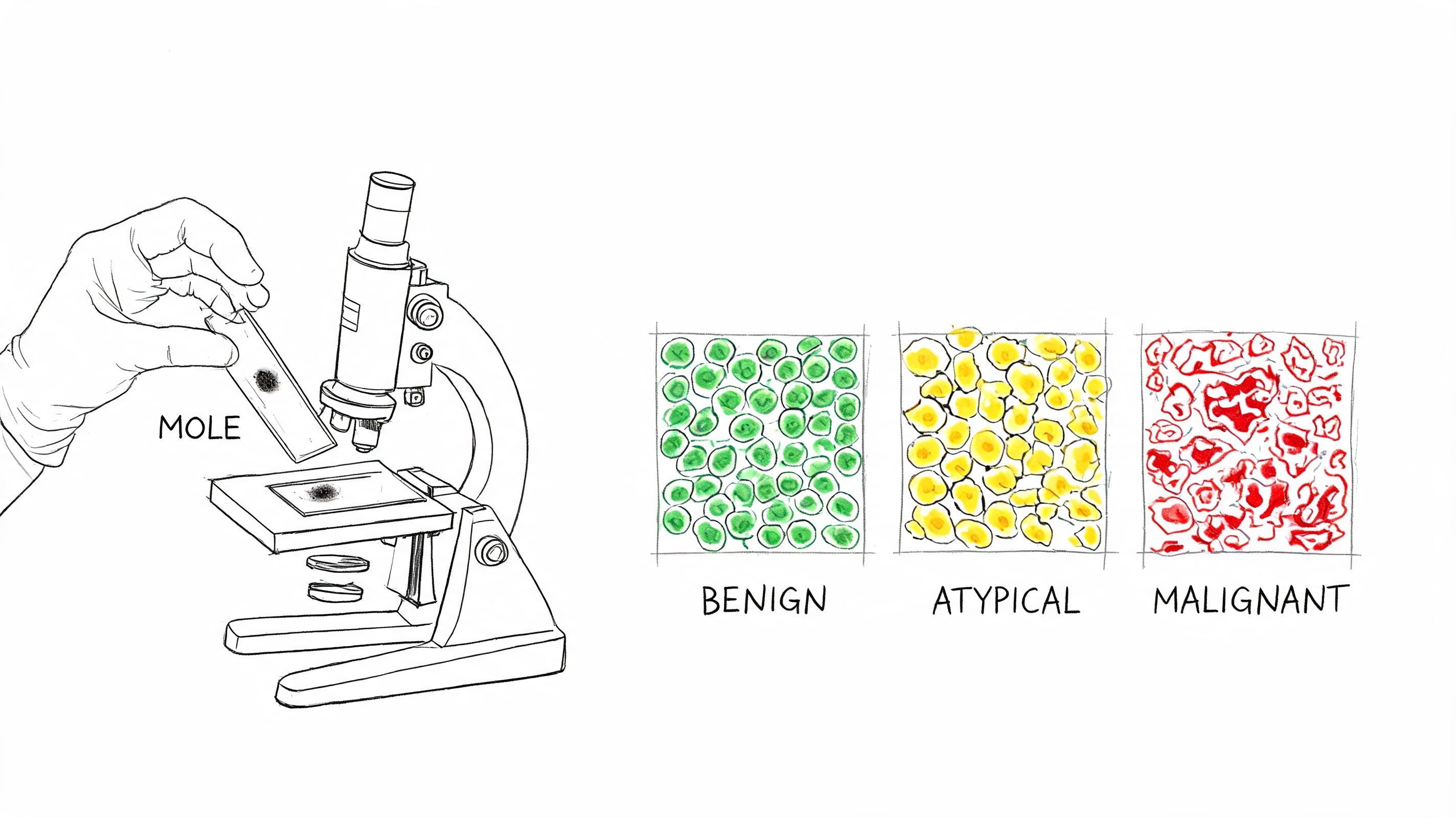
A biopsy gives that answer by turning a visual concern into a tissue diagnosis. Under the microscope, the pathologist can tell whether the spot is benign, melanoma in situ, or an invasive melanoma. If melanoma is present, the sample also needs to show how deep the tumor extends into the skin. That measurement, called Breslow depth, works like a ruler inside the pathology report. It helps determine stage and shapes the next treatment decisions.
Many people get lost in the connection between the procedure and the report. A shave, punch, or excisional biopsy may all sound like small office procedures, but they do not provide the same kind of specimen. The method used on the skin affects what the pathologist can measure later in the lab.
That is why the biopsy is not only about removing a suspicious spot. It is about removing it in a way that preserves the information the pathologist needs. If you have ever wondered how mole biopsy results are interpreted, that link between technique and report is the part that makes the whole process easier to understand.
When a Mole Needs a Closer Look
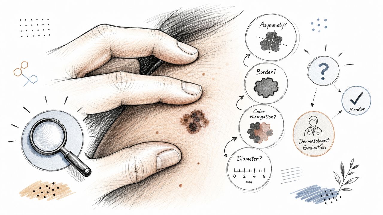
A suspicious mole rarely announces itself dramatically. More often, it's a quiet change. The border seems less even. One half looks different from the other. The color isn't uniform anymore. Sometimes it itches or bleeds, but many concerning spots do neither.
Dermatologists often teach the ABCDE pattern to help patients notice warning signs:
- A for asymmetry. One side doesn't match the other.
- B for border. The edge looks irregular or blurred.
- C for color. There are multiple shades rather than one even tone.
- D for diameter. The spot seems larger than expected.
- E for evolving. It's changing over time.
Why looking isn't the same as diagnosing
The ABCDE rule is useful, but it's still a screening tool. A mole can look worrying and turn out benign. Another can seem subtle and still require treatment. That's why a visual exam, even by an experienced clinician, isn't the final step when melanoma is on the table.
Practical rule: If a spot is changing, looks meaningfully different from your other moles, or concerns your clinician after exam, a biopsy is the step that gives a definite tissue diagnosis.
Screening has also made biopsy a more common part of care. In a prospective analysis of more than 35,000 participants, people who underwent physician-based skin screening had a 29% higher melanoma detection rate and an 85% higher rate of subsequent skin biopsies, according to this British Journal of Dermatology study. That doesn't mean every screened person has cancer. It means screening and biopsy are closely linked in finding melanoma, especially earlier lesions.
If you're trying to decide whether a spot deserves professional attention, practical resources on skin lesion and mole checks can help you understand what clinicians look for before a biopsy is recommended.
Why your doctor may suggest biopsy sooner than you expect
Patients sometimes think a doctor should “watch it for a while” before removing it. Sometimes monitoring is reasonable. Sometimes it isn't. If the lesion has enough concerning features, waiting doesn't add clarity. Tissue does.
If you've already had a sample taken and the wording in the report feels hard to interpret, this guide to mole biopsy results can help translate common pathology terms into plain language.
The Three Main Types of Skin Biopsies
Not all biopsies answer the same question equally well. For melanoma, the central issue is often depth. The pathologist needs enough of the lesion, and enough of the skin beneath it, to measure Breslow thickness accurately. That measurement becomes a key part of staging.
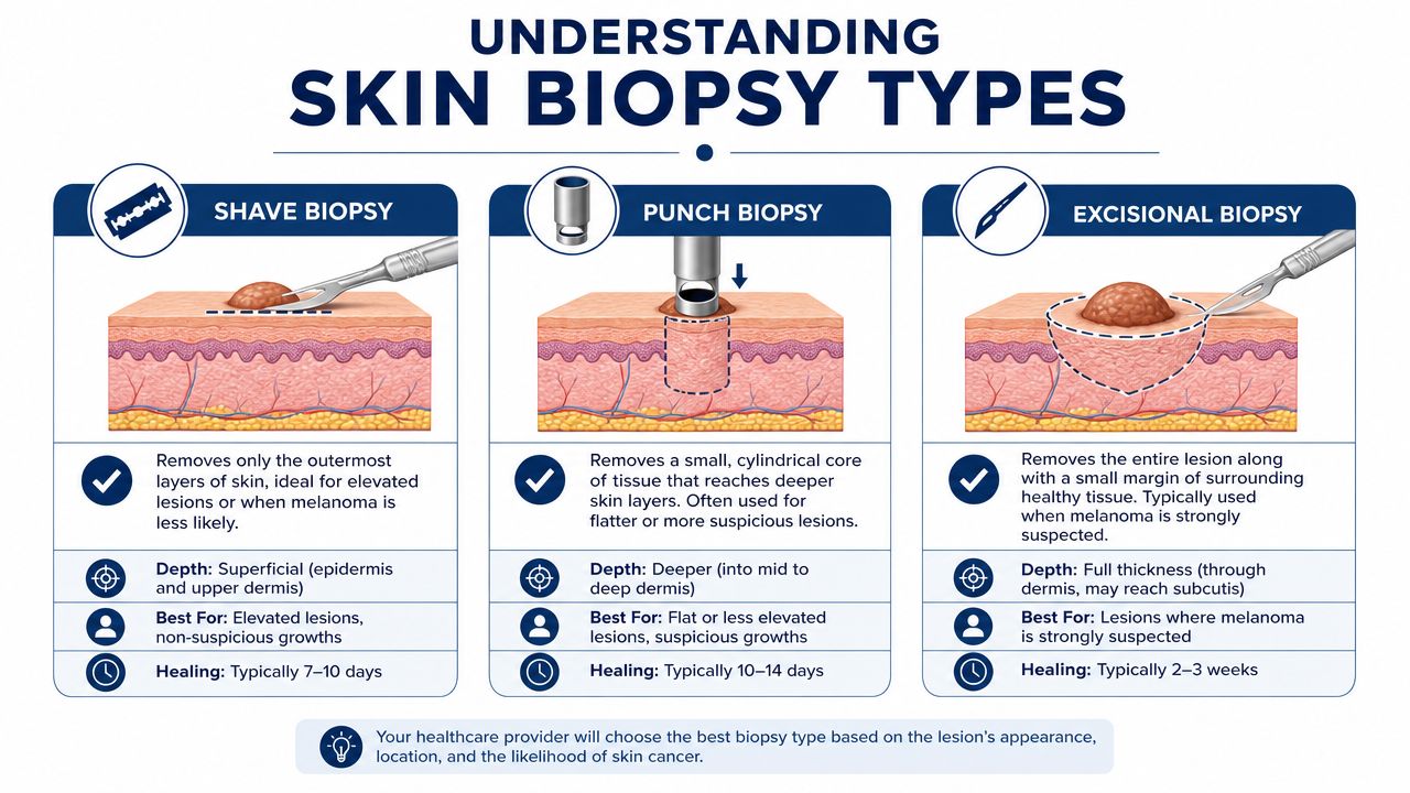
Shave biopsy
A shave biopsy removes the upper portion of the skin lesion. It can be useful for some raised or clearly superficial spots. It is quick, and in many everyday dermatology settings it's a familiar office procedure.
The problem is that a superficial shave may not include the full base of a melanoma. If the deepest part of the lesion isn't present in the sample, the pathologist may not be able to measure Breslow thickness reliably. That can limit staging accuracy.
Punch biopsy
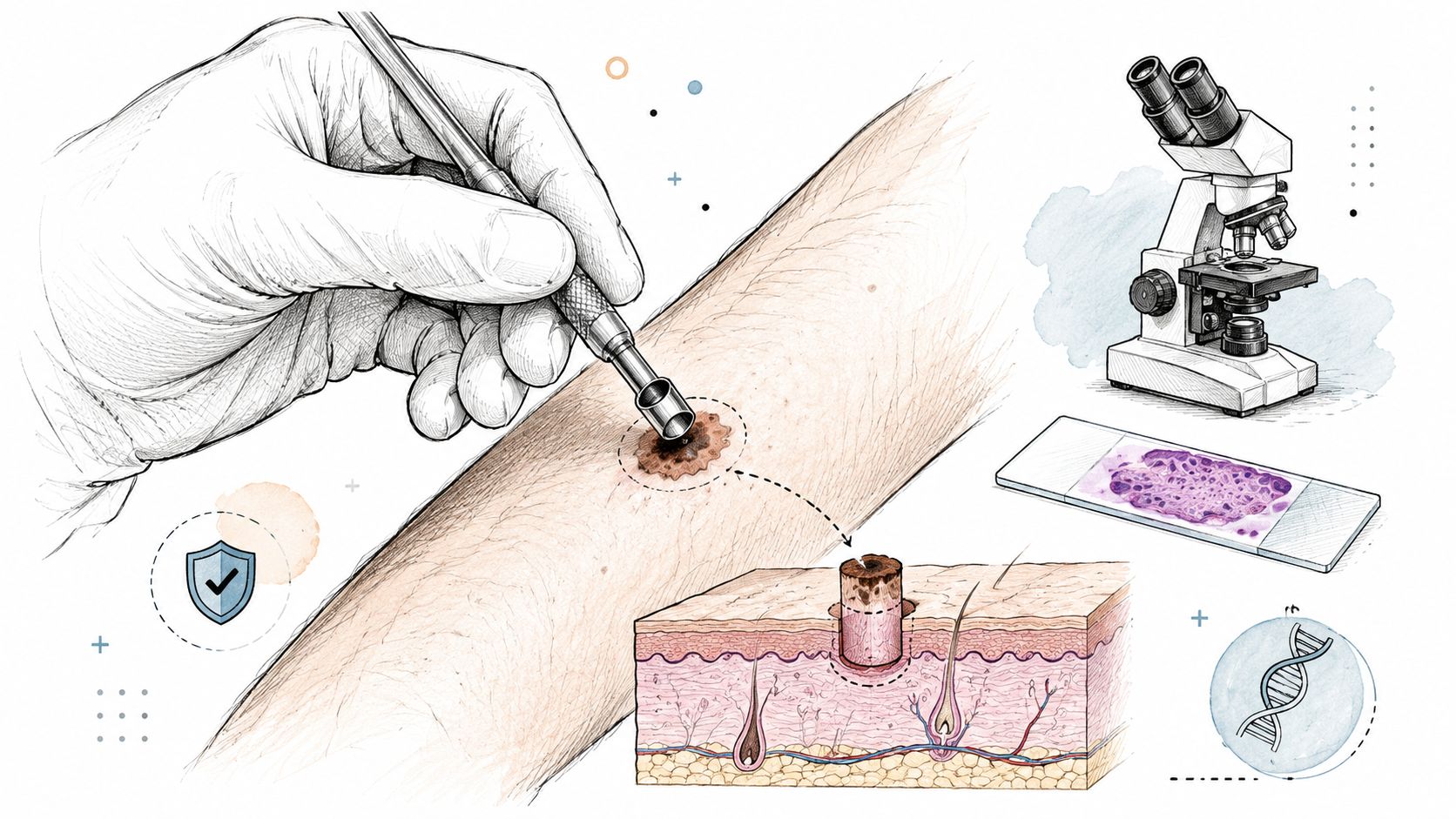
A punch biopsy uses a circular tool to remove a small core of skin. Unlike a superficial shave, it reaches deeper layers. That makes it more informative when tissue depth matters.
Still, a punch biopsy samples only part of the lesion unless the lesion is small enough to be fully removed by the punch. If the sampled area misses the deepest or most diagnostically important region, the pathology report may be incomplete for staging purposes.
Excisional biopsy
An excisional biopsy removes the entire suspicious lesion with a small rim of normal skin. For suspected melanoma, this is generally the highest-value specimen because it gives the pathologist the full lesion and its full depth.
Literature-based guidance recommends an excisional, full-thickness biopsy with about 2 mm lateral clearance and a cuff of subcutaneous fat beneath the tumor, as outlined in this review of melanoma biopsy practice. That specimen gives the pathologist the best chance to assess whether the base is fully seen and to measure Breslow thickness correctly.
A good biopsy for melanoma is not just “enough to tell what it is.” It should also be enough to tell how deep it goes.
Comparison of skin biopsy types for melanoma
| Biopsy Type | Procedure | Best For | Diagnostic Value for Melanoma |
|---|---|---|---|
| Shave biopsy | Removes the top layer or raised portion of a lesion | Some superficial or raised lesions when melanoma is less likely | Limited if the sample does not include the full depth |
| Punch biopsy | Removes a cylindrical core through deeper skin layers | Sampling part of a suspicious flat or deeper lesion | Better depth information than a superficial shave, but may miss the deepest area if only part of the lesion is sampled |
| Excisional biopsy | Removes the entire lesion with a narrow margin | Lesions where melanoma is suspected | Highest value for diagnosis and staging because full thickness can be assessed |
Why doctors choose differently
Patients often assume the least invasive method is always the best method. In melanoma workup, that isn't necessarily true. The “best” biopsy is the one that gives the pathologist the tissue needed to answer the right questions.
If you want a second plain-language explanation of how a core sample differs from other biopsy methods, this overview of skin punch biopsy results may help clarify what information each specimen can and cannot provide.
What to Expect Before During and After Your Biopsy
Most skin biopsies are straightforward office procedures. The unknown is often worse than the procedure itself. When patients know the sequence, anxiety usually drops.
Before the biopsy
At the visit, your clinician examines the lesion, explains why biopsy is recommended, and chooses the method based on the location, size, and level of concern. This is the right time to mention medications, bleeding history, allergies, and whether you've had trouble with local anesthetics or wound healing before.
Helpful questions include:
- What type of biopsy are you recommending and why that method fits this lesion
- Will stitches be needed and what kind of scar to expect
- How should I care for the site afterward
- When and how will results be discussed
Some patients also ask about numbing options before the injection. General patient resources such as this numbing cream guide for Canada can help you understand topical anesthetic products, though your own clinician should tell you whether any pre-procedure product is appropriate for your skin and biopsy plan.
During the biopsy
The area is cleaned first. Then local anesthetic is injected. That brief sting or burn is usually the part people notice most. Once the skin is numb, you may feel pressure or movement, but you shouldn't feel sharp pain.
The lesion is then removed by shave, punch, or excision, depending on the plan. If the sample is deeper or larger, stitches may be placed. A dressing goes on at the end.
This short video gives a simple overview of the office experience.
After the biopsy
Mild soreness, a small amount of spotting on the bandage, and tenderness around the site can be normal. Your clinician will usually give wound care instructions that focus on keeping the area clean, protected, and moist while it heals.
Keep the aftercare sheet. Most problems after biopsy are not emergencies. They're routine healing questions that are easiest to answer if you can compare what you see with the instructions you were given.
If stitches are present, movement that pulls on the area may need to be limited until removal or follow-up. If the site becomes increasingly red, more painful, or begins draining concerning material, contact the treating office.
Decoding Your Pathology Report
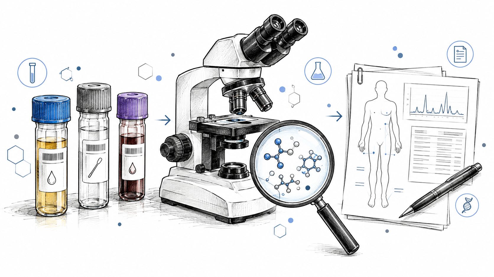
Once the biopsy is done, the tissue goes to a pathologist. This physician examines the specimen under the microscope and issues the report that guides what happens next. For patients, that report can feel like a different language.
The first thing to know is that pathology is highly structured. The report is not a vague opinion. It is a careful description of what is present in the tissue and, for melanoma, how the lesion behaves biologically and anatomically.
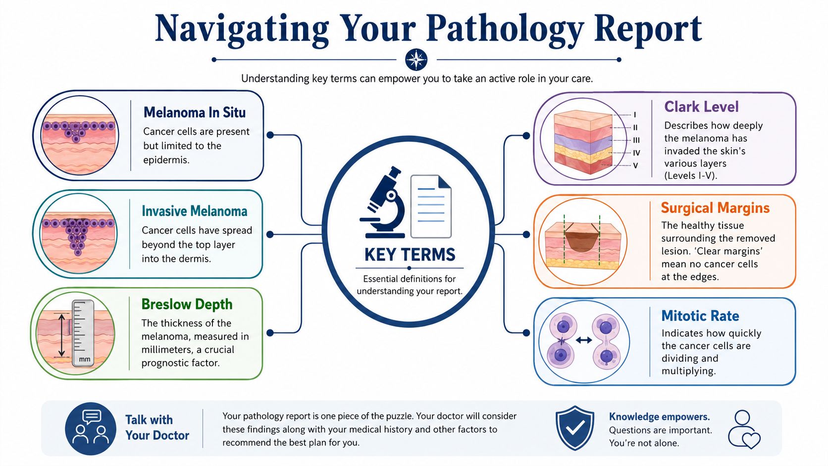
Terms that matter most
Here are the words that usually carry the most weight:
- Melanoma in situ means abnormal melanocytic cells are confined to the top layer of skin. They have not invaded deeper tissue.
- Invasive melanoma means the melanoma extends beyond the top layer into deeper skin.
- Breslow thickness is the measured depth of the melanoma. This is one of the most important pieces of staging information.
- Margins describe whether melanoma extends to the edge of the tissue removed.
- Ulceration refers to whether the skin over the melanoma is broken.
- Mitotic rate reflects how actively tumor cells are dividing.
Why mitotic rate is counted in a standardized way
Mitotic rate can sound abstract, but it is a microscopic count of dividing cells. The key point is that pathologists use a standardized process rather than an eyeball estimate.
The College of American Pathologists directs pathologists to identify the dermal “hot spot” of mitotic activity and count mitoses across adjacent non-overlapping high-power fields until 1 mm² has been assessed, according to the CAP melanoma biopsy protocol. That standardization improves reproducibility and helps convert what is seen on the slide into clinically useful risk stratification.
Why this matters: When the specimen is well taken and well processed, the pathology report becomes much more than a yes-or-no cancer test. It becomes a map for the next treatment decision.
Reading the report without getting overwhelmed
Many reports include technical details beyond what a patient needs to memorize. You do not need to master every line. Focus first on the diagnosis, the depth, and whether the lesion appears fully evaluated in the sample.
If the wording feels dense, a plain-language guide to a biopsy pathology report can help you sort the core findings from the background terminology.
After the Diagnosis Navigating Next Steps
The next step depends entirely on what the biopsy shows.
If the lesion is benign, that is often the end of the immediate problem. The site heals, the pathology is filed in your record, and your doctor may advise continued skin checks, sun protection, and monitoring of other moles over time. Relief is common, but it's still worth paying attention to your skin going forward, especially if you have many atypical moles or a personal history of prior biopsies.
If the report shows melanoma in situ or invasive melanoma, the biopsy is usually the beginning of treatment planning rather than the end of the process. Patients are commonly referred to a dermatologist with surgical expertise, a dermatologic surgeon, or another specialist involved in melanoma care. In many cases, a wider excision is recommended to remove additional surrounding tissue after the diagnosis is confirmed.
That can be confusing for patients who thought the original biopsy “already took it off.” Sometimes it did remove the visible lesion. Even so, treatment planning often requires a formal wider procedure because the goal is not just visible removal. It is complete removal with appropriate margins based on the diagnosis and pathology findings.
A second opinion can also be reasonable in selected cases, especially when the report language is borderline, the biopsy was partial, or the clinical appearance and pathology do not seem to match. In that setting, one option is independent slide review by another pathologist. Texas Autopsy Services publishes educational material that helps readers understand pathology terminology, which can be useful when patients and families are trying to make sense of what they've been told.
Frequently Asked Questions About Skin Biopsies
Does a skin biopsy hurt
Many patients worry most about pain, and that makes sense. In practice, the brief numbing injection is usually the sharpest part. Once the skin is anesthetized, you will usually feel pressure, movement, or a light tugging sensation rather than pain.
Will I have a scar
Yes. Any biopsy that removes skin leaves some kind of mark.
The size and shape of that mark depend on how the sample was taken. A shave biopsy often leaves a flatter, rounder spot. A punch biopsy leaves a small circular scar. An excisional biopsy usually leaves a line, because the doctor removes the full lesion with a margin and then closes the skin. Body location, skin type, and your own healing pattern also affect the final appearance.
Can cutting into a melanoma make it spread
This is one of the most common fears I hear. A biopsy does not cause melanoma to spread. It gives the pathologist the tissue needed to confirm whether melanoma is present and equally to measure features such as Breslow depth when the sample includes the full thickness of the lesion. That information helps determine staging and guides what happens next.
Are newer noninvasive tests replacing biopsy
For diagnosis, biopsy remains the gold standard over newer noninvasive tests. Tools such as imaging or adhesive patch testing may help decide which spot deserves a closer look first, but they do not replace examining actual tissue under the microscope. That direct tissue sample is what lets the pathologist assess architecture, cell behavior, and, in the right biopsy, the depth of invasion that matters for melanoma staging, as explained in this overview of melanoma diagnosis tools.
What if my report is hard to understand
That is very common. Pathology reports are written for medical decision-making, so the language can feel technical and impersonal even when the result is straightforward.
Ask your clinician to go through it line by line. It often helps to focus on a few key questions: Was this benign, atypical, melanoma in situ, or invasive melanoma? Was the whole lesion sampled? Does the biopsy type allow the pathologist to judge depth confidently, or could a partial sample limit that answer? Those details connect the procedure itself to the meaning of the report.
If the wording still does not make sense, a pathology review or second opinion may help clarify the diagnosis and what it means for treatment.
If you're trying to understand a pathology finding after a skin biopsy, or you need help making sense of a medical report in the setting of a death investigation or second-opinion review, Texas Autopsy Services may be a useful starting point for educational guidance and forensic pathology context. Patients and families deserve clear explanations, careful language, and honest answers, especially when the medical details feel difficult to sort through alone.