May 26, 2026 · Texas Autopsy Services
Skin Punch Biopsy Results Explained by a Pathologist
Understand your skin punch biopsy results. A pathologist explains common diagnoses, what the terms mean, and what to do next with your report.
On this page
- What a Pathologist Sees in Your Biopsy Sample
- Decoding the Language of Your Pathology Report
- Understanding Diagnoses Beyond Skin Cancer
- The Journey of Your Biopsy Why Results Take Time
- A Forensic Pathologist's Perspective on Skin Biopsies
- When to Question a Report and Seek a Second Opinion
- Frequently Asked Questions About Biopsy Results
A skin punch biopsy often feels routine on the front end. A dermatologist removes a small piece of skin, sends it to the lab, and the patient waits for answers. The waiting is usually the hard part, especially when the report arrives filled with terms that sound technical, impersonal, and difficult to interpret.
At Texas Autopsy Services, every examination is performed by a board-certified forensic pathologist experienced in medicolegal death investigations. Our work centers on one principle: follow the evidence carefully, explain it plainly, and treat the deceased and their family with respect.
TL;DR
- A punch biopsy samples skin through multiple layers, which gives the pathologist more useful architecture than a surface-only sample can provide.
- Skin punch biopsy results are not instant. Results are often available in 1 to 2 weeks in some settings, but the full process can take 3 to 4 weeks depending on lab work and specialized review, as summarized by MedlinePlus on skin biopsy testing.
- An abnormal result does not automatically mean cancer. Punch biopsies can diagnose inflammatory disease, infection, and malignancy.
- A benign or normal result is not always the end of the story if the lesion still looks suspicious clinically.
- In forensic pathology, skin biopsies can help answer focused questions about injury, infection, and lesion characterization during a cause of death investigation.
What a Pathologist Sees in Your Biopsy Sample
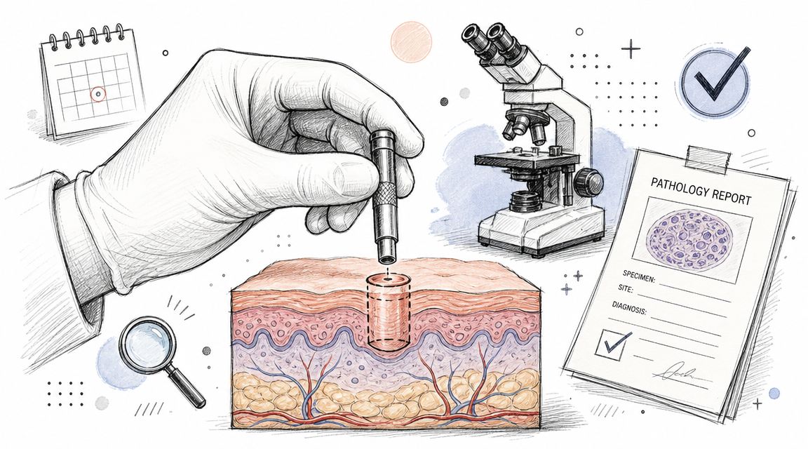
A punch biopsy is useful because it gives me more than cells. It gives me structure. The sample is a cylindrical core that can include the epidermis, dermis, and subcutaneous tissue, which is why it remains a standard method when a full-thickness specimen is needed for diagnosis and treatment planning, as described by the Canadian Cancer Society's explanation of punch biopsy.
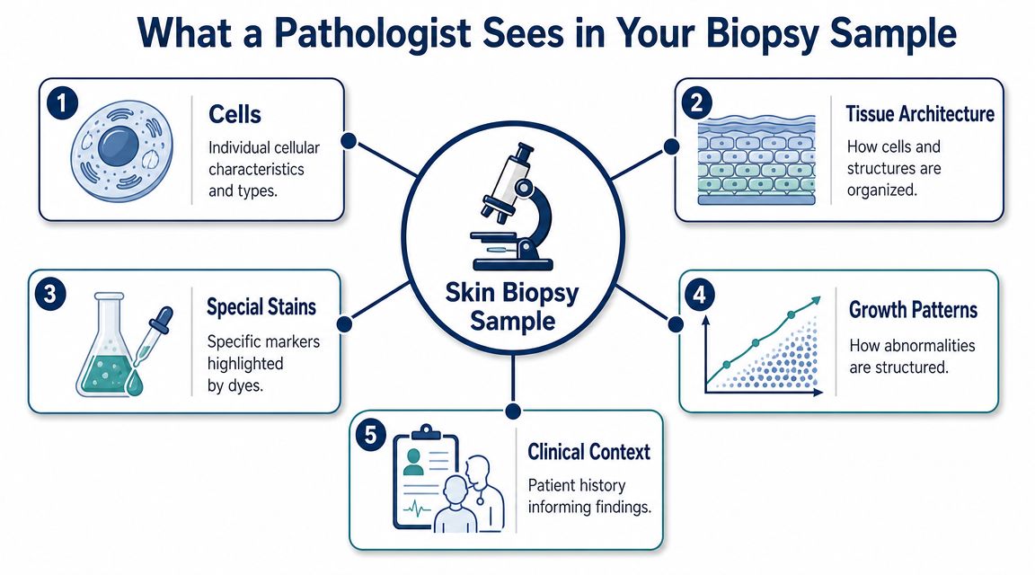
When I examine a punch biopsy, I'm not only asking what the cells look like. I'm asking how the layers relate to each other, where inflammation sits, whether an infection tracks deep, and whether an atypical process is confined or invasive. That architectural view is often what turns a vague rash or suspicious lesion into a workable diagnosis.
Why depth matters
A superficial sample can miss the key finding. A punch biopsy is designed to avoid that by taking a core through the skin layers. In routine dermatopathology, that matters for inflammatory eruptions, infection, and skin cancer because the answer may depend on the relationship between the surface and the deeper tissue.
There is also a practical sizing issue. A 4 mm punch is generally sufficient for many lesions, while 5 mm or larger is preferred when deep inflammation or architectural patterns are important, and very small biopsies risk missing diagnostic features, as outlined in the dermatology review at PMC on punch biopsy technique and adequacy.
Practical rule: A technically perfect biopsy from the wrong spot can still produce a limited answer.
What I'm evaluating under the microscope
A pathologist usually assesses several layers of information at once:
- Cell type and cell behavior. Are the cells mature, inflamed, infected, atypical, or malignant-looking?
- Tissue architecture. Do the epidermis, dermis, and deeper tissue show a pattern that fits psoriasis, eczema, infection, or neoplasia?
- Distribution of the process. Is the abnormality focal, diffuse, superficial, deep, or centered around hair follicles, blood vessels, or the dermal-epidermal junction?
- Specimen quality. Was the tissue crushed, fragmented, or too limited to answer the clinical question cleanly?
In real practice, communication matters too. Many clinicians now use tools that reduce transcription burden and preserve the details of lesion location, duration, and morphology. For physicians interested in cleaner documentation workflows, voice-to-text for medical practitioners is worth understanding because the quality of the clinical history can materially shape the usefulness of the pathology interpretation.
Decoding the Language of Your Pathology Report
Most pathology reports are written for clinicians first. Patients and attorneys often read them second, and that's where confusion starts. The report may be accurate yet still feel opaque because it follows laboratory conventions rather than ordinary speech.
I encourage people to read the report in layers. Don't jump straight to one alarming word. Read the specimen description, the microscopic findings, and the final diagnosis together. If you want a broader framework for pathology documents in general, Texas Autopsy Services has a separate guide on how to read a pathology report.
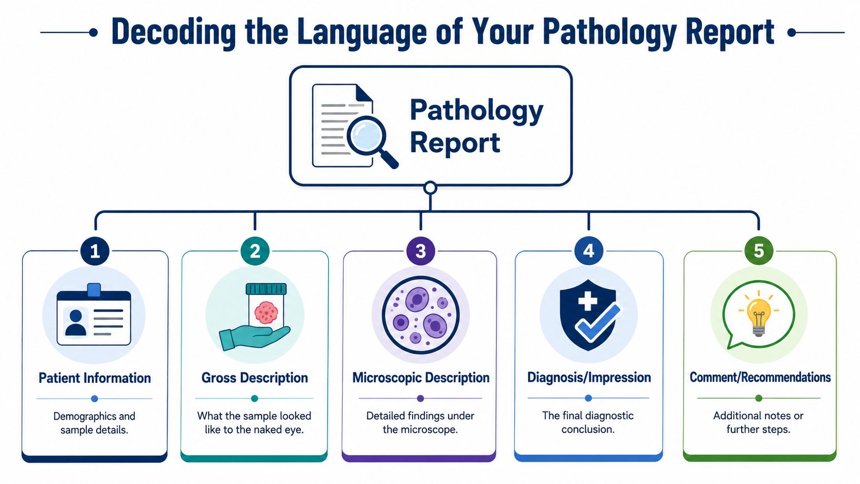
The main parts of the report
Most skin punch biopsy reports include familiar sections, even if the wording varies by laboratory.
| Section | What it usually means |
|---|---|
| Patient information | Confirms whose specimen was examined and what site was submitted |
| Gross description | What the tissue looked like before microscopic processing |
| Microscopic description | The observed histologic pattern under the microscope |
| Diagnosis | The final interpretive conclusion |
| Comment | Added context, limitations, or clinicopathologic correlation |
Terms that often worry people
Some words sound worse than they are. Others sound mild but require close follow-up.
- Lesion means an area of abnormal tissue. It does not by itself mean cancer.
- Margins refer to whether the sampled edges contain the process being described. In a small punch, that may or may not answer whether the whole lesion is removed.
- Inflammatory infiltrate means inflammatory cells are present in the tissue.
- Atypia means abnormal features are present. It is not identical to malignancy, but it may be significant depending on the context.
A report is strongest when the microscope findings and the clinical appearance make sense together.
Categories people look for
Readers often want the result sorted into plain language. That's reasonable. In broad terms, reports may point toward:
- Benign findings such as a non-cancerous inflammatory or reactive process
- Abnormal but not clearly malignant findings where the wording may be descriptive and further clinical correlation matters
- Malignant findings such as basal cell carcinoma, squamous cell carcinoma, or melanoma
Those categories help, but they don't replace interpretation by the treating clinician. A descriptive report can still be very useful when it narrows the field and guides the next step.
Understanding Diagnoses Beyond Skin Cancer
A patient comes in worried about melanoma. The biopsy comes back with no cancer at all, yet the report is still full of unfamiliar terms. That situation is common in practice. A skin punch biopsy often answers a different question than the one the patient expected.
Many punch biopsies are performed to sort out rashes, drug reactions, infections, autoimmune disease, and other inflammatory processes. In those cases, the pathologist may identify a histologic pattern before naming one final disease. That is careful reporting, not uncertainty for its own sake.
Cleveland Clinic explains that a skin biopsy can help diagnose conditions beyond cancer, including inflammatory disorders and infections, and that the result may reflect only the portion of tissue sampled in the punch, as described in Cleveland Clinic's skin biopsy overview.
When abnormal does not mean malignant
An abnormal result means the tissue is not normal. It does not automatically mean cancer.
In dermatopathology, pattern-based diagnoses are common because the skin reacts in a limited number of ways to many different injuries. Eczema, a medication eruption, allergic contact dermatitis, and an arthropod reaction can overlap under the microscope. A report may therefore describe the pattern with discipline and leave room for clinical correlation.
That same principle matters in forensic pathology. After death, skin can still answer focused questions, but only if the limits of the sample are respected. A post-mortem skin biopsy may help distinguish true inflammation from artifact, document a blistering disorder, or assess whether a lesion reflects prior disease rather than trauma. The microscope remains useful. The context changes.
How to read pattern-based language
When I sign out an inflammatory skin biopsy, I am translating structure into probability. Some terms sound imprecise to patients because they are not everyday clinical language. To a pathologist, they are specific descriptions of where the injury sits and how the skin is reacting.
A few examples:
- Spongiotic dermatitis describes intercellular edema in the epidermis, a pattern often seen in eczematous conditions.
- Interface dermatitis means injury is centered at the junction of the epidermis and dermis. Lupus, a drug reaction, viral eruptions, and other entities can produce that pattern.
- Psoriasiform hyperplasia refers to a patterned thickening of the epidermis that may support psoriasis but is not exclusive to it.
- Nonspecific inflammation means real abnormality is present, but the sample does not contain enough distinctive features for one confident label.
A good pathology report sometimes narrows the field instead of forcing a diagnosis the slide cannot support.
That distinction matters clinically. It matters in forensic work as well. In a living patient, a broad but accurate diagnosis may guide treatment, repeat sampling, or referral. In a death investigation, the same honest restraint can prevent overcalling trauma, infection, or neglect when the tissue does not prove that conclusion.
What makes these results more or less useful
Biopsy interpretation depends heavily on where the punch was taken and how the lesion was evolving at that moment. An early rash, an excoriated plaque, and a partially treated eruption may all look different under the microscope even if they belong to the same disease process. The report has to account for that trade-off.
The most useful result usually comes from a representative site chosen with a specific question in mind. For suspected vasculitis, blistering disease, connective tissue disease, or infection, site selection can change what the pathologist can reasonably say. If timing, sampling, or clinicopathologic fit is off, another biopsy may be the right next step, not a sign that the first one was mishandled.
Patients often ask why the report cannot always give a single plain-language answer immediately. Part of the reason is that skin pathology is pattern recognition tied to history, distribution, medications, and gross appearance. Part of the reason is laboratory workflow, which you can review in this guide to how long a pathology report takes.
The practical takeaway is simple. A punch biopsy can diagnose much more than skin cancer, but some of its most valuable results are narrower and more technical. They help the clinician decide what fits, what does not, and whether the next step is treatment, added testing, or a second look at the tissue.
The Journey of Your Biopsy Why Results Take Time
A patient has a punch biopsy on Tuesday and expects an answer by Friday. From the bedside, that expectation makes sense. The sample is small. Under the microscope, small does not mean simple.
Before I can sign out a skin biopsy, the tissue has to be preserved, processed, cut, stained, matched to the clinical question, and reviewed for patterns that may be subtle or incomplete on the first slides. In some cases, the diagnosis is straightforward. In others, the first pass raises a narrower and more technical question that needs additional work before the report is safe to release.
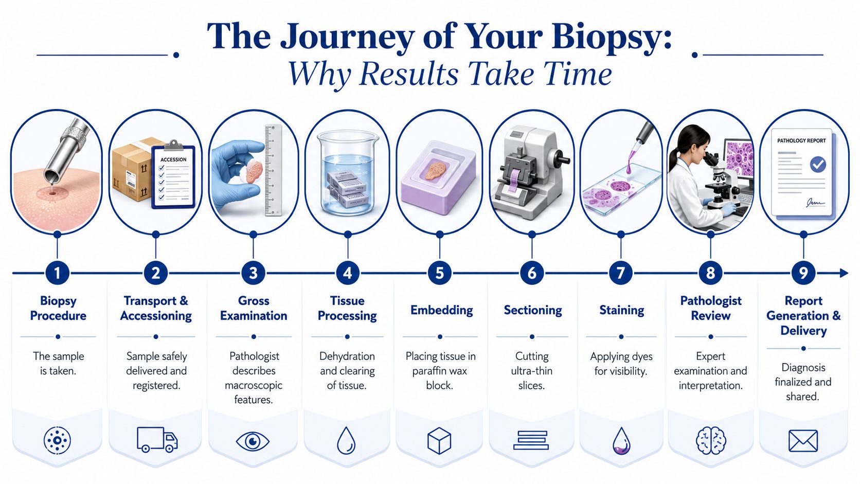
What happens after the specimen leaves the clinic
The biopsy enters a lab workflow that is methodical for a reason.
- Fixation preserves the tissue and limits post-procedure breakdown.
- Embedding places the specimen in paraffin so it can be cut in a controlled way.
- Sectioning produces very thin slices for glass slides.
- Staining highlights cells, connective tissue, organisms, pigment, and other diagnostic features.
- Microscopic review is where the pathologist decides whether the findings answer the clinical question or whether more testing is needed.
Yale Medicine explains this process in practical terms and points out a detail clinicians know well. Handling during removal matters. Crush artifact can distort the specimen and make interpretation harder, which is why careful technique during collection is part of getting a usable result, as described in Yale Medicine's discussion of skin biopsy processing and technique.
Patients who want more context on laboratory turnaround can review this explanation of how long a pathology report takes.
A brief visual overview can help make the workflow easier to follow.
Why the timeline varies
Some skin biopsies can be signed out quickly. Others need deeper levels, special stains, immunofluorescence, or correlation with photographs, medications, distribution, and prior pathology. A tiny core can generate a long differential diagnosis.
I see the same principle in forensic work. A small skin sample collected after death may look limited to a non-pathologist, yet the processing standards are still exacting because the question can carry medical, legal, or investigative weight. Clinical biopsies and forensic biopsies differ in purpose, but both depend on careful preparation and disciplined interpretation.
Waiting is frustrating. In pathology, that interval usually reflects active work in the lab, not delay for its own sake.
A Forensic Pathologist's Perspective on Skin Biopsies
Most punch biopsies are performed on living patients in dermatology or primary care settings. The same tool can also matter after death, which surprises many people. In forensic pathology, small samples of skin can answer focused questions that the external exam alone cannot settle.
That overlap between clinical pathology and forensic pathology is one reason I value precise tissue interpretation. If you want a broader overview of the profession itself, this explanation of what a pathologist does gives helpful context.
Where punch biopsies fit after death
A postmortem skin biopsy is not a routine answer to every question. It is a targeted tool. I use that kind of sampling when the histology may help clarify what a mark, lesion, or suspicious area represents.
Examples include:
- Possible injury sites where microscopic examination may help characterize tissue reaction
- Suspected infection when skin findings may connect to a broader disease process
- Uncertain lesions where the appearance alone does not resolve whether the process is inflammatory, infectious, or neoplastic
Why forensic context changes the interpretation
In a clinic, the question is often “What diagnosis explains this lesion, and what treatment follows?” In a death investigation, the question may be different. I may need to know whether a finding is incidental, contributed to death, reflects prior medical care, or supports or contradicts the history provided.
That is where forensic discipline matters. Cause of death means the disease or injury that started the lethal sequence. Manner of death is the classification of how the death occurred, such as natural, accident, suicide, homicide, or undetermined. Chain of custody means documented control of evidence from collection through analysis so the integrity of the material can be defended.
For families seeking more clarity after a death, a skin finding may be one part of a larger review that includes records, toxicology, and full postmortem examination. In some circumstances, that may include a private autopsy in Texas.
In forensic work, the tissue slide is rarely interpreted alone. It has to fit the scene, the history, the autopsy findings, and the medical records.
When to Question a Report and Seek a Second Opinion
A pathology report carries real weight, but it is not immune to the limits of sampling. A punch biopsy examines the tissue that was submitted, not necessarily the entire biology of a larger or uneven lesion. That distinction matters when the report says one thing and the clinical appearance suggests another.
Mayo Clinic's patient guidance underscores an important point. A normal result does not always end the workup if the lesion remains suspicious, because sampling limitations can create false reassurance and may justify repeat biopsy or second review, as noted in Mayo Clinic's skin biopsy overview.
Situations where questions are reasonable
I encourage patients, attorneys, and clinicians to slow down and ask for clarification when any of the following occur:
- The lesion still looks concerning even though the report sounds benign
- The pathology wording is descriptive but noncommittal, and the clinical stakes are high
- The sample was very small or from a difficult site, where representativeness may be limited
- The lesion changes after the biopsy rather than settling or resolving
None of that means the first report was wrong. It means the report answered the question the submitted tissue allowed it to answer.
What a productive second opinion looks like
A useful second opinion is not just “send the slides somewhere else and hope.” It works best when the reviewing pathologist has the pathology material, the clinician's description, lesion photographs if available, and the reason the original interpretation does not fit the clinical picture.
For families and attorneys in the postmortem setting, the same logic applies. A focused record review or independent postmortem consultation can be appropriate when the pathology, clinical course, and final conclusions do not align cleanly. One option in that setting is Texas Autopsy Services, which provides independent autopsy and forensic pathology review in Texas as part of a broader cause of death investigation.
What does not help
What does not help is treating a reassuring report as untouchable when the clinical appearance remains discordant. The reverse is also true. A frightening phrase in a report should not be interpreted without the clinician who knows the lesion, the patient, and the reason the biopsy was done.
The most reliable answers come from clinical-pathologic correlation. That phrase can sound abstract, but it means the microscope findings must make sense for the patient.
Frequently Asked Questions About Biopsy Results
A common scene in practice goes like this. The biopsy site is healing, the report has posted to the patient portal, and one phrase in the diagnosis line becomes the entire focus of the conversation. Patients want plain language. Clinicians want to know whether the microscopy fits the lesion they saw. In forensic review, families and attorneys often ask a related question: what can this small piece of skin prove, and what can it never prove?
Those are different settings, but the core issue is the same. A skin punch biopsy gives a pathologist a limited tissue sample, and the value of the report depends on how well the microscopic findings fit the clinical or investigative context.
FAQ on Skin Biopsy Results
| Question | Answer |
|---|---|
| How long do skin punch biopsy results take? | Many routine cases are reported within several business days to two weeks. Results can take longer when the tissue needs deeper sections, special stains, immunohistochemistry, outside consultation, or correlation with added clinical information. |
| Does an abnormal result mean cancer? | No. Many abnormal reports describe inflammation, infection, scarring, drug reaction, or another non-malignant process. The diagnosis line has to be read together with the microscopic description and the clinical impression. |
| What does “spongiotic dermatitis” mean? | It describes a reaction pattern under the microscope, usually one seen in eczematous conditions. It is a histologic description, not always a final disease name by itself. |
| Can a benign or normal biopsy still miss something important? | Yes. A punch biopsy samples one part of a lesion. If the clinical appearance remains concerning, the next step may be another biopsy, a different biopsy site, or complete removal for fuller evaluation. |
| Can biopsy findings matter in a death investigation? | Yes. In selected forensic cases, skin histology helps sort out whether a lesion reflects disease, infection, treatment effect, artifact, or injury. The same slide-reading skills used in clinic become highly consequential after death, where the question may affect cause and manner of death conclusions. |
Practical next steps after a report
The next step depends on who is reading the report and why.
- For patients and families: Ask two specific questions. What is the diagnosis, and does it match what the lesion looked like on the skin?
- For attorneys: Obtain the full pathology report, including the microscopic description and comment section. The final diagnosis alone may leave out uncertainty, sampling limits, or differential considerations.
- For clinicians: Re-examine the site, review the differential diagnosis, and contact the pathologist if the report and the lesion do not fit each other.
- For postmortem reviewers: Separate observation from interpretation. Histology may support a conclusion, but the tissue cannot answer questions it was never suited to answer.
I tell clinicians this often: a pathology report is not the end of the case. It is a piece of the case.
If you're facing unanswered questions about pathology, skin findings, or a death investigation in Texas, careful review matters. Families, attorneys, and healthcare professionals can contact Texas Autopsy Services for respectful guidance on whether an independent forensic review, record analysis, or private autopsy is appropriate under Texas law.