May 16, 2026 · Texas Autopsy Services
Skin Lesion Biopsy Results: A Pathologist's Guide
A pathologist explains how to read your skin lesion biopsy results, what diagnoses like BCC or melanoma mean, and what questions to ask your doctor.
On this page
- The Wait Is Over Now What
- From Your Skin to My Microscope The Biopsy Process
- How to Read Your Pathology Report Section by Section
- Common Diagnoses Explained Benign Malignant and In-Between
- What Margins and Staging Numbers Mean for Treatment
- After the Report Questions for Your Doctor and Next Steps
- Advanced Considerations Second Opinions and Medico-Legal Use
When the phone rings and someone says your skin lesion biopsy results are back, patients often don't hear much after that. They hear one word, or think they might hear one word, and anxiety fills in the rest.
I understand that reaction. I'm our forensic pathology team, a board-certified pathologist. In my daily work, I write reports that patients later read through a screen, a portal message, or a printed sheet handed across an exam-room desk. By the time that report reaches you, it can feel cold, compressed, and harder to understand than it should be.
A pathology report is not written to frighten you. It's written to answer a specific medical question using tissue, microscope findings, and clinical context. Still, even a straightforward report can look cryptic. Terms like atypical, margin, in situ, or transected carry real meaning, and they deserve plain explanation.
That matters especially when a report is unclear, limited, or doesn't seem to match what you were told in the clinic. If that is your situation, my discussion of inconclusive biopsy results may also help frame why some reports leave room for follow-up rather than finality.
The Wait Is Over Now What
The moment after you receive skin lesion biopsy results is usually not dramatic. It's quieter than that. You read a few lines, search for the diagnosis, and then start scanning for words you recognize. Benign. Atypical. Carcinoma. Melanoma. Margins. Suddenly the report feels like a test you were never taught how to take.
Most patients approach the report as if the diagnosis line is the whole story. It isn't. That line matters most, but it rests on everything above it, including what tissue was sampled, where it was taken from, what your clinician suspected, and whether the specimen answered the question cleanly.
Why the wording feels so technical
Pathology language is compressed on purpose. The report has to communicate accurately with clinicians, surgeons, and other pathologists. That means it often favors precision over comfort.
A phrase like “consistent with” may reflect a strong fit between what was sampled and what I see under the microscope. A phrase like “cannot exclude” signals caution. It doesn't mean disaster. It means the tissue or the pattern leaves some uncertainty.
A pathology report is a medical interpretation of a sample, not a personality test for your lesion and not a prediction of your future.
What I want patients to know first
If you're reading your report alone, start with three grounding points:
- The biopsy was done to answer a question: Suspicion is not the same as diagnosis.
- The sample has limits: A report can only describe the tissue that was removed.
- The next step depends on both pathology and clinic findings: Your doctor and pathologist are supposed to connect those pieces.
That last point is easy to miss. The lesion on your skin has a clinical appearance. The tissue on my slide has a microscopic appearance. Good medicine happens when those two views agree, or when they don't agree and someone notices.
The emotional side is real
Even benign results can leave people unsettled. They may still wonder whether the biopsy missed something, whether the spot will come back, or whether another procedure is coming. Those concerns are reasonable.
I've learned that patients do better when the report is translated instead of merely delivered. The medical truth should stay precise, but the explanation should still sound human. That's what I'm aiming to give you here.
From Your Skin to My Microscope The Biopsy Process
A skin biopsy may take only a short visit in the clinic, but the tissue then begins a much longer path. By the time skin lesion biopsy results appear in your chart, several technical steps have already happened behind the scenes.
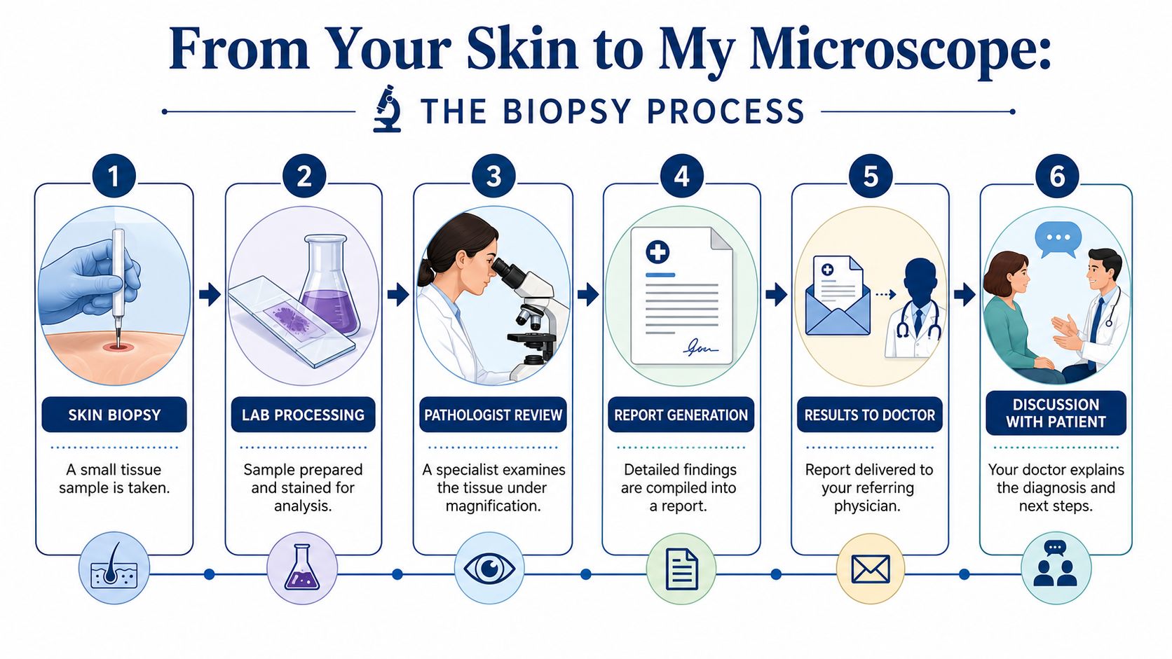
What happens after the biopsy
Once the clinician removes the tissue, the specimen is placed in a preservative so the cells and tissue structures don't degrade. In routine practice, that preservation step lets the lab process the sample without losing the architecture that makes diagnosis possible.
The specimen is then labeled, logged, and tracked. In pathology, and especially in forensic work, chain of custody means there is a documented path showing who handled the specimen and how it was identified. In a hospital or outpatient lab, the purpose is practical and essential. The right tissue must stay connected to the right patient from procedure room to final report.
How the lab turns tissue into slides
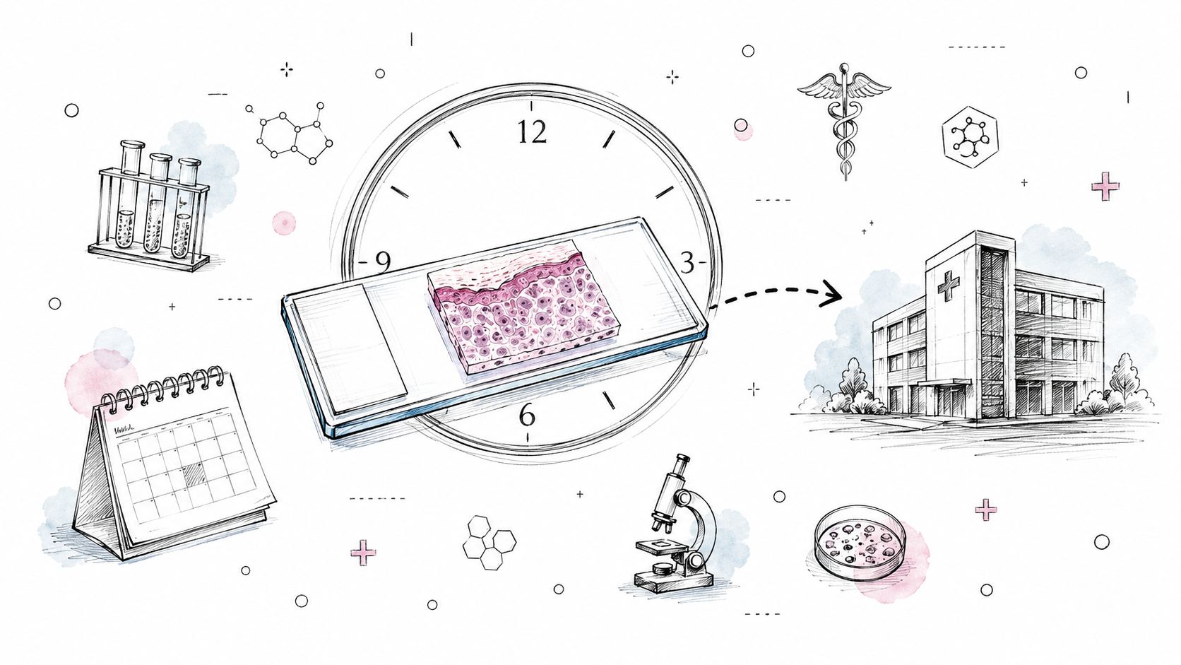
The preserved tissue is processed into a paraffin block, which is a wax block that supports the sample so it can be cut into very thin sections. Those sections are placed on glass slides and stained so nuclei, connective tissue, pigment, inflammation, and invasion patterns can be evaluated.
Here is the simple version of that workflow:
- Accessioning: The lab receives the specimen and confirms identifiers.
- Gross examination: A pathologist or pathology assistant describes the tissue by sight and touch.
- Processing: The tissue is dehydrated, embedded in wax, and prepared for cutting.
- Sectioning and staining: Thin slices go onto slides for microscopic review.
- Interpretation: A pathologist studies the slides and writes the diagnosis.
Why timing varies
Best practices indicate that most skin biopsy results return within a few days to a week, though special stains or other added studies can take longer, as summarized by the RACGP discussion of skin biopsy interpretation and timing. Waiting feels passive, but the delay usually reflects technical work, not neglect.
That also helps explain an important clinical reality. In a 2024 study of 12,852 skin biopsy specimens submitted with language explicitly referring to melanoma suspicion, 1,724 cases, or 13.41%, were histologically confirmed as melanoma, meaning most biopsies performed to rule out melanoma in that setting did not show melanoma, according to the retrospective study in SKIN The Journal of Cutaneous Medicine.
Practical rule: A biopsy is often a triage tool. It investigates suspicion. It does not mean the clinician already knows the lesion is cancer.
What the pathologist is really looking for
Under the microscope, I'm not just asking, “Is this cancer?” I'm also asking whether the sample is deep enough, broad enough, and representative enough to support a reliable conclusion. A technically perfect report can still be limited by a technically limited sample.
That distinction matters more than most patients are told.
How to Read Your Pathology Report Section by Section
The fastest way to make sense of skin lesion biopsy results is to stop treating the report as one block of text. It's a structured medical document. Each part has a job.
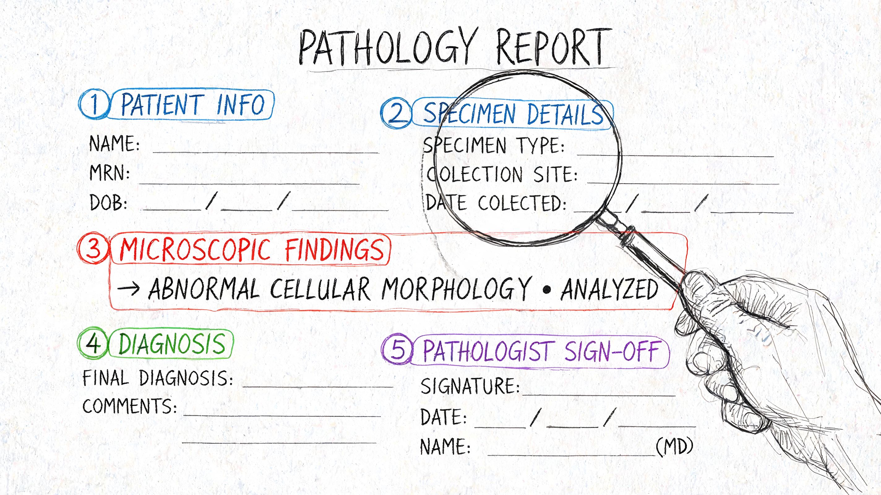
If you've never seen one explained line by line, my guide on how to read a pathology report gives a broader framework. For skin lesions, a few sections carry most of the practical weight.
Patient and specimen identifiers
The top portion usually lists your name, date of birth, specimen source, and often the body site. This is not filler. It confirms exactly what tissue was submitted.
A report that says “left shoulder shave biopsy” is answering a narrower question than one that says “right calf excision.” If you had more than one lesion sampled, make sure each diagnosis matches the correct site.
Clinical history or clinical impression
This part records what the clinician suspected or why the biopsy was done. It might say something brief like “rule out melanoma,” “pigmented lesion,” or “nonhealing papule.”
That line matters because pathology interpretation does not happen in isolation. The tissue findings need to be matched to the lesion's appearance and location, which is why clinical-pathologic correlation is standard practice. If the history is sparse, the report may still be diagnosable, but the context is thinner.
Gross description
The gross description is what the specimen looked like before it was turned into slides. It may include dimensions, color, whether the tissue was fragmented, and whether the specimen appeared oriented.
Patients often skip this section. Sometimes that's fine. Sometimes it contains clues that matter, especially if the sample was small, superficial, or submitted in pieces.
Microscopic description
At this point, the technical language usually becomes dense. The pathologist describes what the tissue architecture and cells are doing under magnification.
You might see words such as these:
| Term | Plain meaning |
|---|---|
| Atypia | Cells look abnormal in some way |
| Nests | Groups of similar cells clustered together |
| Maturation | Whether cells change in the expected way as they extend deeper |
| Inflammation | The immune system is reacting in the tissue |
| Ulceration | Surface loss or breakdown of skin |
| Transection | The lesion extends to the cut edge of the biopsy sample |
These terms are building blocks. They are not the final answer by themselves.
Final diagnosis
This is the line most patients search for first, and understandably so. It may be short or qualified. It may include whether the lesion is benign, malignant, dysplastic, inflamed, incompletely removed, or requires correlation with the clinical picture.
Read this section with the rest of the report in mind. A diagnosis of a benign lesion is reassuring, but if the sample was partial or the lesion remains clinically worrisome, the conversation may not be over.
The report is strongest when the microscope, the biopsy method, and the clinician's impression all point in the same direction.
Comment section and why it matters
Some reports include a comment that explains uncertainty, suggests re-excision, or notes a limitation. Don't treat that as optional language. In many skin cases, the comment contains the most practical guidance in the whole report.
For patients, a useful reading order is often this:
- Start with the diagnosis: Find the main conclusion first.
- Check the specimen site: Make sure you're reading the correct lesion.
- Look for comments on margins or limitations: These often determine next steps.
- Read the clinical history: It tells you what question the biopsy was meant to answer.
That sequence usually turns a confusing report into a manageable one.
Common Diagnoses Explained Benign Malignant and In-Between
The diagnosis line can carry very different levels of concern. Some lesions are harmless. Some are cancers that are usually managed effectively when identified. Others sit in a gray zone where the cells are not normal, but the report is not saying invasive cancer.
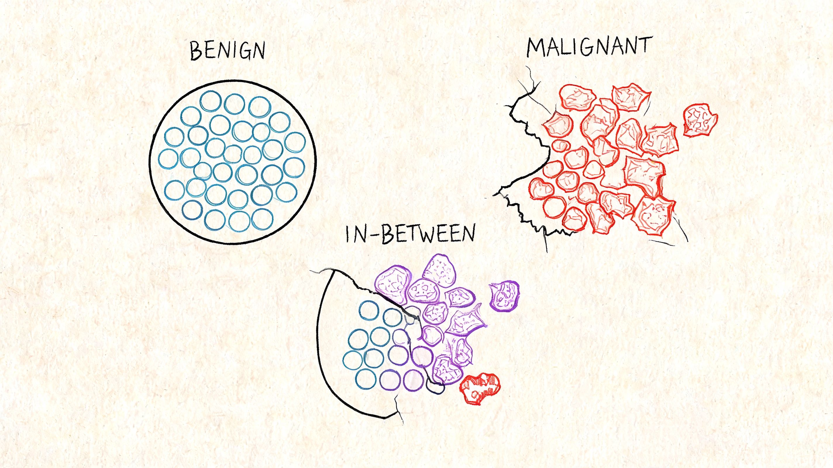
One reason biopsy is so common is that suspicious-looking lesions often turn out not to be melanoma. In a large analysis of about 80,368 skin biopsies, only 23% of diagnoses were melanocytic lesions, and among those melanocytic lesions 83% were Class I benign nevi and other benign proliferations, with smaller portions falling into higher-risk categories, as summarized by the University of Washington report on most mole biopsies being benign.
Benign diagnoses
A benign diagnosis means the lesion is not cancer. In practical terms, that often includes ordinary moles, harmless overgrowths, or reactive changes.
Benign does not always mean meaningless. A benign lesion may still have caused symptoms, looked irregular, or required removal because of location, irritation, or cosmetic concerns. It means the microscopic features do not support malignancy.
Common benign report language may include:
- Nevus: A mole.
- Seborrheic keratosis: A common noncancerous skin growth.
- Dermal nevus or compound nevus: Variants of mole location within skin layers.
- Lentigo or solar lentigo: A flat pigmented lesion often related to sun exposure.
The in-between category
Many patients face difficulty at this stage. Terms like dysplastic nevus, atypical melanocytic proliferation, or actinic keratosis sound ominous because they are describing abnormality, not normalcy.
A useful way to think about dysplasia is this: the cells are breaking some of the usual architectural rules, but they have not crossed the threshold for invasive cancer. That threshold matters. Medicine often lives in thresholds.
For some people, seeing “moderate atypia” on a report is more stressful than reading a clearly benign diagnosis because uncertainty is harder to sit with than a clean yes or no.
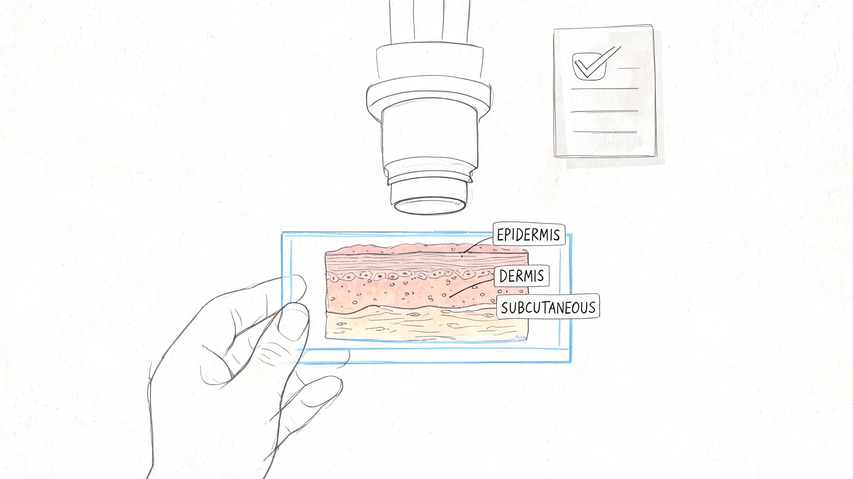
A brief visual explanation can help if these categories still feel abstract:
Malignant diagnoses
The three cancer terms many patients encounter are basal cell carcinoma, squamous cell carcinoma, and melanoma.
- Basal cell carcinoma: Usually managed by complete removal. It tends to matter most for local growth and tissue damage if untreated.
- Squamous cell carcinoma: Also generally requires complete removal and can raise additional concerns depending on depth and other features.
- Melanoma: This diagnosis carries greater staging implications, so details like depth become especially important.
If pathology shows basal cell carcinoma or squamous cell carcinoma, the next usual step is confirming complete removal. If it shows melanoma, additional surgery or staging workup may be needed, based on accepted clinical practice described in the earlier cited RACGP guidance.
Why wording can look cautious
You may see phrasing such as “favor,” “compatible with,” or “at least.” That wording does not mean the pathologist is guessing casually. It indicates that a partial tissue sample can show enough to raise concern without revealing the lesion's full extent.
That is one of the central truths of skin pathology. The tissue answers the question only as completely as the sample allows.
What Margins and Staging Numbers Mean for Treatment
After the diagnosis, most patients focus on one practical issue. Is this over, or do I need more treatment?
That question often turns on margins. In pathology, a margin is the edge of the removed tissue. If tumor extends to that edge, the lesion may not have been completely removed in the sample submitted.
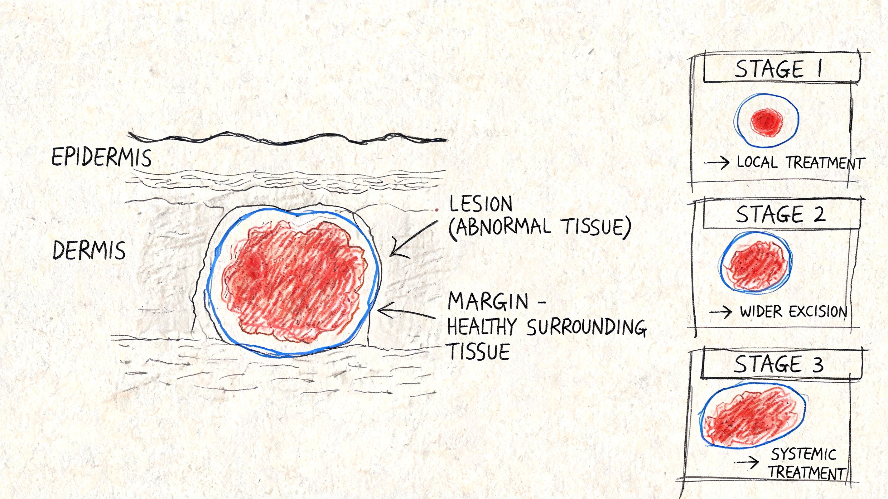
How to think about margins
A garden analogy works well here. If you pull a weed and the root is entirely out, you have a cleaner result. If part of the root remains in the soil, you may need to go back and remove more.
That's broadly how clear margins and involved margins function in skin lesion biopsy results.
| Margin wording | What it usually means |
|---|---|
| Clear / negative | The lesion does not appear to extend to the specimen edge examined |
| Positive / involved | The lesion reaches the cut edge, so residual lesion may remain |
| Close | The lesion comes near the edge, and management depends on diagnosis and context |
Why biopsy type matters so much
For suspected melanoma, the biopsy method directly affects what the pathologist can say with confidence. According to the NCBI StatPearls review on skin biopsy, excisional biopsy is preferred for suspected melanoma when size and location permit because it allows more accurate assessment of Breslow depth, while a partial punch or shave biopsy may not provide enough information for a complete diagnosis.
That is not just a technical preference. It changes treatment planning.
Breslow depth in plain language
Breslow depth measures how far into the skin melanoma cells extend. If margins tell you whether the lesion may still be present at the edges, Breslow depth helps show how far downward the melanoma has traveled.
Deeper is generally more consequential than shallower because depth is tied to staging and treatment decisions. A superficial sample can underestimate that depth if the base of the lesion was not fully captured.
A melanoma report can be accurate about the tissue on the slide and still incomplete about the whole lesion if the biopsy only sampled part of it.
Practical takeaways for treatment discussions
If your report mentions margins or depth, ask your doctor to tie those findings to the treatment plan. The pathology language becomes less intimidating when translated into action.
Focus on these points:
- Was the lesion completely removed in the biopsy sample
- If not, is a wider excision recommended
- If melanoma is present, was depth fully assessable
- Does the report show any limitation caused by the biopsy type
Those questions move the discussion from terminology to decisions.
After the Report Questions for Your Doctor and Next Steps
The most useful follow-up visit is the one where you arrive with specific questions. Patients often feel they should wait passively for the doctor's interpretation. I don't think that serves them well.
A pathology report is a technical document. Your job is not to decode it perfectly on your own. Your job is to leave the appointment understanding what was found, what remains uncertain, and what happens next.
Questions worth asking directly
Take the report with you and ask clear, concrete questions:
- What is the exact diagnosis in plain English: Ask your doctor to translate the diagnosis line without abbreviations.
- Was the sample complete enough to answer the question: This is especially important if the result sounds reassuring but the lesion looked highly suspicious.
- Do the pathology findings match what you saw on my skin: That question forces clinical-pathologic correlation.
- Are the margins clear, involved, or not applicable: This often determines whether another procedure is needed.
- Do I need more treatment or only follow-up: The management plan should be stated plainly.
- What changes should prompt me to come back sooner: A benign report does not mean you should ignore a lesion that keeps changing.
What not to do after reading the report
The least helpful response is to isolate one phrase and build the entire future around it. Words like atypical and moderate are not self-interpreting. They only become meaningful in the context of diagnosis, margin status, biopsy method, and exam findings.
Another mistake is assuming a benign result ends the issue even if the lesion continues to evolve. If the clinical picture and the pathology report don't fit together, the mismatch deserves attention.
A practical note for the appointment
Bring either a printed copy or a screenshot of the report. If there are multiple lesions, mark which one concerns you. If the procedure note is available, bring that too.
This is also the stage where another review can become useful. If a case is unusually complex, or if the findings are critical and the report language is limited, a slide review by another pathologist may be reasonable. In some settings, that can include consultation through an independent pathology or forensic practice. For postmortem pathology and related expert review, Texas Autopsy Services provides independent pathology-focused services, though clinical skin lesion management remains in the treating physician's domain.
The goal of the follow-up visit
You should leave with three things:
- A diagnosis you understand
- A treatment or surveillance plan
- An explanation of any remaining uncertainty
If one of those is missing, ask again. That isn't being difficult. That is good medical communication.
Advanced Considerations Second Opinions and Medico-Legal Use
Some reports are not cleanly final. A lesion may be called atypical, suggestive, or limited by sample. That can happen because biopsy accuracy is sometimes constrained by the tissue itself. Guidance from OHSU notes that false-negative or uninformative results can occur when the biopsy comes from a non-representative area, including crusted, scarred, or otherwise poor sampling sites, as discussed in the OHSU review of skin biopsy issues in specific diseases.
That is one reason second opinions on the slides themselves can be appropriate. In pathology, a second opinion does not imply wrongdoing. It recognizes that difficult cases benefit from another expert looking at the same tissue, the same levels, and the same wording.
A pathology report is also a legal medical record. In my forensic work, that matters. The report may later be reviewed in insurance disputes, malpractice review, disability claims, or litigation involving delayed diagnosis. If someone is trying to understand the legal side of suing for late skin cancer diagnosis, the key document is often the pathology itself, along with the clinical timeline surrounding it. In litigation settings, the standards for review are different from routine patient counseling, which is why a pathologist expert witness may be involved.
FAQ
How long do skin lesion biopsy results usually take?
Many results return within a few days to a week. Some take longer if special studies are needed.
Does a biopsy mean my doctor thinks I have cancer?
Not necessarily. A biopsy investigates a concern. Many suspicious lesions do not prove to be melanoma or another malignancy.
Can a skin biopsy miss part of the problem?
Yes. A partial or poorly targeted sample can be limited, especially in heterogeneous lesions or crusted areas.
Should I ask for a second opinion on the slides?
It can be reasonable when the diagnosis is atypical, inconclusive, high-stakes, or hard to reconcile with the clinical picture.
What does chain of custody mean in pathology?
It means the specimen is tracked and identified through handling, processing, and reporting so the tissue remains correctly linked to the patient.
If you're facing a pathology question that overlaps with forensic review, record interpretation, or the need for an independent medical explanation, I encourage you to contact Texas Autopsy Services. My work is grounded in careful documentation, clear communication, and respect for the seriousness of medical records when families, attorneys, and professionals need answers they can understand.