June 3, 2026 · Texas Autopsy Services
Pathology Report Melanoma: Explained for You
Understand your pathology report melanoma with expert guidance. Learn about Breslow depth, staging, and prognosis implications from our 2026 guide.
On this page
Pathology Report Melanoma Explained for You
- Your melanoma pathology report is the document that confirms the diagnosis and provides the details your treatment team uses to plan next steps.
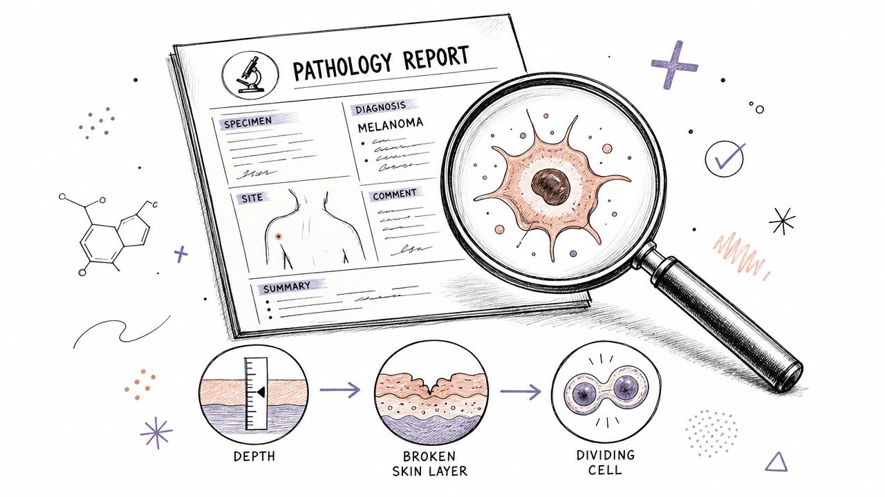
- The most important findings for a primary melanoma are Breslow thickness, ulceration, and mitotic rate.
- A pathology report usually tells the tumor part of staging best. It does not by itself determine whether melanoma is stage III or IV.
- Some report terms matter more than others. Clark level may still appear, but it is not used in current AJCC pT staging.
- A second pathology review can be reasonable when the diagnosis is borderline, the wording is unclear, or major treatment decisions depend on a small detail.
When people open a melanoma pathology report for the first time, they often see a page full of technical language and stop at the first unfamiliar word. That reaction is normal. Most families I speak with are not struggling because they are careless. They are struggling because pathology reports are written for clinicians first, while patients are expected to live with the consequences of every line.
At Texas Autopsy Services, every examination is performed by a board-certified forensic pathologist experienced in medicolegal death investigations. Our work centers on one principle: follow the evidence carefully, explain it plainly, and treat the deceased and their family with respect.
The Critical Role of Your Melanoma Pathology Report
The hardest stretch for many patients is the time between the biopsy and the report. A lesion has been removed. People know melanoma is serious. Then they are asked to wait while someone they may never meet studies the sample and writes the document that will shape the rest of care.
That document matters because melanoma remains a major cancer burden. The SEER Cancer Stat Facts page projects 112,000 new cases and 8,510 deaths in the United States in 2026, and estimates melanoma will account for 5.3% of all new cancer cases in the U.S. (SEER melanoma statistics). Those numbers help explain why pathology reports are treated so seriously in dermatology, surgery, and oncology.
What the pathologist actually does
A pathologist examines the biopsy or excision specimen, confirms whether melanoma is present, and describes the microscopic features that affect prognosis and management. The report is where the lesion is identified as a primary melanoma or, in some cases, a metastatic melanoma deposit. That distinction is not academic. It changes how doctors think about staging and treatment planning.
The report also becomes the reference point for later decisions. Surgeons use it when deciding about excision margins. Clinicians use it when considering whether a sentinel lymph node biopsy should be discussed. Oncologists use it to place the pathology in the larger context of staging, risk, and follow-up.
A melanoma pathology report is best understood as a blueprint. If the blueprint is incomplete or unclear, every later decision becomes harder.
Why families should keep this report accessible
I encourage patients and families to save the full report, not just the portal summary or diagnosis line. The details below the diagnosis often matter just as much as the headline. Keeping organized records makes follow-up visits easier, especially if care moves between dermatology, surgery, oncology, and outside review. A simple system for managing family health records can prevent a lot of avoidable confusion.
If you'd like a broader clinical foundation before looking at the melanoma-specific terms, this background guide on understanding a cancer pathology report can help.
How to Read a Sample Pathology Report
Most melanoma reports follow a recognizable structure. Once you know the layout, the document becomes much less intimidating.
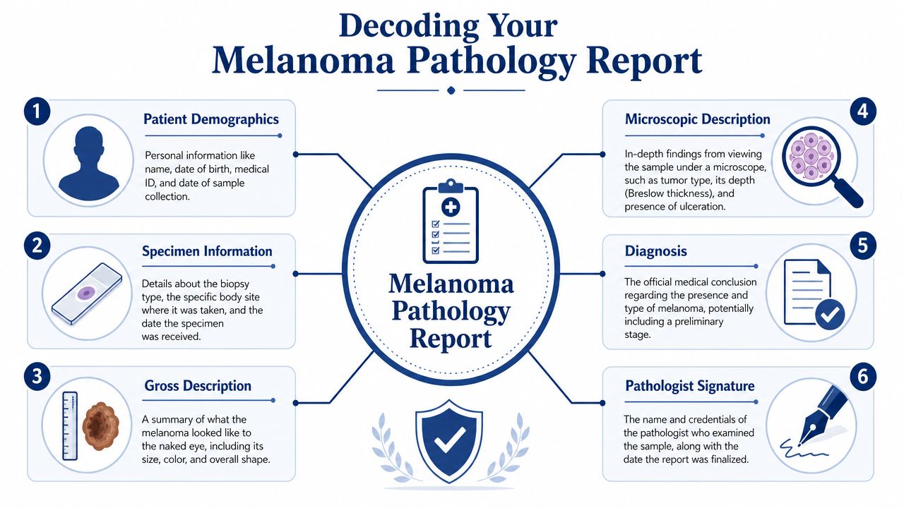
The top of the report
The first section usually contains patient demographics and specimen information. That includes your name, date of birth, the specimen source, the procedure type, and when the laboratory received the sample. This may seem routine, but it is part of basic identification and chain of documentation.
Then you may see a short clinical history. This is the dermatologist's or surgeon's description of why the sample was taken. It might mention a pigmented lesion, changing mole, or concern for melanoma. This portion provides context, but it does not replace the microscopic diagnosis.
Gross description and microscopic description
The gross description records what the specimen looked like to the naked eye. It may describe size, shape, color, and whether the tissue arrived as a shave biopsy, punch biopsy, or excision. Gross description helps correlate the physical specimen with the biopsy site and confirms what was submitted.
The microscopic description is where the pathologist explains what was seen on the slides. This can include melanoma subtype, depth, ulceration, mitotic activity, margin involvement, and other pathologic findings. For patients, this is often the densest part of the report.
A simple way to think about the report is this:
| Report section | What it answers |
|---|---|
| Patient and specimen data | Is this the correct patient and correct sample? |
| Clinical history | Why was the lesion sampled? |
| Gross description | What did the tissue look like before slide preparation? |
| Microscopic description | What did the pathologist see under the microscope? |
| Diagnosis | What is the official conclusion? |
| Signature | Who finalized the report and when? |
Why synoptic reporting matters
Many modern melanoma reports use structured or synoptic reporting rather than free-form narrative alone. That change became important because older narrative reports often omitted findings clinicians needed. A pathology review noted that fewer than 50% of older reports on primary melanomas documented all key prognostic details required for treatment planning, which is one reason structured reporting became a quality issue (Journal of Clinical and Aesthetic Dermatology review).
Practical rule: If your report looks brief but includes clearly labeled fields for depth, ulceration, margins, and related findings, that is often a strength, not a weakness.
If you're comparing biopsy methods and report wording, this guide to a skin biopsy for melanoma gives useful context.
The Three Most Important Prognostic Factors
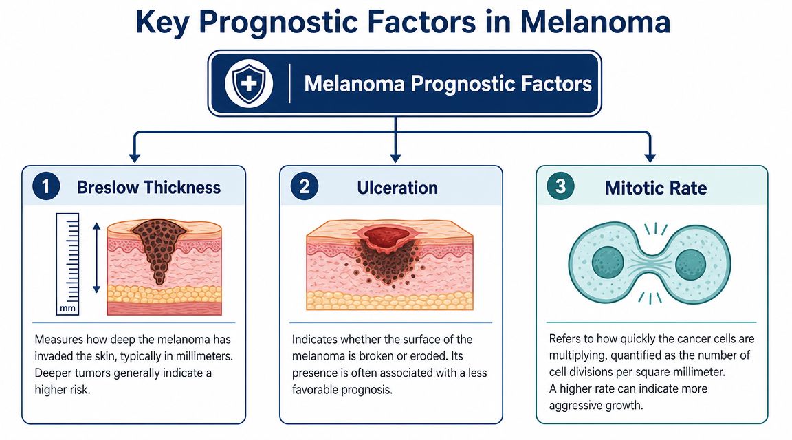
When a report contains many unfamiliar terms, patients often assume every line carries equal weight. It doesn't. For localized melanoma, the three findings that most strongly shape prognosis are Breslow thickness, ulceration, and mitotic rate.
Breslow thickness
Breslow thickness is the measured depth of the melanoma in millimeters. Picture it as the depth of roots beneath the surface rather than the width of leaves above ground. Two lesions can look somewhat similar on the skin but differ significantly in the depth to which tumor cells extend.
Pathology guidance is very specific here. The report should document Breslow thickness to the nearest 0.1 mm, and together with ulceration this is the minimum information needed to assign the pathologic T category (NCBI Bookshelf melanoma pathology guidance).
Ulceration
Ulceration means the surface over the melanoma is absent or broken in a way that reflects the biology of the tumor, not just a scratch or handling artifact. Its presence matters because it is associated with a less favorable outlook and affects the T category.
This is one reason patients should be careful about overreading the visual appearance of a lesion. A lesion can look small and still carry more concern if ulceration is present.
Mitotic rate
Mitotic rate refers to how actively the tumor cells are dividing. In pathology language, it is reported as mitoses per mm². Higher mitotic activity points to faster cellular growth and can add important context when doctors discuss prognosis and management.
For families, I often translate it this way:
- Breslow thickness tells how deep the melanoma has gone.
- Ulceration tells whether the surface barrier has been lost.
- Mitotic rate tells how busy the tumor appears biologically.
If you remember only a few terms from the pathology report melanoma process, remember these three first.
Why small measurement differences can matter
Patients sometimes focus on whether a number seems “close enough.” In pathology, a small difference may still matter because treatment discussions follow defined staging categories and clinical thresholds. That does not mean one decimal place predicts an individual future with certainty. It means the report must be precise because the clinical team uses precise categories.
What does not work well is trying to interpret one factor in isolation. A thin lesion, an ulcerated lesion, and a lesion with active mitoses are not understood the same way, even if only one feature changes.
Connecting Your Report to Melanoma Staging
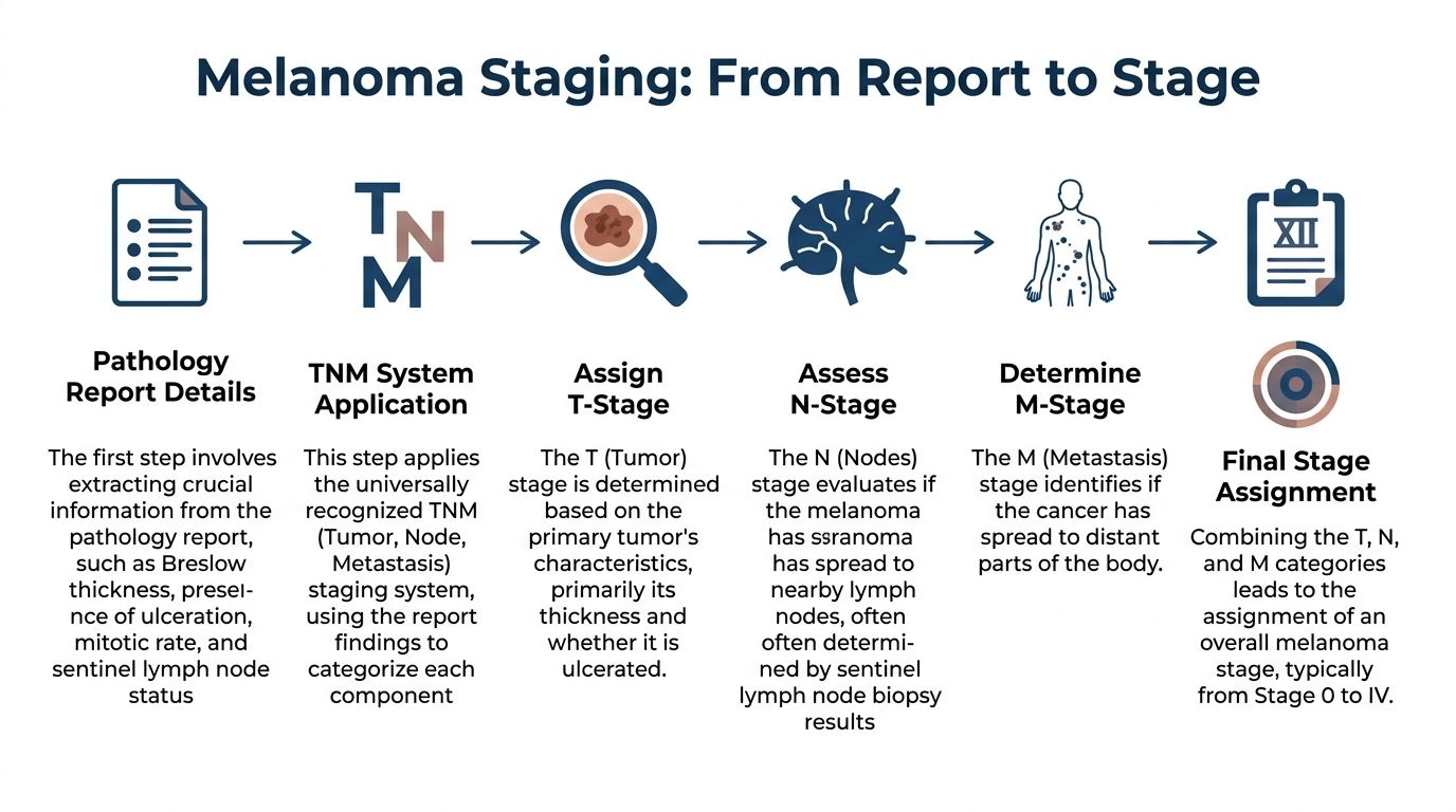
A pathology report and a cancer stage are related, but they are not identical. This is one of the most common sources of confusion.
What the report does define
The pathology report is strongest at defining the T, or tumor, part of the TNM system. That is where findings from the primary lesion, especially depth and ulceration, come into play. In practical terms, the report describes the tumor itself.
That is why the pathologist's wording matters so much. If the report clearly documents the core tumor findings, the treating team has a reliable starting point for staging discussions.
A brief visual explanation can help if you prefer to hear the concept discussed aloud:
What the report does not determine by itself
The pathology report on the primary lesion does not by itself tell you whether a patient is stage III or IV. That requires information about nodes and metastasis, which comes from additional assessment such as lymph node evaluation, sentinel lymph node biopsy, imaging, or other clinical workup.
Patients often see a severe-sounding diagnosis line and assume the full stage has already been decided. It usually has not. The pathology report is a major piece of the staging process, but it is not the entire process.
A useful way to frame it
Think of the report as answering, “What is this tumor doing in the tissue we removed?” Full staging asks a larger question: “Has this disease spread beyond that site?” Those are related questions, but they are not the same question.
Decoding Other Terms on Your Report
Once the diagnosis and major prognostic factors are clear, most patients move to the remaining lines and start searching for clues. Some of those terms are important. Some are supplementary. Some are legacy terms that still appear because reporting systems and habits evolve over time.
Margins and related findings
Margin status tells whether melanoma extends to the edge of the specimen examined by the pathologist. If a margin is involved, the lesion may not have been fully removed in that specimen. If the margin is clear, that means no melanoma was seen at that examined edge.
Other terms may include:
Lymphovascular invasion
Tumor cells are seen in lymphatic or blood vessel spaces. This is a finding clinicians take seriously because it speaks to a route by which tumor cells may spread.Microsatellites
Small nests of melanoma cells are present apart from the main invasive tumor. This can affect staging and management.Neurotropism
Melanoma cells show growth around or along nerves. This is not common in every melanoma, but when present it is worth attention.
Primary versus metastatic melanoma
One subtle but important job of the pathologist is distinguishing a primary melanoma from a metastatic melanoma deposit when the presentation is difficult. Morphology may answer that directly in straightforward cases. In harder cases, pathologists may use immunohistochemical stains such as S100, Melan-A, and HMB-45 to improve diagnostic confidence when routine appearance alone is not enough. These stains support interpretation. They do not replace careful review of the tissue architecture and clinical context.
A report can contain many technical descriptors, but the practical question remains simple. Which findings change management, and which ones are mainly descriptive?
Clark level and why it causes confusion
Clark level still appears on some reports, and patients often fixate on it because it sounds formal and numbered. Current pathology guidance is more cautious. The CAP protocol notes that Clark level is less reproducible among pathologists and is not used in current AJCC pT staging (CAP melanoma protocol PDF).
That doesn't mean the report is wrong if Clark level appears. It means you should not treat it as the main driver of modern staging. If a patient is overwhelmed by extra fields, I usually advise focusing first on depth, ulceration, mitotic activity, and margins.
When to Seek a Second Opinion or Further Review
A second opinion in pathology does not mean distrust. It means you understand that a tissue diagnosis can carry major consequences, and sometimes another expert review is appropriate before treatment moves forward.
Situations where a second review makes sense
Some reasons are clinical, and some are practical.
Borderline or unusual wording
If the report uses terms like atypical, indeterminate, suspicious, or difficult to classify, another review may help clarify the diagnosis.A major treatment decision depends on a narrow point
If management may change based on a subtle depth measurement, ulceration call, or distinction between in situ and invasive disease, an outside review can be reasonable.The pathology and the clinical picture don't fit well
If the lesion looked one way to the treating physician but the report suggests something unexpected, correlation is important.You need peace of mind before surgery or oncology treatment
This is a valid reason. Patients do not need to apologize for wanting confidence in a serious diagnosis.
What a second pathology opinion usually involves
In most cases, another pathologist reviews the original slides and, if needed, the paraffin blocks or additional sections. Sometimes the second reviewer agrees completely. Sometimes the interpretation is refined. Sometimes the wording changes in a way that helps the treating team understand uncertainty more clearly.
What does not help is ordering random repeat testing without a defined question. The best second opinions begin with a focused concern: Is this definitely invasive melanoma? Is ulceration present? Are the margins involved? Is this primary or metastatic?
If legal review, medical review, or formal expert analysis becomes necessary, a pathologist may also be asked to provide an opinion in the context of records review or testimony. I discuss that process in this overview of a pathologist expert witness.
Where forensic pathology fits
My perspective is somewhat different from that of a purely clinic-based consultant. In forensic practice, I am trained to assess evidence carefully, document uncertainty accurately, and explain findings in a way that can withstand scrutiny. That same discipline is useful when diagnosis is disputed or when a family needs a clear explanation after death.
In rare and tragic situations, melanoma is discovered or clarified only after death. In those cases, private autopsies and independent postmortem review may help establish the extent of disease and the role it played in the death. At Texas Autopsy Services, that kind of review is part of the broader work of cause of death investigation, not a substitute for the person's treating doctors.
Frequently Asked Questions About Pathology
Patients usually have a second round of questions after they've read the report once, then read it again more slowly. These are the questions I hear most often.
Who should explain my melanoma pathology report to me
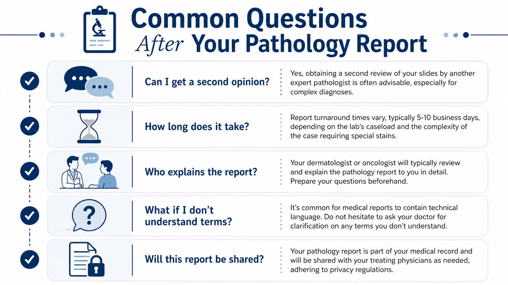
Your dermatologist, surgeon, or oncologist is usually the person who explains how the pathology findings apply to treatment. The pathologist writes the report, but the treating clinician places it in the context of surgery, imaging, lymph node evaluation, and follow-up.
Bring the full report to the visit if you can. A patient portal summary is often too abbreviated.
Can I ask for a copy of the actual report
Yes. The pathology report is part of your medical record. Ask for the complete final report, not just the diagnosis line.
If your care involves dictated notes, consult letters, and multiple versions of reports, accurate documentation matters. Practices that use tools for HIPAA compliant transcription for healthcare can sometimes produce cleaner records, but patients should still verify that they have the final signed pathology report.
Does a pathology report tell me my full melanoma stage
Not by itself. The report usually gives the tissue findings needed for the primary tumor assessment. Full stage assignment may require nodal or distant spread evaluation by your clinical team.
A report can strongly shape the staging conversation without finishing it on its own.
What if I don't understand a term on the report
Ask for clarification, and be specific. Instead of saying “I don't understand any of this,” ask “What does ulceration mean in my case?” or “Were my margins clear?” or “Is this invasive melanoma or melanoma in situ?” Specific questions usually get better answers.
I also encourage families to separate terms into two groups:
| Focus first | Ask about later if needed |
|---|---|
| Diagnosis | Subtype details |
| Breslow thickness | Legacy fields |
| Ulceration | Supplementary descriptive comments |
| Mitotic rate | Technical stain lists |
| Margins | Formatting language |
Are pathology reports ever revised
Yes. An addendum may be issued if more sections are reviewed, special stains are completed, or a second look changes part of the interpretation. This is not automatically a sign that something went wrong. Pathology is a process, and some cases become clearer as additional information is added.
When should a family consider forensic review after death
That question comes up when melanoma may have contributed to death, when the diagnosis was uncertain during life, or when relatives need a clear independent explanation of what happened medically. A forensic or postmortem review can describe cause of death and manner of death in plain language.
For clarity, cause of death means the disease or injury that started the fatal sequence. Manner of death is the category used in death investigation, such as natural or accident. Chain of custody means documented control over specimens or evidence so their handling can be traced and verified.
Those definitions matter most in medicolegal and postmortem settings, but families often find them helpful because they show how pathology fits into the larger record.
If you're facing a melanoma diagnosis, or if your family is trying to understand whether melanoma played a role after a death, careful review of the pathology can bring needed clarity. If an independent postmortem examination or forensic pathology review would help answer those questions, you can learn more through Texas Autopsy Services.