April 3, 2026 · Texas Autopsy Services
Understanding Pathology Reports for Cancer
A guide to understanding pathology reports for cancer. Learn to decode key sections and terms from a board-certified pathologist.
On this page
When you or someone you love is diagnosed with cancer, a single document suddenly becomes the most important piece of paper in your life: the pathology report. This isn't just a confirmation of a diagnosis; it's the detailed map that will guide every single decision on the road ahead.
Your Guide to Understanding a Cancer Diagnosis
As a board-certified forensic pathologist at Texas Autopsy Services, I spend my days examining critical medical documents. None are more pivotal to a patient's future than a cancer pathology report. My name is our forensic pathology team, and I want to walk you through why this report is the foundation of a patient's care, whether you're the patient, a concerned family member, or a legal professional.
This guide is designed to make the report's purpose clear and simple. When you understand what's in this document, you're better equipped to face the journey ahead. It's about empowering you with knowledge when you need it most.
The Pathologist’s Role in Your Care
Your report is prepared by a pathologist a medical doctor who specializes in looking at tissue under a microscope to diagnose disease. Their analysis is definitive. Think of the pathologist as the intelligence officer in the fight against cancer. They identify the specific "enemy" so the oncology team knows which "weapons" will be most effective.
Essentially, their work answers the most urgent questions:
- What exact type of cancer is it?
- How aggressive does it seem to be?
- Did the surgeon get it all?
"A pathology report is the scientific cornerstone of a cancer diagnosis. It translates what is seen under the microscope into a clear, actionable plan for oncologists, surgeons, and patients."
Why This Report Matters to Everyone
The details locked inside pathology reports for cancer are what determine the best path forward. A precise diagnosis is what tells doctors whether surgery, chemotherapy, radiation, or a targeted therapy is the right call.
For families, having a grasp on the report can make the whole process less intimidating and help you ask the right questions. For attorneys handling medical-legal cases, this document is the source of objective, scientific truth.
My goal here is to give you a clear, compassionate perspective, built on the same commitment to precision that guides my work in private autopsies. The information can feel overwhelming, but its importance can't be overstated. By breaking it down piece by piece, we can replace some of the fear and uncertainty with knowledge and a clear path forward. This report is your starting point.
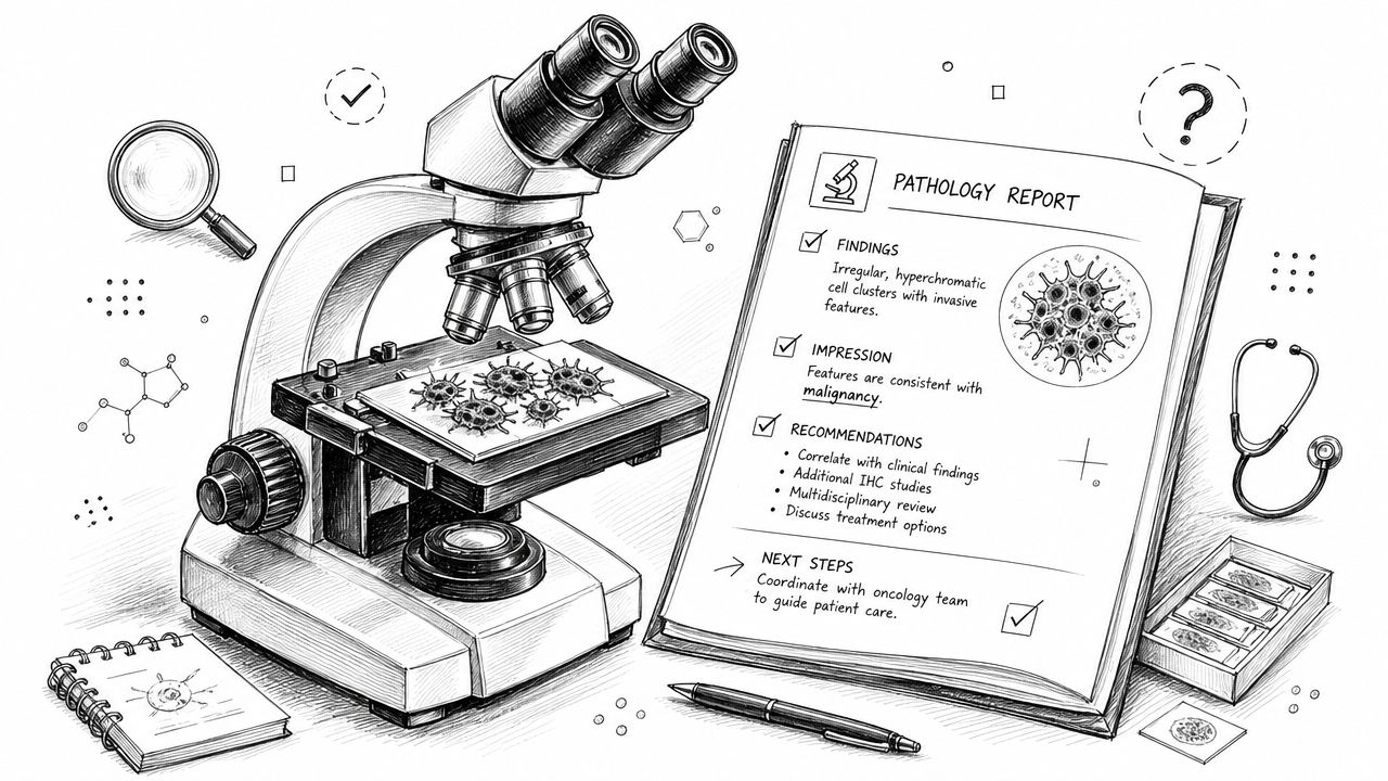
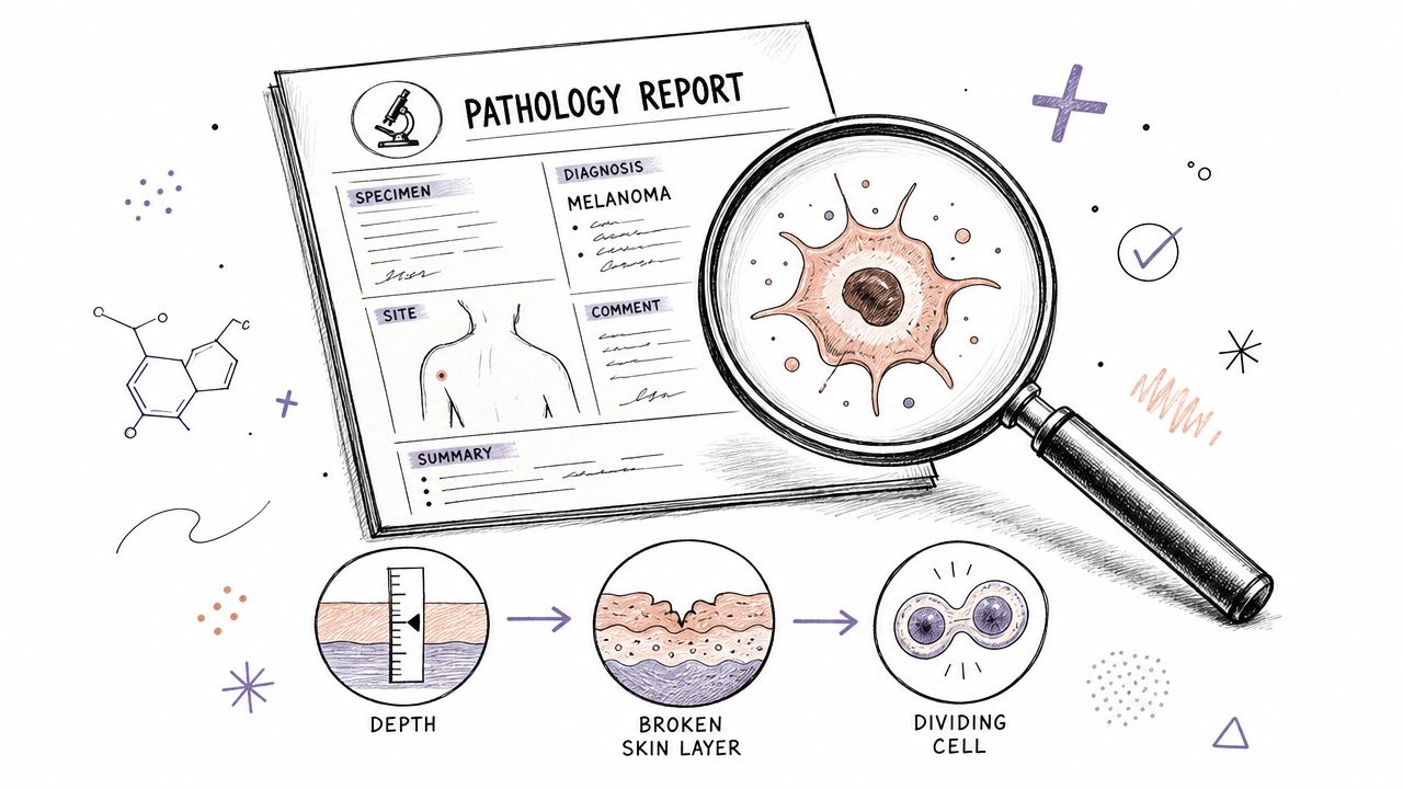
When you first get a pathology report, it’s easy to feel overwhelmed. The document is packed with medical terminology and looks more like a scientific paper than something meant for a patient. Let’s break it down together, section by section, so you can understand what it all means.
Think of it as a story about the tissue sample that was taken from you. Each part of the report adds another layer to that story, building toward the final conclusion the diagnosis. Once you get the hang of the structure, these reports become much less intimidating.
This flow is the foundation of your cancer journey. The report’s findings are what guide your doctors in creating a plan specifically for you.
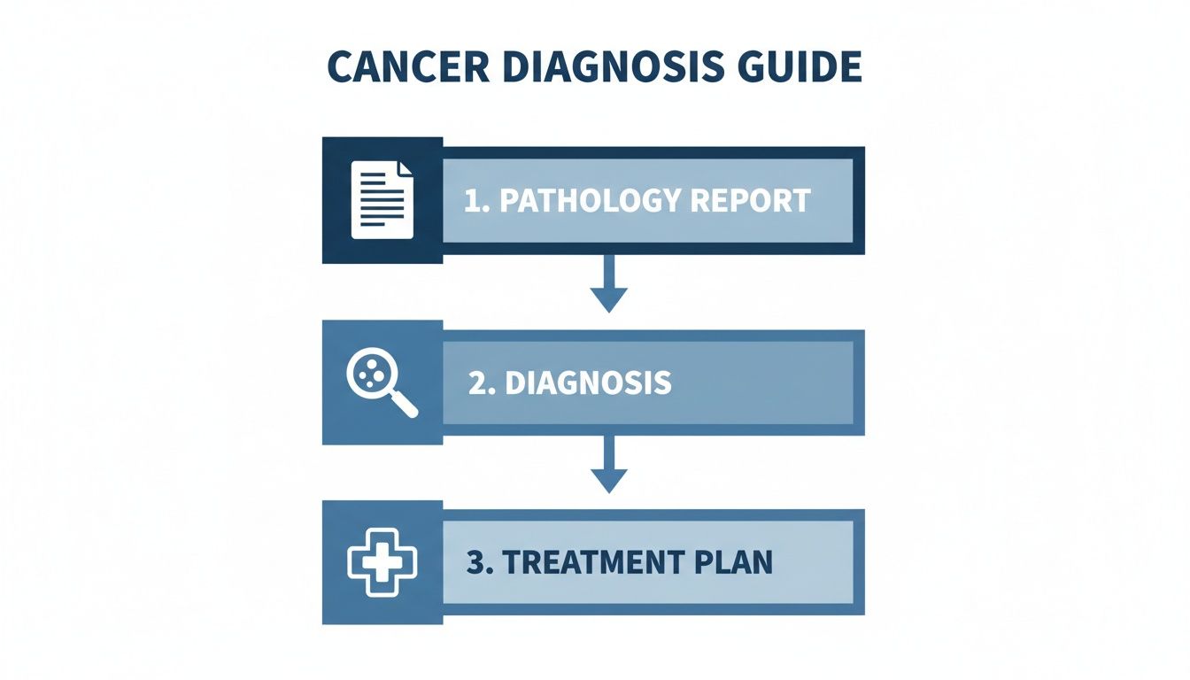
As you can see, everything starts here. The science in this report leads directly to the diagnosis, which then points the way toward the right treatment.
To get started, we've put together a quick overview of the most common sections you'll find in any cancer pathology report.
Key Sections of a Cancer Pathology Report
This table summarizes the standard sections in a cancer pathology report and explains the crucial role each one plays in the diagnostic process.
| Section Name | What It Tells You | Why It's Important |
|---|---|---|
| Patient & Clinical Info | Your personal details (name, DOB) and a brief medical summary from your doctor. | Ensures the report is for the right person and gives the pathologist vital context about your case. |
| Specimen | A list of every tissue sample sent to the lab, each with a unique identifier. | Guarantees that every piece of tissue is tracked and analyzed correctly, preventing mix-ups. |
| Gross Description | What the tissue looks like to the naked eye its size, weight, color, and texture. | Provides the initial "big picture" assessment before diving into the cellular level. |
| Microscopic Description | The pathologist's detailed findings from looking at the tissue under a microscope. | This is where cancer is identified and its characteristics (like cell type) are described. |
| Final Diagnosis | The definitive conclusion, summarizing all the findings into a single diagnosis. | This is the most critical section, stating clearly whether cancer is present and, if so, what kind. |
| Margins | Whether cancer cells are found at the edge of the removed tissue. | Determines if the entire tumor was likely removed or if some cancer cells were left behind. |
| Staging (pTNM) | The tumor's size (T), lymph node involvement (N), and metastasis (M). | Provides the official stage of the cancer, which is essential for prognosis and treatment planning. |
| Biomarkers | Results from special tests that look for specific proteins or genes in the cancer cells. | Helps predict how the cancer might behave and identifies targets for specific therapies. |
Understanding these components will empower you to follow the narrative of your report from start to finish.
Your Personal and Clinical Information
Every report starts with the basics to make sure there are no mix-ups. You'll see your name, date of birth, and medical record number right at the top, along with the doctor who ordered the test and the pathologist who reviewed it.
Just below that, you’ll find a section called Clinical History. This is a short note from your doctor to the pathologist, giving them crucial context. It might say something like, "62-year-old female with a suspicious mass in the left breast" or "Follow-up on a known colon polyp." This helps the pathologist know exactly what they're looking for.
The Specimen Description
This part is all about logistics. It’s a simple, clear list of every single piece of tissue that arrived at the lab for examination. For instance, you might see "A: Left breast, core needle biopsy" and "B: Left axillary lymph node, fine needle aspiration." Each sample gets its own unique letter or number, so nothing gets lost or confused.
The meticulous notes in a pathology report are often transcribed and organized by skilled medical transcriptionists, who are essential for maintaining the accuracy of these complex documents.
A pathology report is a story told in two parts: what the pathologist sees with their own eyes and what they discover under the microscope. Both are equally crucial for an accurate diagnosis.
Gross and Microscopic Descriptions
Now we get to the core of the analysis. This is where the pathologist documents what they found, and it’s split into two main parts.
Gross Description: In medicine, "gross" simply means "large scale" or what's visible without a microscope. The pathologist will describe the tissue's size, weight, color, and feel. A note might read, "a 2.5 cm firm, white, irregular mass," giving a physical description of the tumor.
Microscopic Description: This is the deep dive. The pathologist takes incredibly thin slices of the tissue, stains them with special dyes to highlight different cell structures, and examines them under a powerful microscope. Here, they describe the cells themselves what kind they are, how they’re arranged, and whether they look cancerous compared to the healthy tissue around them.
This two-step process is fundamental. The gross description provides the physical context, and the microscopic examination reveals the cellular truth that ultimately leads to a diagnosis. If you'd like to explore this topic further, our detailed guide on how to read a pathology report offers an even deeper look.
Getting to the Heart of the Report: Diagnosis and Staging
Once you've navigated the first few sections of a pathology report, you arrive at its core the final diagnosis and staging. This is where all the microscopic observations come together into a definitive conclusion. Frankly, this is the part of the report that matters most, as it directly informs the treatment plan your oncologist will build.
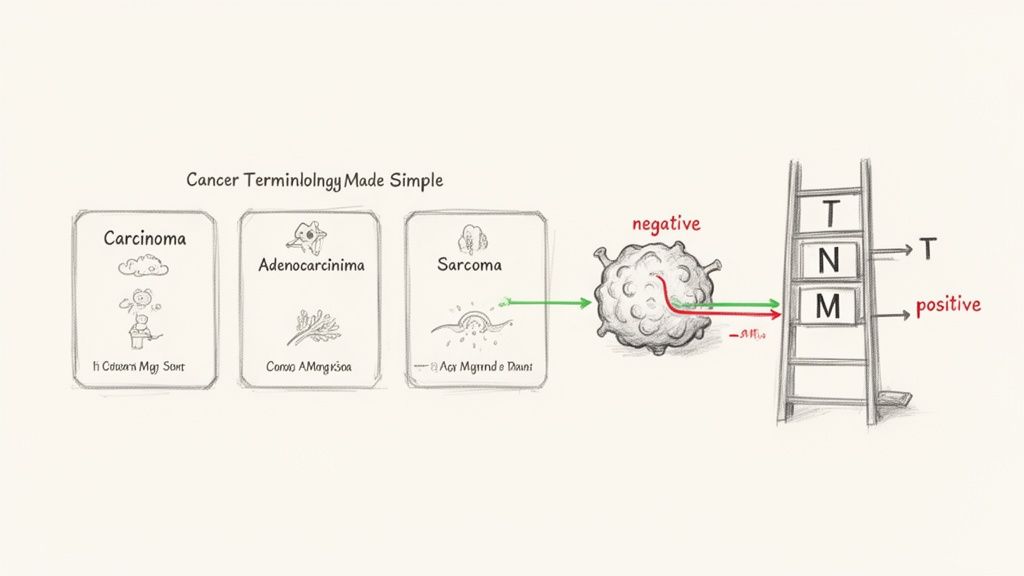
As a pathologist, I see my role as a translator, turning complex cellular data into clear answers. Let's walk through the key terms you'll find here, because understanding this language is the first step in understanding the path ahead.
What Is the Final Diagnosis?
The Final Diagnosis is the bottom line. It’s the pathologist’s official identification of the cancer found in the tissue sample. The first thing you'll see is a name that classifies the cancer based on the cell it came from.
Here are a few of the most common families of cancer you'll encounter:
- Carcinoma: This is by far the most common type. It starts in the cells lining the surfaces of the body, like the skin, breast ducts, or lungs. An adenocarcinoma is a specific type that grows from glandular cells.
- Sarcoma: A much rarer cancer that forms in bone and soft tissues think muscle, fat, or cartilage.
- Leukemia: This is a cancer of the blood and bone marrow, which causes the body to produce abnormal white blood cells.
- Lymphoma: This cancer originates in the immune system, specifically in cells called lymphocytes.
Of course, these broad categories are just the starting point. The report will get much more specific, which brings us to the tumor's grade.
Understanding Tumor Grade
Tumor grade tells us about the cancer's personality. It describes how abnormal the cancer cells look under the microscope when compared to healthy, normal cells. Essentially, it’s a measure of the cancer’s potential aggressiveness.
- Well-differentiated (Low Grade - Grade 1): The cancer cells look a lot like normal cells. They tend to be slow-growing and better-behaved.
- Moderately-differentiated (Intermediate Grade - Grade 2): The cells are in a middle ground they look somewhat abnormal and are growing at a moderate speed.
- Poorly-differentiated (High Grade - Grade 3): These cells look very little like normal cells. They are often fast-growing and more aggressive.
A lower grade often points to a better prognosis, while a higher grade might call for a more aggressive treatment plan. It’s one of the first things an oncologist looks at to predict how a cancer might act.
Why Tumor Margins Are So Critical
When a surgeon removes a tumor, they don't just take the lump itself. They also take a small buffer of healthy-looking tissue around it. The edge of this entire piece of removed tissue is what we pathologists call the margin. Examining it is one of our most important jobs.
The report will classify the margin status in one of three ways:
- Negative (or Clean): Great news. This means no cancer cells were found at the very edge of the tissue, suggesting the surgeon got the entire tumor out.
- Positive: This means cancer cells were touching the edge of the removed tissue. It’s a red flag that some cancer might have been left behind, possibly requiring another surgery or treatment.
- Close: The cancer cells are near the edge, but not quite touching. Your surgeon will carefully consider this to decide if more action is needed.
The margin status is incredibly important because it often dictates the very next step in a patient's care.
Making Sense of the TNM Staging System
Staging is the process of figuring out just how far the cancer has spread. The gold standard for this is the TNM system, a universal language that helps doctors everywhere describe the extent of a cancer. These reports are also vital for public health, helping experts track the 2,041,910 new cancer cases projected for 2026 in the U.S. This data reveals crucial patterns for instance, that breast cancer comprises 32% of new diagnoses in women, while prostate cancer accounts for 30% in men and guides everything from national prevention strategies to individual patient care.
So what does TNM actually stand for?
- T (Tumor): This describes the size and reach of the original tumor. It's typically numbered 1 to 4, with higher numbers indicating a larger or more invasive tumor.
- N (Nodes): This tells us if the cancer has escaped into nearby lymph nodes. N0 means the nodes are clear, while N1, N2, or N3 indicate an increasing amount of lymph node involvement.
- M (Metastasis): This is the big one. It states whether the cancer has spread to distant parts of the body. M0 means no distant spread was found, but M1 confirms that the cancer has metastasized.
After determining the T, N, and M values, they are all put together to assign an overall stage, usually from Stage 0 (a pre-cancer) to Stage IV (advanced, metastatic cancer). This final stage is arguably the single most important factor for shaping a patient's prognosis and treatment roadmap.
While most reports lay this out in a narrative format, some institutions are moving toward more structured checklists. If you're interested in that, you might want to read about synoptic pathology reports in our article, which explains this clear, standardized format.
Biomarkers: The Modern Roadmap for Cancer Treatment
It wasn't that long ago that a pathology report's main job was to name the cancer, grade how aggressive it looked, and map out how far it had spread. That information is still the bedrock of any diagnosis, but today's reports go much deeper. They now include a crucial layer of detail that has completely changed the game: biomarker testing.
We're essentially looking for specific genetic or protein signatures on the cancer cells themselves.
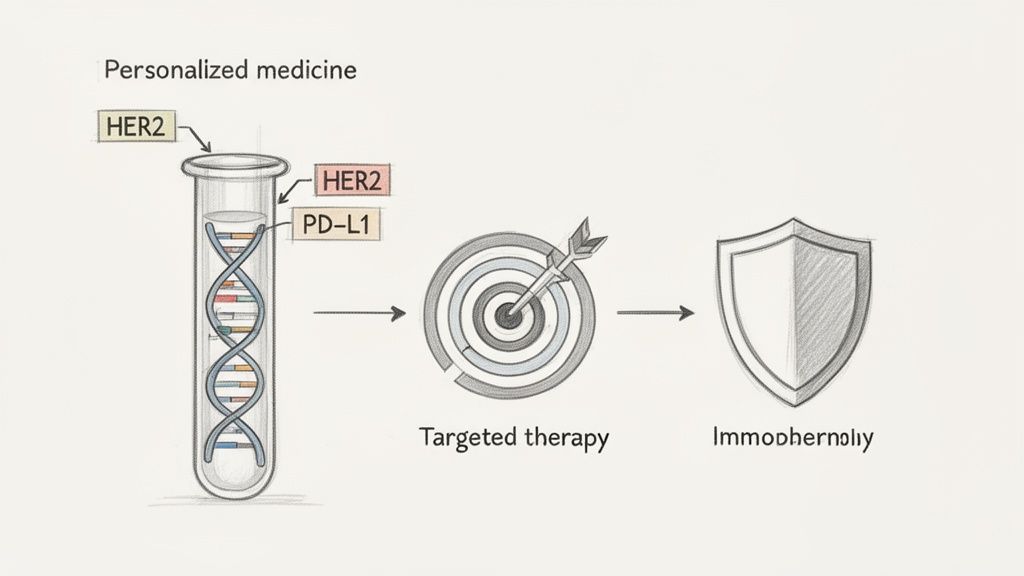
I like to think of a biomarker as a unique flag or even a secret code that a cancer cell carries. My job as the pathologist is to crack that code. We're no longer just identifying the enemy; we're searching for its specific vulnerabilities. This section of the report is what gives oncologists the power to move past one-size-fits-all approaches and into the world of truly personalized medicine.
Common Biomarkers and What They Tell Us
In a modern pathology report for cancer, you’ll find a section dedicated to these results. Two of the most common examples you might see are HER2 in breast cancer and PD-L1, which is found in a range of cancers like lung and melanoma.
HER2 (Human Epidermal Growth Factor Receptor 2): This is a protein that acts like a gas pedal for cancer, making cells grow and divide at high speed. When a breast cancer is "HER2-positive," it means it’s making way too much of this protein. Finding this opens the door to targeted therapies that are specifically designed to block HER2, often with incredible results. You can see how this looks on a real report in our detailed breast cancer pathology report sample.
PD-L1 (Programmed Death-Ligand 1): Think of this protein as an invisibility cloak that cancer cells use to hide from the body's own immune system. A "PD-L1 positive" result means the cancer is using this trick. This is a key piece of intelligence for the oncologist, signaling that the patient could be a great candidate for immunotherapy a type of treatment that unmasks the cancer and helps the immune system hunt it down.
And these are just two of many. We also look for hormone receptors (ER/PR) in breast cancer, gene mutations like EGFR in lung cancer, or MGMT in brain tumors. Each one provides another clue for cracking the cancer's code.
From Lab Bench to Better Patient Care
Every biomarker finding is a critical piece of intelligence. It gives the oncology team a way to choose a therapy with a much higher chance of working for that specific person's tumor. Just as importantly, it helps them avoid treatments that are unlikely to work, sparing the patient from unnecessary and harsh side effects.
"Biomarker analysis transforms the pathology report from a diagnostic summary into a strategic blueprint for personalized cancer treatment."
The number of these reports, and the complexity within them, is only growing. The American Cancer Society estimates that in 2026, pathologists will be involved in roughly 2,041,910 new cancer diagnoses. Of these, breast cancer is projected to make up about 319,750 cases and prostate cancer 306,870 cases.
The detailed biomarker data within each of those reports is absolutely essential for creating the precise, individualized treatment plans needed. It’s a fundamental shift in medicine, drawing a direct line from a detailed lab finding to a more hopeful and effective outcome for the patient.
When to Seek a Second Opinion or Postmortem Review
Receiving a cancer diagnosis is a moment that changes everything. That diagnosis is built upon a single, critical document: the pathology report. As a patient or a concerned family member, you have every right to feel 100% confident in that report.
The truth is, the vast majority of pathology reports are accurate. But medicine is incredibly complex. Asking for a second opinion isn't a sign of distrust in your medical team; it's a way of ensuring certainty during a very uncertain time.
As a forensic pathologist, I'm often brought in to provide objective answers and clarity. This need for truth is just as vital for a living patient's diagnosis as it is for understanding what happened after a loved one has passed away.
Second Opinions on Pathology Reports for Cancer
It is completely normal and reasonable to ask for another expert pathologist to review the original tissue slides. This is a common request, and your medical team should be supportive.
A second opinion can go one of two ways: it might confirm the original diagnosis, which brings invaluable peace of mind. Or, it could refine the details sometimes in ways that can significantly change the recommended treatment plan.
Consider asking for a second look in these situations:
- Rare or Unusual Cancers: If the diagnosis is uncommon, getting it confirmed by a pathologist who specializes in that specific type of cancer is just plain smart.
- Conflicting or Ambiguous Results: Sometimes the report's language is unclear, or the findings don't seem to match the clinical symptoms. A fresh set of expert eyes can cut through the confusion.
- High-Stakes Treatment Decisions: Before you commit to major surgery, aggressive chemotherapy, or other intensive treatments, you want to be absolutely sure you're on the right path. Confirming the diagnosis provides that solid foundation.
Getting a second opinion is a standard procedure. Your oncologist can help coordinate sending your tissue slides and the original report to another pathologist or institution. It's all done with one goal in mind: ensuring you get the best possible care.
The Role of Postmortem Review in Cancer Cases
Much of my work at Texas Autopsy Services involves helping families find answers after a loved one has passed away from cancer. This is a different kind of second opinion a final, detailed review through a private autopsy. While it can't alter what happened, it can provide profound clarity and closure.
Families often request a postmortem review for several critical reasons:
- To Confirm the Primary Cancer: Sometimes, cancer spreads so widely (metastasizes) that doctors are never sure where it started. An autopsy can often locate the primary tumor, providing a complete and final diagnostic picture.
- To Understand the Extent of the Disease: A postmortem exam creates a definitive map of where the cancer had spread. This is often more detailed than what imaging scans could show, helping families understand the true burden of the illness.
- To Evaluate Treatment Effectiveness: An autopsy can reveal how tumors responded to treatments like chemotherapy and radiation. Did they shrink? Did they become resistant? These are questions that can be answered.
- To Identify Hereditary Cancer Risks: For some cancers, a postmortem exam provides an opportunity for genetic analysis of tissue. This can identify inherited mutations that could be crucial for the health of children, grandchildren, and other relatives.
An independent review, whether it's looking at a biopsy slide or performing a postmortem exam, is ultimately about one thing: the pursuit of objective truth. It empowers patients and families with the most accurate information possible.
This relentless pursuit of accuracy is what drives progress. Think about this: the remarkable 34% decline in cancer death rates from 1991 to 2022 is directly linked to the growing precision of pathology. That progress, which has averted nearly 4.5 million deaths, rests on the foundation of earlier detection and more effective treatments both of which depend entirely on getting the pathology right. You can read more about how better pathology has dramatically improved cancer survival rates and what challenges still remain.
Your Questions About Cancer Pathology, Answered
After years of working with families and medical professionals, I’ve found that the same questions about pathology reports pop up again and again. When a diagnosis like cancer enters the picture, confusion and anxiety are completely normal. Let's walk through some of the most common concerns I hear, and I'll give you the straightforward answers I share in my own practice.
How Do I Get a Copy of My Pathology Report?
It’s your health information, and you have a legal right to it. The most direct route is simply to ask your doctor usually your oncologist or the physician who ordered the test. Their office can get you a copy, often through an online patient portal or as a printout you can pick up.
You can also go straight to the source: the medical records department of the hospital or clinic where the biopsy was processed. You’ll probably have to fill out a standard release form and show your ID, but it's a routine request they handle every day.
What Should I Do if I Can't Make Sense of My Report?
First, take a deep breath. These reports are dense, technical documents written by one doctor for another. You are not meant to understand every word, so don't feel overwhelmed if it reads like a foreign language.
Your own doctor or a nurse navigator is your best guide. Set aside time to go over the report with them, page by page. It is perfectly okay and expected for you to say, "I don't understand what this means. Can you explain it in simple terms?"
To make that conversation as productive as possible, do a little prep work:
- Circle or highlight the words and phrases that you find most confusing or concerning.
- Write down your questions before the meeting. Think about things like, "What does 'poorly differentiated' actually mean for me?" or "Why does the report talk so much about the 'margins'?"
- Bring a trusted friend or family member to your appointment. A second person can help listen, take notes, and think of questions you might miss.
While this guide can give you a solid foundation, nothing beats a one-on-one conversation about your specific results with your care team.
Can a Pathology Report Be Wrong?
This is a tough but fair question. The short answer is yes, but it is very rare. Pathologists are highly trained specialists, but because the process relies on human expertise and interpretation, mistakes can happen.
When we talk about an "error," it could mean a few things:
- A Diagnostic Misinterpretation: The pathologist may see the cells under the microscope and come to the wrong conclusion. This is most likely with extremely rare cancers or when a tumor has very unusual features.
- A Sampling Issue: The needle biopsy might have just missed the small area of cancer, resulting in a "false negative." Or, the piece of tissue that was removed might not be representative of the most aggressive part of the tumor.
- A Clerical Mistake: Things like mixing up patient samples or a typo in the data entry are exceptionally rare because of the strict safety protocols in modern labs, but they aren't impossible.
If your gut tells you something is off, or you simply want the reassurance of a confirmation, getting a second opinion is a completely valid and proactive step. This involves having an independent pathologist at another institution review your original tissue slides to give their own assessment.
How Long Does It Take to Get Pathology Results?
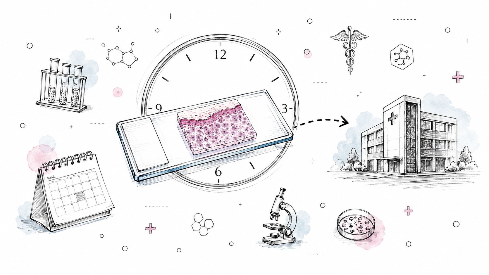
Waiting for results is often the hardest part. Generally, you can expect a final report within two to ten business days, but the timeline can vary quite a bit depending on the complexity of the case.
Many people don't realize just how many steps are involved after the biopsy is taken:
- Transport and Lab Prep (1-2 days): Your tissue sample is meticulously labeled, logged, and sent to the lab. There, it's preserved in formaldehyde (formalin) and carefully embedded into a small block of paraffin wax.
- Sectioning and Staining (1 day): A technician uses a special instrument called a microtome to cut paper-thin ribbons from the wax block. These delicate slices are mounted on glass slides and treated with dyes that highlight different cell structures, making them visible under a microscope.
- Pathologist Review (1-3 days): This is where the magic happens. The pathologist examines the slides, identifies the cell types, makes a diagnosis, grades the tumor, and checks the margins. It's the most intensive part of the process.
- Specialized Testing (3-10+ days): If your doctor ordered additional tests for biomarkers (like HER2, ER, PR, or PD-L1), that adds more time. These tests require different techniques and are often performed after the initial diagnosis is confirmed, sometimes taking another week or more.
- Final Report (1 day): Once every analysis is complete, the pathologist compiles all the findings into the final, detailed report and transmits it securely to your doctor.
If the case is particularly complex or the tissue has to be sent to an outside lab for special genetic testing, the wait can be longer. Your doctor's office should be able to give you a rough idea of when to expect your results.
Navigating a medical diagnosis is challenging, and getting clear, reliable information is essential. If you are a family member or legal professional in Texas seeking definitive answers that only a postmortem examination can provide, my team and I are here to offer clarity.
At Texas Autopsy Services, we provide objective, medically precise, and compassionate postmortem examinations. Whether the goal is to understand the true cause of death, confirm a clinical diagnosis, or provide peace of mind, we are dedicated to delivering the answers you deserve. Please contact our office for a confidential consultation at texasautopsyservices.com.