March 11, 2026 · Texas Autopsy Services
What Families Should Know About a Breast Cancer Pathology Report
I'll help you decode your breast cancer pathology report sample. This guide explains key terms, receptor status, margins, and what it all means for you.

On this page
- Understanding Patient and Specimen Information
- Interpreting the Gross and Microscopic Descriptions
- Understanding Tumor Grade, Size, and Surgical Margins
- Understanding Your Hormone Receptor and HER2 Status
- Understanding Lymph Node Status and Cancer Stage
- Questions to Ask Your Doctor About Your Pathology Report
- Common Questions About Your Pathology Report
As a board-certified pathologist, I understand that a medical document can define a person's future. A breast cancer pathology report is one of the most critical—it's the blueprint that guides every treatment decision. It's often filled with technical terms that can feel overwhelming when you need clarity most. That’s why I want to walk you through a breast cancer pathology report sample.
- A pathology report provides the definitive diagnosis and key details about a breast tumor.
- It identifies the cancer type, grade, size, and whether it has been fully removed (margins).
- Biomarker tests (ER, PR, HER2) determine if the cancer is sensitive to targeted therapies.
- The report is used to assign a cancer stage, which guides the overall treatment plan.
- Understanding your report helps you have more productive conversations with your oncologist.
My goal is simple: to help you understand what your report says so you can have more informed conversations with your doctors. Think of this guide as a reference. We’ll break down a sample report, piece by piece, from basic patient details to the critical biomarkers that define your treatment options.
You're not alone in this. An estimated 2.3 million women worldwide were diagnosed with breast cancer in 2022. The good news is that about 66% of cases are discovered when the cancer is still localized. This is when your pathology report is most powerful, as it gives your medical team the precise information needed to choose the most effective treatment.
Decoding the Language of Your Report
Your report isn't just a diagnosis; it’s a detailed map of the cancer. To read it, you need to understand its language. Getting a general sense of the layout is helpful. For a broader overview, some resources explain how to read a pathology report in general terms.
While this guide focuses on breast cancer, the basic structure is similar for other pathology reports. We also have a general guide on how to read a pathology report that you might find useful.
Here’s a quick look at the key sections we'll cover:
- Patient and Specimen Information: The first step is to confirm it's your report and understand where the tissue sample came from.
- Gross and Microscopic Descriptions: This is what the pathologist saw, first with the naked eye and then under the microscope.
- Tumor Grade, Size, and Margins: These details tell you about the cancer’s characteristics—how aggressive it is, how big it is, and whether the surgeon removed it all.
- Biomarker Status (ER, PR, HER2): This is the genetic fingerprint of the cancer, which is crucial for targeted therapies.
- Lymph Node Status and Staging: This section answers the critical question of whether the cancer has spread.
Understanding Patient and Specimen Information
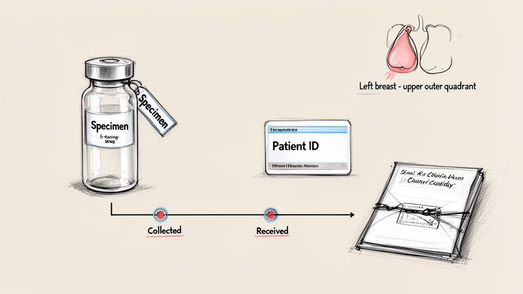
Before a pathologist begins their analysis, the first step is getting the paperwork right. The top section of your pathology report is about administrative confirmation. It’s designed to confirm the report is yours and that the tissue sample was handled correctly from the moment it left your body. Think of it as the foundation for the entire diagnosis.
You’ll see basic identifiers like your full name, date of birth, and medical record number. Every lab has a meticulous process for this data entry to prevent mix-ups. This section also documents the tissue's journey, something we call the “chain of custody,” which guarantees its integrity from collection to analysis.
Specimen Source and Procedure Details
The report gets very specific about where the tissue came from and how it was removed. You can expect to see:
- Specimen Source: Details like “left breast, upper outer quadrant” pinpoint the exact location.
- Procedure Type: This names the procedure, whether it was a “core needle biopsy” or a “lumpectomy.”
- Key Dates: The date the tissue was collected and the date it arrived at the lab are always recorded.
Knowing how the sample was collected adds important context. For instance, a procedure like a stereotactic breast biopsy uses a specific technique to get the tissue, and this information helps the pathologist orient the sample correctly for evaluation.
This part of the report is also essential for any legal or secondary reviews. For families or attorneys seeking a second opinion autopsy, this section provides verifiable proof that the results belong to the right person and that the sample was managed under strict, documented protocols. This administrative precision is the first sign of a trustworthy diagnostic process.
Interpreting the Gross and Microscopic Descriptions
This is where my analysis begins. When I receive a tissue sample, I start by describing what I see with my own eyes, then move to the high-powered view under the microscope. These two parts, the gross and microscopic descriptions, are the building blocks of your diagnosis.
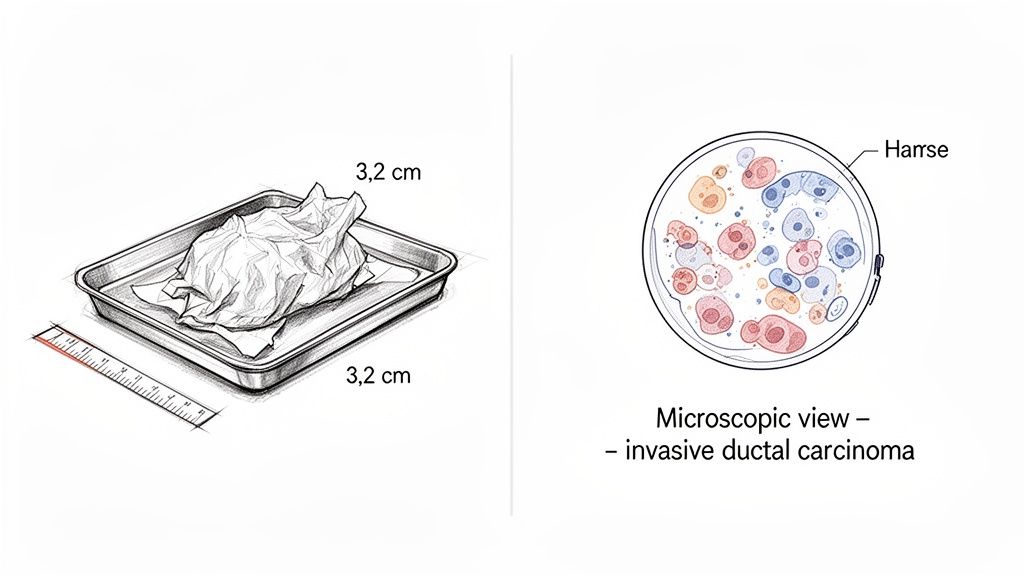
The Gross Description is a summary of the tissue as a whole, before it’s processed for the microscope. I’m looking at its physical traits and making notes.
- Size and Weight: I'll measure the dimensions of the entire tissue specimen, usually in centimeters (cm).
- Color and Texture: I note whether the tissue feels firm, soft, rubbery, or has other notable qualities.
- Visible Tumor: If I can see a distinct tumor, I’ll measure it. My note might say, "A 3.2 cm firm, white, irregular mass is identified."
This initial overview acts like a map. It helps me get my bearings and decide which specific areas of the tissue are most important to examine up close.
From Tissue to Diagnosis
After the gross exam, we get to the Microscopic Description. This is the core of the process. We take very thin slices of the tissue, stain them to make the cells visible, and put them on glass slides. Looking through the microscope is where the true story of the cells is told.
This detailed view confirms if cancer cells are present and, if so, how they’re behaving. I describe their shape, how they are arranged, and how they relate to the normal tissue around them. All of this information leads to the Final Diagnosis.
The Final Diagnosis is the most critical line in the report. It's a clear, concise statement that names the cancer, such as "Invasive Ductal Carcinoma." This is the definitive conclusion and the foundation for every treatment decision that comes next.
Understanding what happens under the microscope is key. The technical process of preparing and analyzing these slides is called histology, a precise science. If you're curious about the detailed lab work involved, you can learn more about what a histology report is from one of our other guides.
When you’re looking at a breast cancer pathology report sample, knowing the difference between the gross and microscopic sections helps you see the path a pathologist takes—from a piece of tissue to a life-changing diagnosis.
Understanding Tumor Grade, Size, and Surgical Margins
After a biopsy confirms cancer, a pathologist's work truly begins. We examine the cancer's characteristics to figure out how it’s likely to behave. Three of the most important factors are the tumor's grade, its size, and the surgical margins. These details are fundamental because they help your care team map out the most effective treatment plan.
What Is Tumor Grade?
People often mix up tumor grade and cancer stage, but they tell us different things. Staging tells us where the cancer is located and how far it has spread, while the grade describes what the cancer cells themselves look like under a microscope. I compare the cancer cells to normal, healthy breast cells to see how abnormal they've become.
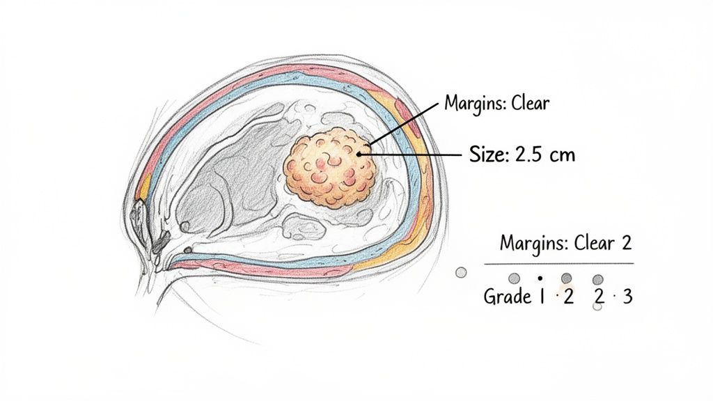
Based on this analysis, I assign a grade from 1 to 3:
- Grade 1 (Well-Differentiated): These cancer cells still look a lot like normal breast tissue. They are typically organized and slow-growing.
- Grade 2 (Moderately-Differentiated): The cells are in a middle ground. They look less like normal cells and are growing at a slightly faster, more moderate pace.
- Grade 3 (Poorly-Differentiated): These cells look very different from healthy cells. They are disorganized and tend to grow and divide more quickly, which can make them more aggressive.
Think of the grade as a snapshot of the tumor's personality. It gives your oncologist a good indication of how the cancer might act over time.
Measuring the Tumor and Its Removal
Your report will also list the tumor size, which is a measurement of the largest dimension of the invasive cancer, usually given in centimeters (cm) or millimeters (mm). For example, a tumor might be measured as 2.5 cm. Tumor size is a crucial part of the staging process, but it’s just one piece of the puzzle.
Finally, we look at the surgical margins. When a surgeon removes a tumor (a lumpectomy or mastectomy), they also take out a small border of what appears to be normal tissue surrounding it. My job is to carefully examine this border, or margin, under the microscope to see if any cancer cells are present at the very edge.
This part of the report tells us how successful the surgery was at removing the entire tumor. The goal is always to have "negative" or "clean" margins. This means there are no cancer cells at the edge of the removed tissue, giving confidence that all the cancer was taken out.
Sometimes, the report will state the margins are "positive." This means cancer cells were found right at the edge, suggesting some cancer might have been left behind. In most cases, this will mean another surgery is needed to get clean margins. You might also see the term "close margin," which means the cancer cells are very near the edge but not touching it. Your team will discuss whether more surgery is necessary.
To help you keep these terms straight, here is a quick-reference table.
Tumor Grade, Size, and Margin Interpretation
| Factor | What It Means | Common Classifications |
|---|---|---|
| Tumor Grade | How abnormal the cancer cells look and how fast they are growing. | • Grade 1: Slow-growing, look more like normal cells. • Grade 2: Moderate growth, look somewhat abnormal. • Grade 3: Fast-growing, look very abnormal. |
| Tumor Size | The largest dimension of the invasive portion of the tumor, usually in cm or mm. | Measured and used as a key factor in determining the overall cancer stage. |
| Surgical Margin | The rim of tissue removed around the tumor to check for remaining cancer cells. | • Negative/Clean: No cancer at the edge. • Positive: Cancer cells are at the edge; more surgery may be needed. • Close: Cancer cells are near the edge. |
Each of these findings on a breast cancer pathology report sample is a critical data point. Your doctor will review them with you to explain what they mean in the context of your specific diagnosis and plan the next steps in your care.
Understanding Your Hormone Receptor and HER2 Status
Once your report has covered the cancer's type, grade, and size, it looks at what makes the tumor tick—its biological signature. This part of the report examines specific proteins, called receptors, on the surface of the cancer cells. Think of these as the "on" switches that tell the cancer to grow. Figuring out which switches are active tells your doctors what's fueling the cancer and how to shut it down.
The three main biomarkers your report will look at are the Estrogen Receptor (ER), the Progesterone Receptor (PR), and the HER2 protein. For each one, you'll see a result listed as "positive" or "negative."
Estrogen and Progesterone Receptors (ER/PR)
Many breast cancers are fueled by the body’s natural hormones, estrogen and progesterone. The pathology report checks to see if your cancer cells have receptors for these hormones.
- Estrogen Receptor (ER)-Positive: This means the cancer cells have receptors that estrogen can attach to, which signals the cells to grow and divide.
- Progesterone Receptor (PR)-Positive: In the same way, this means the cells have receptors for progesterone.
If your cancer comes back ER-positive, PR-positive, or both, it is often good news. This means the cancer is "hormone-sensitive," and we have effective treatments called hormone-blocking therapies. These drugs work by cutting off the cancer's fuel supply, which lowers the risk of it coming back.
HER2 Status and Ki-67
Another major piece of the puzzle is the HER2 status. HER2 is a protein that helps control how breast cells grow.
Some breast cancers make too many copies of this HER2 protein. When this happens, the cancer is called HER2-positive. All that extra HER2 can make the cancer grow faster and more aggressively. While that might sound scary, the discovery of HER2-positive cancer led to the development of targeted therapies that specifically attack these cells. Today, this is a very treatable subtype.
Your report might also mention a Ki-67 score. This isn't a receptor but a protein that tells us how many cancer cells are actively dividing. A higher Ki-67 percentage (over 20%, for instance) is a sign of a faster-growing tumor.
Getting this part of the pathology right is critical, especially when we consider that disparities in breast cancer outcomes still exist. Black women, for example, face a 40% higher mortality rate and are more likely to be diagnosed with aggressive triple-negative breast cancer (TNBC), which is negative for all three receptors (ER, PR, and HER2). With more than four million women in the U.S. having a history of breast cancer, these numbers show why every detail in a pathology report matters.
Understanding what these biomarkers mean is a powerful first step. If you're looking for a broader overview, our general guide on reading a cancer pathology report may be helpful. Putting all these results together gives your oncology team a detailed roadmap to create the most precise and effective treatment plan for you.
Understanding Lymph Node Status and Cancer Stage
One of the most important sections in any breast cancer pathology report deals with the lymph nodes. This part of the report answers a crucial question: has the cancer started to spread beyond the breast? To figure this out, a pathologist like me examines the lymph nodes that were removed from the armpit area, known as the axillary nodes.
My report will clearly state how many lymph nodes were removed and checked and, more importantly, how many were "positive" for cancer cells. Finding cancer in even a single lymph node is significant. It's the first sign the cancer has learned how to travel, which means it has the potential to reach other parts of the body.
From Lymph Nodes to Cancer Stage
The lymph node status is a key piece of the puzzle that helps determine the cancer’s overall stage. Staging is a standardized system that gives your entire medical team a common language to describe the extent of the cancer. This is essential for mapping out the best treatment plan.
We pull all the details together using the TNM staging system:
- T (Tumor): This describes the size and extent of the main tumor in the breast.
- N (Nodes): This tells us whether the cancer has spread to any nearby lymph nodes.
- M (Metastasis): This indicates if the cancer has spread to distant organs, like the bones, liver, or lungs.
By combining these three factors (T, N, and M), we arrive at a stage, usually ranging from Stage 0 (non-invasive cancer) to Stage IV (metastatic cancer). A higher stage number generally means the cancer is more advanced and will likely require more aggressive treatment, such as chemotherapy or radiation.
The flowchart below shows how the biomarker results from your report—like ER/PR, HER2, and Ki-67 status—also play a huge role in guiding your specific treatment path.
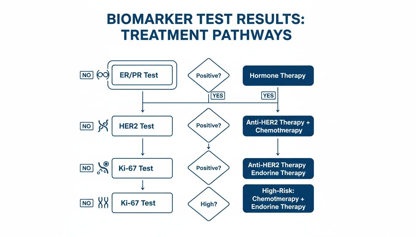
As you can see, a positive or negative result for each of these markers points your doctors toward very specific therapies designed to target your exact type of cancer.
Your final stage isn't just a number; it's a complete summary of your diagnosis. It pulls together the tumor's size, grade, biomarker profile, and lymph node status into one clear framework that directs your entire treatment journey.
The global impact of breast cancer underscores why this detailed pathology work is so critical. While nearly 50% of cases are found in less developed countries, the mortality rate in high-income nations dropped by 40% between the 1980s and 2020. This progress is largely thanks to early detection programs and the precise information provided by pathology services. You can learn more about these global statistics on breast cancer to see the bigger picture.
Questions to Ask Your Doctor About Your Pathology Report
Your pathology report has arrived. It’s dense, filled with medical terms, and it’s completely normal to feel overwhelmed. This document is the foundation of your treatment, and your next conversation with your oncologist is where it all starts to make sense.
Going into that appointment prepared with questions can make a world of difference. It shifts the conversation from your doctor simply telling you the results to a genuine discussion about your health. The goal isn't to become a pathology expert overnight but to understand your diagnosis and feel confident about the road ahead.
As a pathologist, I can tell you that the most empowered patients are the ones who ask questions. When you translate the technical jargon from a report into real-world knowledge, you become a true partner in your own care.
Key Questions for Your Oncologist
To help you get the conversation started, here are some essential questions to bring to your appointment. These are based on the sections you’ll find in a typical breast cancer pathology report sample.
- What specific type of cancer do I have (like invasive ductal or lobular), and what is its grade?
- Were the surgical margins clear? If not, what do we do next?
- What were my ER, PR, and HER2 results? How will those results shape my treatment plan?
- Did the cancer spread to my lymph nodes? What does that mean for my prognosis and treatment?
- Based on all this information, what is the final stage of my cancer?
Turning a Report into a Plan
Asking these questions helps you connect the lab findings directly to your treatment. It’s how you get from a piece of paper to a concrete plan of action.
For example, learning your cancer is ER-positive is the reason your doctor might recommend hormone therapy. In the same way, finding out your lymph node status helps clarify why chemotherapy may—or may not—be necessary. This dialogue ensures you understand not just what the plan is, but why it’s the right one for you.
Common Questions About Your Pathology Report
It’s completely normal to have questions after reading your breast cancer pathology report. The language is technical, and the information is life-changing. I've found one of the most important things I can do as a pathologist is help translate these complex findings into clear, straightforward answers.
Let's walk through some of the questions I hear most often from patients, so you can feel more confident and informed.
What’s the Difference Between Invasive Cancer and DCIS?
This is one of the most critical distinctions in your report. You’ll see terms like Ductal Carcinoma In Situ (DCIS), which means abnormal cells were found inside a milk duct but haven’t broken through the duct's wall. Think of it as being contained. That’s why DCIS is considered non-invasive or pre-invasive breast cancer.
On the other hand, invasive cancer (like Invasive Ductal Carcinoma or IDC) means the cancer cells have moved out of the duct and started growing into the surrounding breast tissue. This difference is fundamental because it directly shapes your staging and what treatments your doctors will recommend.
What Does Lymphovascular Invasion Mean?
Your report might note the presence or absence of lymphovascular invasion (LVI). This isn't something you can see or feel; it’s a finding under the microscope. It means the pathologist saw cancer cells inside the tiny blood vessels or lymph channels within the tumor.
Why does this matter? If LVI is present, it signals a higher risk that the cancer could have traveled to the lymph nodes or other parts of the body. Your oncologist will use this information as a key factor in deciding whether treatments like chemotherapy are necessary.
Can My Pathology Results Change Over Time?
Your initial biopsy report gives the definitive diagnosis—it tells you if you have cancer and what kind. But the story doesn't end there. If you have surgery to remove the tumor, the pathology done on that larger piece of tissue will add crucial details, like the final tumor size and whether the surgical margins are clear.
It’s also worth knowing that if cancer ever comes back (a recurrence), its biology can sometimes change. For example, a new tumor might have a different hormone receptor or HER2 status than the first one. This is why doctors almost always perform a new biopsy on a recurrent cancer to make sure the treatment plan is right for the new cells.
When Should I Get a Second Opinion on My Pathology?
You always have the right to ask for a second opinion, and you should never feel hesitant about it. Many patients seek one simply for peace of mind before beginning treatment. As an independent pathologist at Texas Autopsy Services, performing these expert reviews is a big part of what I do.
Getting another set of eyes on your slides is especially a good idea if:
- Your diagnosis is for a very rare type of cancer.
- Any of the findings are listed as "unclear" or "equivocal."
- You simply want confirmation before moving forward with a major treatment plan.
A second opinion helps ensure your treatment is based on the most accurate information possible, which is something every patient deserves.