March 6, 2026 · Texas Autopsy Services
Understanding the Histology Report in an Autopsy
Learn what is a histology report and how this microscopic tissue analysis helps families and attorneys in Texas understand the true cause of death.
On this page
When a loved one passes away, families often turn to me for answers. They're not just seeking peace of mind; they need medical certainty for legal clarity and to understand potential risks to their own health. A private autopsy provides a great deal of information, but the complete story is rarely visible to the naked eye. To find it, I must look much closer.
That is where the histology report comes in. It’s a detailed narrative told by the body's own cells, and it's essential for a conclusive investigation.
Finding Definitive Answers One Cell at a Time
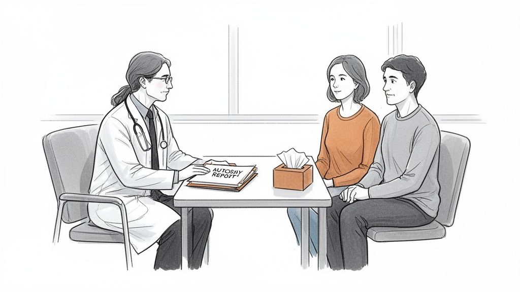
When I perform an autopsy, my entire focus is on delivering a clear, medically sound conclusion. The initial physical examination gives me a roadmap, pointing out areas of concern. However, the most critical questions are often answered at the microscopic level.
Think of it this way: the main autopsy might identify a problem with an organ, but histology explains why that organ failed on a cellular level. It’s the difference between knowing a bridge collapsed and knowing that rusted bolts were the cause.
Why a Microscopic Exam Is So Crucial
At Texas Autopsy Services, I depend on histology to connect all the dots. The process is meticulous. I carefully prepare small tissue samples and analyze them under a high-powered microscope. What I find there is essential for determining the ultimate cause of death.
For example, a visual examination might reveal an enlarged heart. That’s an important clue, but it's not the final answer. The histology report is what tells us if the enlargement was caused by:
- Microscopic scarring from a “silent” heart attack.
- Widespread inflammation of the heart muscle (myocarditis).
- Unusual protein deposits that made the heart tissue stiff and inefficient.
Each of those findings tells a completely different story. They point to different causes of death and can have significant implications for the health of surviving family members. This detailed microscopic analysis is the cornerstone of any truly thorough investigation. It's how we move from uncertainty to clarity. You can learn more about how all these elements fit together by reading about my forensic pathology service.
What a Histology Report Reveals
A histology report is, quite simply, the story the body's tissues tell when I put them under a microscope. It’s where the smallest clues can answer the biggest questions.
During the autopsy, I carefully select small tissue samples from vital organs like the heart, lungs, and brain. These samples are preserved, sliced into sections thinner than a human hair, and stained with special dyes. These dyes act like highlighters, making different parts of the cells stand out so any abnormalities become clear.
Uncovering the Microscopic Truth
When I examine these slides, I’m searching for the microscopic evidence that is completely invisible to the naked eye. More often than not, this is where the final, definitive answer about the cause of death is found.
I’m looking for specific signs:
- Inflammation: Are there white blood cells rushing to a specific area? This can point directly to a hidden infection, an injury, or an autoimmune disease.
- Oxygen Deprivation: Cells starved of oxygen leave behind a distinct pattern of damage. This is how I can confirm a heart attack or stroke, even when the organ looks normal at first glance.
- Abnormal Cells: This is where I can spot cancer cells that may have spread or identify other strange cell structures that signal a chronic, underlying disease.
- Hidden Infections: Sometimes, I can physically see the bacteria, viruses, or fungi that caused a fatal illness, right there inside the tissue.
This detailed look builds a clear picture of what was happening inside the body at the cellular level. It can either confirm what I suspected or, in many cases, reveal a completely unexpected condition that was the true cause of death. For a closer look at how this fits into the complete picture, see our guide defining a pathology report.
The histology report is what turns a strong suspicion into scientific fact. It's where I can see the faint beginnings of a heart attack, the subtle scarring from a long-term illness, or the body’s cellular fight against a fatal infection.
How We Create the Histology Report
A histology report isn't the result of a quick peek under a microscope. It’s the final product of a careful, hands-on process that begins during the autopsy. This process takes both scientific skill and patience, as each step is designed to unlock the cellular secrets hidden within the body's tissues.
It all starts with collecting small tissue samples from key organs and any areas that look unusual. These samples, often no larger than a postage stamp, are immediately placed in a preservative fluid, usually formalin. This step, known as fixation, is absolutely critical. It stops all natural decay and essentially freezes the tissue's structure in time, preserving it exactly as it was.
From Tissue to Slide
Once fixed, the tissue is too soft to be sliced. First, it goes through a process where all the water is removed and replaced with hot paraffin wax. As the wax cools and hardens, it creates a firm, stable block with the tissue safely embedded inside.
This wax block is then secured in a special instrument called a microtome. This device is a high-precision slicer, capable of cutting the block into incredibly thin ribbons. We're talking sections that are only 4 to 5 microns thick, about ten times thinner than a single human hair.
Each transparent, paper-thin section is then floated on a warm water bath to smooth out any wrinkles before being carefully transferred onto a glass slide. At this stage, the cells are mostly invisible. To bring them to life, I have to stain them.
The standard method for this is a staining combination called Hematoxylin and Eosin (H&E). Hematoxylin stains the core of the cell (the nucleus) a deep blue or purple, while Eosin colors the rest of the cell and surrounding tissues in various shades of pink. This contrast is what allows me, as a pathologist, to clearly see a cell’s layout and spot anything out of the ordinary.
This visual guide shows the basic workflow from collecting the tissue sample to identifying the crucial cellular clues that form the basis of the histology report.
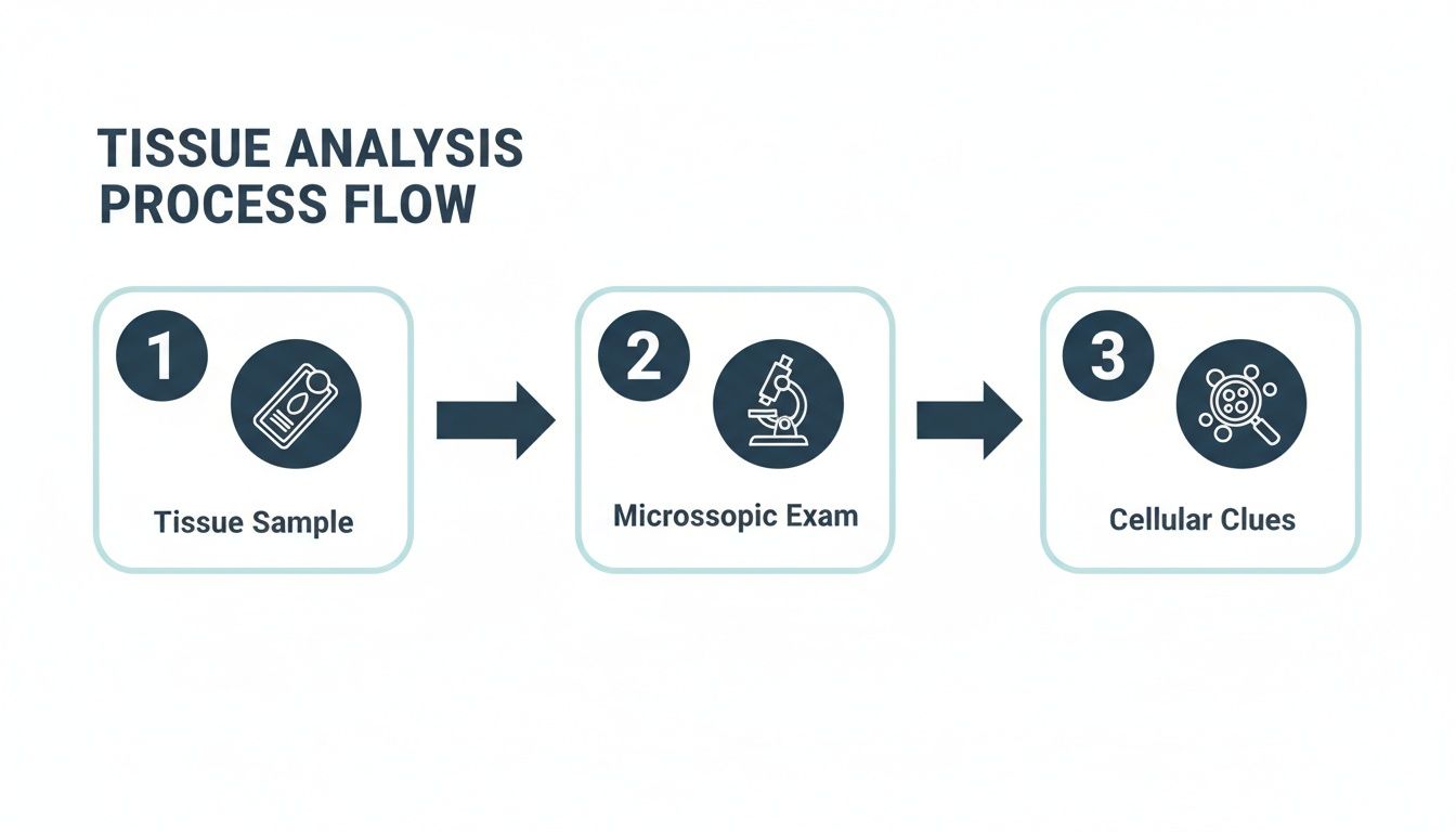
Following this systematic approach guarantees that what I see under the microscope is an accurate and verifiable representation of the tissue.
The Final Microscopic Examination
Only after all this preparation is complete does the final analysis begin. I examine each stained slide under a high-powered microscope, looking for tell-tale signs of disease, injury, or infection. These are details that are impossible to see with the naked eye and provide the answers we're searching for.
This careful interpretation of the cellular story is what ultimately becomes the histology report. It's a detailed process from start to finish, and understanding how it fits into the bigger picture is key. You can learn more about the entire process by reading our guide on the steps to an autopsy.
Connecting Microscopic Findings to the Cause of Death
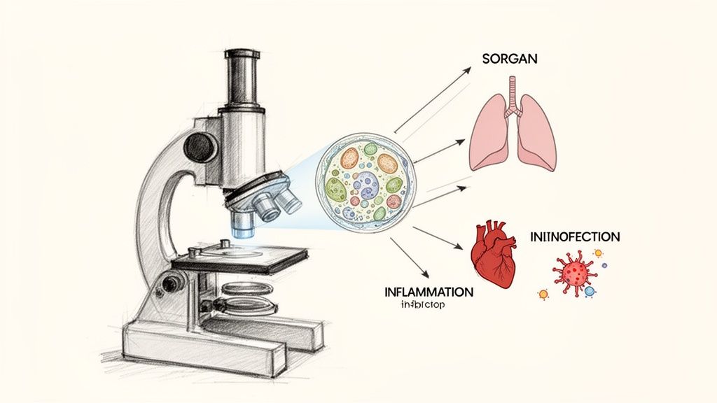
The histology report is where everything comes together. It’s the final, crucial link between what I observe during the physical autopsy and the underlying cellular reason for death. This is often where the most definitive answers are found.
It's not uncommon for an organ to look perfectly normal to the naked eye, but the story under the microscope can be entirely different. Histology is the tool that lets a forensic pathologist see the subtle, hidden damage that confirms a diagnosis.
Where the Real Story Is Often Found
I’ve seen countless cases where histology provides the answer. Take a sudden, unexplained death where the heart appears structurally sound during the initial examination. The histology report can reveal microscopic damage to individual heart muscle cells, the earliest signs of a heart attack that were completely invisible otherwise.
The same goes for infections. If someone passes away from what appears to be a severe infection, histology gives us the proof. By examining lung tissue under the microscope, I can identify the specific type of bacteria or fungus that caused a fatal pneumonia, providing concrete evidence to back up the initial theory.
These cellular findings aren't just minor details; they are often the definitive evidence. They allow me to either confirm the suspected cause of death or rule it out entirely.
As a board-certified forensic pathologist, my primary role is to put all the pieces of the puzzle together. I combine the gross findings from the autopsy, the toxicology results, and the microscopic details from the histology report to build a single, coherent narrative that is both medically accurate and legally sound.
Identifying Contributing Factors
But the histology report does more than just pinpoint the immediate cause of death. It also uncovers contributing conditions that played a significant role in the person's health decline over time.
For instance, the report might reveal:
- Chronic kidney disease: Microscopic scarring in the kidneys shows us the long-term damage that compromised their function.
- Cirrhosis of the liver: Widespread scarring and abnormal cell structure in the liver point directly to chronic disease that impacted the body’s ability to function.
- Unsuspected diabetes: Subtle changes in the body’s small blood vessels can be a clear indicator of long-standing high blood sugar the person may not have known about.
This information is invaluable. It helps explain not just how a person died, but also sheds light on the chronic health issues that gradually weakened their body. For families, this complete picture provides a deeper understanding and a sense of closure. For legal matters, it establishes a comprehensive and undeniable medical record of a person's health leading up to their death. This is the real purpose of asking what is a histology report. It’s about finding the full story, written in the body's own cells.
How Families and Attorneys Use the Report
When I talk with families, the first thing they want is to understand why. A histology report can finally give them that answer, cutting through the uncertainty to provide a clear, scientific reason for their loss.
But it’s about more than just closure. Sometimes, what I find under the microscope can be a genuine gift to the living. We might uncover a hereditary heart condition or a genetic disorder, giving surviving family members crucial information that could protect their own health for years to come.
A Tool for Legal Certainty in Texas
For an attorney, a histology report is about facts. It’s a powerful piece of objective evidence that can be pivotal in a case. In legal battles across Texas, from medical malpractice and wrongful death claims to complex insurance disputes, the findings from our lab can prove or disprove an argument with scientific certainty.
A lawyer can use my report to show a jury that a specific medical event, visible only at the cellular level, was the direct cause of death. A large part of my role as a board-certified forensic pathologist is ensuring that information is understood. At Texas Autopsy Services, my work isn’t done until I can explain complex medical findings in plain language, whether for a family member or a courtroom.
Because these reports contain sensitive and private medical details, it's critical that families and legal professionals protect this information. Adhering to HIPAA compliant document sharing practices is essential when transmitting or storing a histology report to maintain privacy and legal compliance.
In the end, whether it’s for a family seeking peace of mind or a lawyer building a case, the histology report tells a story based on unbiased, cellular-level facts.
Your Path to Answers
For families and attorneys all across Texas, from major cities to the most rural counties, this report can be an essential document. It might be what brings personal closure, or it could become the foundation of a legal case. The evidence inside is unbiased, based entirely on facts at the cellular level.
If you are trying to understand what happened to a loved one, please feel free to contact my office for a confidential consultation. I am here to help you find the answers you and your family deserve.
Frequently Asked Questions About Histology
As a forensic pathologist, I've sat down with countless families and attorneys over the years. Naturally, you have questions. Here are the straightforward answers to some of the most common ones I hear at my practice, Texas Autopsy Services.
How Long Does It Take to Get Histology Results?
I know the waiting is the hardest part, but this is one step in the process that simply can't be rushed. Getting the final histology results typically takes several weeks.
The tissue has to be carefully fixed, processed, thinly sliced, and stained before I can even begin my analysis under the microscope. Rushing any part of that would compromise the quality of the findings. My commitment is to provide the clearest, most accurate answers possible, and that takes time.
Will Histology Always Find a Cause of Death?
Most of the time, yes. Histology is an incredibly powerful tool for uncovering the truth, especially when the cause of death isn't obvious from the external examination. It’s how we find silent, microscopic clues like a hidden heart attack, a subtle infection, or an early-stage cancer that left no other trace.
However, in a small number of cases, a definitive cause of death might still be "undetermined" even after a full autopsy and histology review. Even then, the report provides immense value by systematically ruling out dozens of other possibilities, narrowing the focus and giving you as much clarity as science allows.
Can a Histology Report Be Used in Court in Texas?
Absolutely. When a histology report is part of a complete autopsy performed by a board-certified forensic pathologist like myself, it's considered expert evidence in Texas courts.
The findings aren't opinion; they're objective facts based on what the cells themselves show. This kind of microscopic evidence can be the deciding factor in civil and criminal cases, including medical malpractice claims, insurance disputes, and wrongful death lawsuits, where it can prove or disprove a legal argument with scientific authority.
At Texas Autopsy Services, my entire focus is on providing clear, scientifically sound answers to help you find closure and certainty. For a confidential consultation to discuss your specific needs, please get in touch with our team.