August 2, 2026 · Texas Autopsy Services
Biopsy Atypical Cells What the Finding Means for You
Learn what biopsy atypical cells means, how pathologists interpret it, common causes, next steps, and when a second opinion review can help your family.
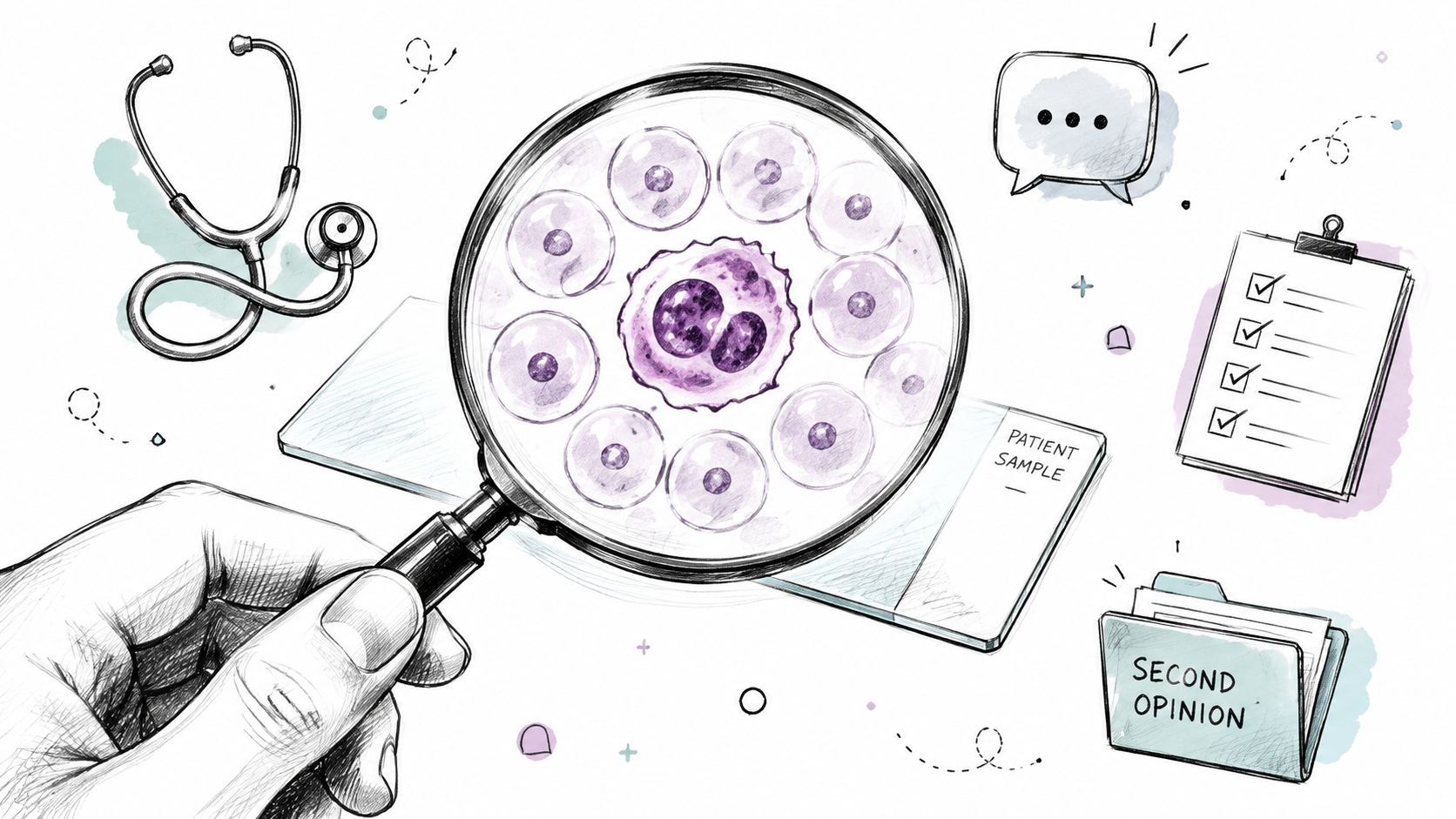
On this page
- When a Biopsy Report Says Atypical Cells
- What Atypical Cells Actually Means Under the Microscope
- How the Same Word Means Different Things in Different Organs
- What Clinicians Usually Do Next
- When a Second-Opinion Review or Forensic Pathology Consultation Helps
- Talking With Your Clinician and Your Family
- Key Takeaways and How to Reach Our Team
A pathology report that says atypical cells can leave a family stuck between two reactions, relief that the word is not cancer, and fear that it still might be. That tension is real because the term lives in a gray zone, it describes cells that don't look normal under the microscope, but it does not, by itself, settle the diagnosis. For readers who are staring at a report and trying to make sense of the language, a plain-English guide can lower the panic and show what usually happens next.
When a Biopsy Report Says Atypical Cells
A family opens a report after a biopsy and sees a word they weren't expecting. The tissue was taken because something looked off on imaging or during an exam, and now the paper says atypical cells. That single word can feel vague, but in pathology it has a specific meaning, and it usually signals that the sample needs context, not instant reassurance and not instant alarm.
Pathology reports are easier to read when the terms are unpacked in order. A useful general guide for that process is what your pathology report means, because it shows how pathologists build conclusions from microscope findings, sample quality, and clinical context. In the same spirit, this article stays focused on what biopsy atypical cells means, how the label changes by organ, and why follow-up often matters more than the word itself.
Practical rule: Atypical does not mean benign, and it does not mean cancer. It means the cells looked unusual enough that the pathologist couldn't call them normal without more information.
That distinction matters because the same term can sit in very different places across lung, breast, thyroid, and urinary tract pathology. A biopsy from one site may trigger closer imaging, while another may prompt repeat sampling or a second read of the slides. Families often hear the term once and assume it carries one universal meaning, but pathology doesn't work that way.
For readers comparing their report with the broader medical record, our own discussion of biopsy pathology report interpretation may help frame the language. Texas Autopsy Services writes from a forensic pathology perspective, so our role here is educational. We're not replacing the treating clinician, we're helping you understand how a pathologist thinks about the finding.
What Atypical Cells Actually Means Under the Microscope
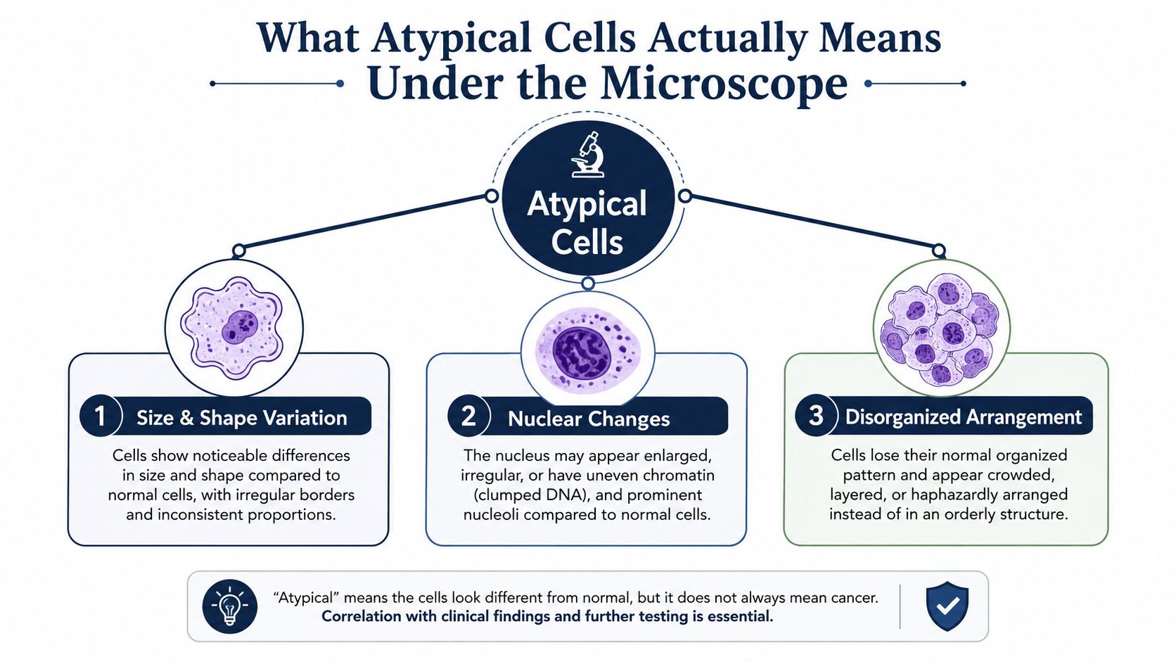
The microscopic changes pathologists look for
Under the microscope, atypia is a morphologic diagnosis. It's based on what the cells look like, including enlarged or irregular nuclei, coarse chromatin, irregular nuclear membranes, higher nuclear-to-cytoplasmic ratio, abnormal crowding or overlap, and pleomorphism, which means variation in size and shape (pathology review). In respiratory cytology, the label is used when cells clearly exceed benign or reactive change but still fall short of “suspicious for malignancy” (respiratory cytology review).
A simple way to picture it is to think of a row of houses. Normal cells have a predictable roofline, windows, and spacing. Atypical cells still look like houses, but some have crooked windows, uneven walls, or a cluttered layout. The structure isn't normal, yet it isn't clearly collapsed.
The term also includes a gray zone between reactive change and true dysplasia. Inflammation, infection, mechanical irritation, and other noncancerous processes can produce microscopic changes that resemble atypia, which is why a report can't be read in isolation. The biopsy site, the sampling method, and the imaging all matter.
Plain-language translation: atypia is a warning label on the microscope slide, not a final verdict on the person.
Why the report is one piece of a larger puzzle
That's why pathologists don't treat the term as a standalone diagnosis. A tissue sample can show cells that look off because the area was inflamed, under-sampled, or precancerous. Without the rest of the story, the slide can't answer every question.
This is also why a family can hear one clinician say the finding is concerning and another say it's indeterminate. Both can be correct, depending on the organ and the rest of the data. The microscope tells part of the story, not the whole thing.
How the Same Word Means Different Things in Different Organs
A pathology report can use atypical cells in more than one way, and the organ system changes the meaning. In the lungs, the term often sits much closer to cancer risk, especially when the imaging or sampling pattern already raises concern. In the breast, the word may describe a finding that is important but far less predictive on its own. In thyroid and urine cytology, the same label is tied to different follow-up pathways, which is why the report has to be read with the organ, the specimen type, and the clinical question in mind.
Lung biopsy literature is the clearest example of that difference. A UCLA review notes that prior studies found 75% to 90% of patients with atypical cells on lung biopsy were later diagnosed with cancer, and one cited series reported atypia in 28% of nondiagnostic biopsies with 66% malignancy within 2 years (UCLA review). In more recent endobronchial ultrasound-guided transbronchial biopsy data, atypical cells appeared in 165 of 2,291 patients, 7.2%, and 120 of 165, 73% were ultimately malignant while 45, 27% were benign (same review).
Four organ systems, four very different conversations
Breast pathology behaves differently. A Yale review found 327 atypia cases among 3,898 biopsies, 8%, with 38 cancers on excision and an overall malignancy rate of 13% across all atypias (Yale review). Long-term data matter too, because one cohort found 143 of 698 women, 20.4%, developed breast cancer over a mean of 12.5 years, and cumulative incidence reached 29% at 25 years after atypia biopsy (Yale review). A finding like that can look modest on the page, yet it carries real weight when the tissue architecture and the patient's history are placed side by side.
Thyroid and urine cytology use the term in a more structured way. For thyroid nodules, AUS/FLUS is commonly followed by repeat fine-needle aspiration after 3 or more months (thyroid review). In voided urine cytology, one follow-up study reported 49 of 72 samples, 68%, had a positive histologic diagnosis of urothelial malignancy, and 55% of those malignant cases were high-grade urothelial carcinoma (urine cytology study). That is why a family may hear the same word in very different clinical settings and receive very different advice about what it means next.
| Organ System | Atypical Frequency | Malignancy on Follow-Up | Typical Next Step |
|---|---|---|---|
| Lung | 7.2% in one biopsy cohort (UCLA review) | 73% malignant in that cohort | Closer imaging, repeat sampling, or additional pathology review |
| Breast | 8% in one core biopsy review (Yale review) | 13% overall malignancy across atypias (Yale review) | Correlation with imaging and long-term risk assessment |
| Thyroid | Indeterminate category rather than a simple frequency figure | Repeat FNA after 3 or more months (thyroid review) | Repeat biopsy after the observation window |
| Urinary tract | Reported as an atypical cytology category | 68% positive histology in one follow-up study (urine cytology study) | Urologic follow-up and histologic correlation |
The point is not that one organ is worse than another. It is that pretest probability changes the meaning of the same word. A lung lesion with concerning imaging is read differently than a thyroid nodule with an indeterminate cytology pattern, and a family that wants a second set of eyes can also ask whether a second-opinion review or a private forensic review is appropriate, including the kind of documentation and chain-of-custody standards used in Texas autopsy and postmortem work, as discussed in our guide to inconclusive biopsy results and when families seek clarity.
What Clinicians Usually Do Next
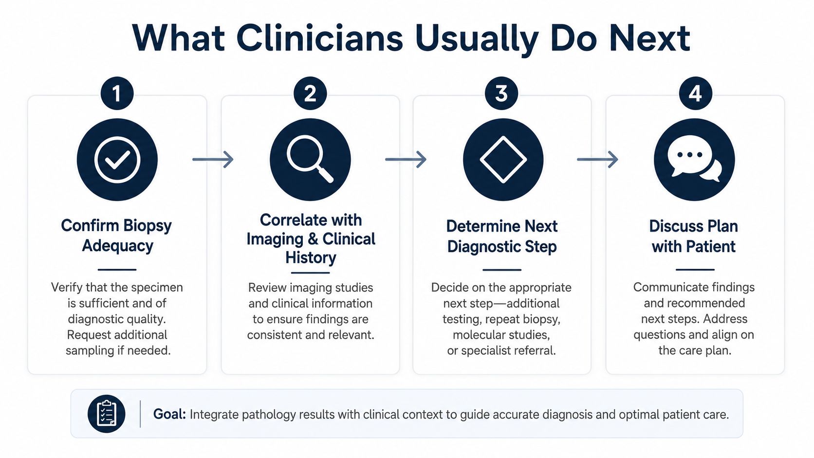
An atypical biopsy result usually leads to a short series of checks, not a single reflex action. Clinicians first ask whether the sample was adequate, because a limited sample can leave the pathologist with just enough tissue to raise concern but not enough to settle the question. Then they compare the pathology with imaging and the clinical story, since a small benign-looking sample can still miss the more important part of a lesion.
The usual decision points
One lung biopsy study found that among 106 patients initially diagnosed with atypia, 80 patients, 75%, were ultimately diagnosed with lung cancer, and more than three-quarters of those cancers were identified within 6 months (lung biopsy study). That kind of result is why clinicians often move to repeat biopsy, surgical sampling, or close radiologic surveillance when imaging is concerning.
The same study found that PET SUV ≥ 2.5, nodule size > 3.5 cm, and mixed ground-glass opacities increased lung-cancer risk (lung biopsy study). Those are the kinds of details that can shift a report from “watch carefully” to “sample again.”
Clinical reality: Atypia is often a decision point, not an endpoint.
For thyroid nodules, the common pattern is different. AUS/FLUS results are often rechecked after at least 3 months, because some atypical changes can settle once inflammation from the first procedure has faded (thyroid review). Families sometimes expect an immediate answer, but timing is part of the method.
If you're trying to make sense of an inconclusive report in a broader medical record, our page on inconclusive biopsy results explains why repeat sampling is so common. That logic is especially important when the lesion's appearance and the pathology report don't line up cleanly.
When a Second-Opinion Review or Forensic Pathology Consultation Helps
A biopsy report that says “atypical” can leave families with a record that feels incomplete. The slide may show changed cells, but the key question is whether those changes fit irritation, sampling limits, or something more serious. A second-opinion pathology review gives another pathologist a chance to look at the same tissue, compare it with the imaging, and read the clinical notes the way a case file is meant to be read, piece by piece.
A review like that usually includes slide re-examination, a fresh look at the prior report, and correlation with the rest of the medical record. That matters because pathology is rarely judged in isolation. A lesion can look suspicious on imaging and still be under-sampled on biopsy, or a report can sound cautious because the specimen only captured the edge of the problem. For families and attorneys who need clarity, the goal is to reduce ambiguity without changing what the tissue already shows.
A representative case of resolving ambiguity
A family may receive one report that uses guarded wording while the treating clinician remains concerned because the imaging does not line up with the pathology. In that situation, an independent review can help answer a practical question: was the first sample too small, did inflammation explain the atypia, or was the lesion probably not fully captured? The point is not to second-guess the care team, but to make the evidence easier to trust.
Before a review can begin, the records have to be assembled carefully. A practical guide for family caregivers can help families request pathology slides, imaging, and related notes without overlooking important documents. If another pathologist is going to read the case, that pathologist needs the same record trail the original clinician had, not just a single report page.
Forensic standard: if the chain of custody is unclear, the conclusion may still be clinically useful, but it may carry less evidentiary weight in a legal setting.
Why chain of custody matters
Chain of custody is the documented record of who handled the specimen, when it changed hands, and how it was stored. In plain language, it is the paper trail that shows the tissue or slides were not lost, mixed up, or altered in a way that would weaken confidence in the result. That standard is routine in forensic pathology, and it also matters when a review may later be used in court or in a formal death investigation.
Texas law and forensic practice place real weight on that documentation. Under Texas-specific rules and Health and Safety Code requirements, a review that may be used beyond the bedside needs a clean record of specimen handling. Texas Autopsy Services provides independent second-opinion review and forensic pathology consultation with chain-of-custody procedures aligned to that setting. For families and counsel who need a structured review path, our pathology consultation services can help organize the record when the report and the rest of the file do not fully align.
Talking With Your Clinician and Your Family
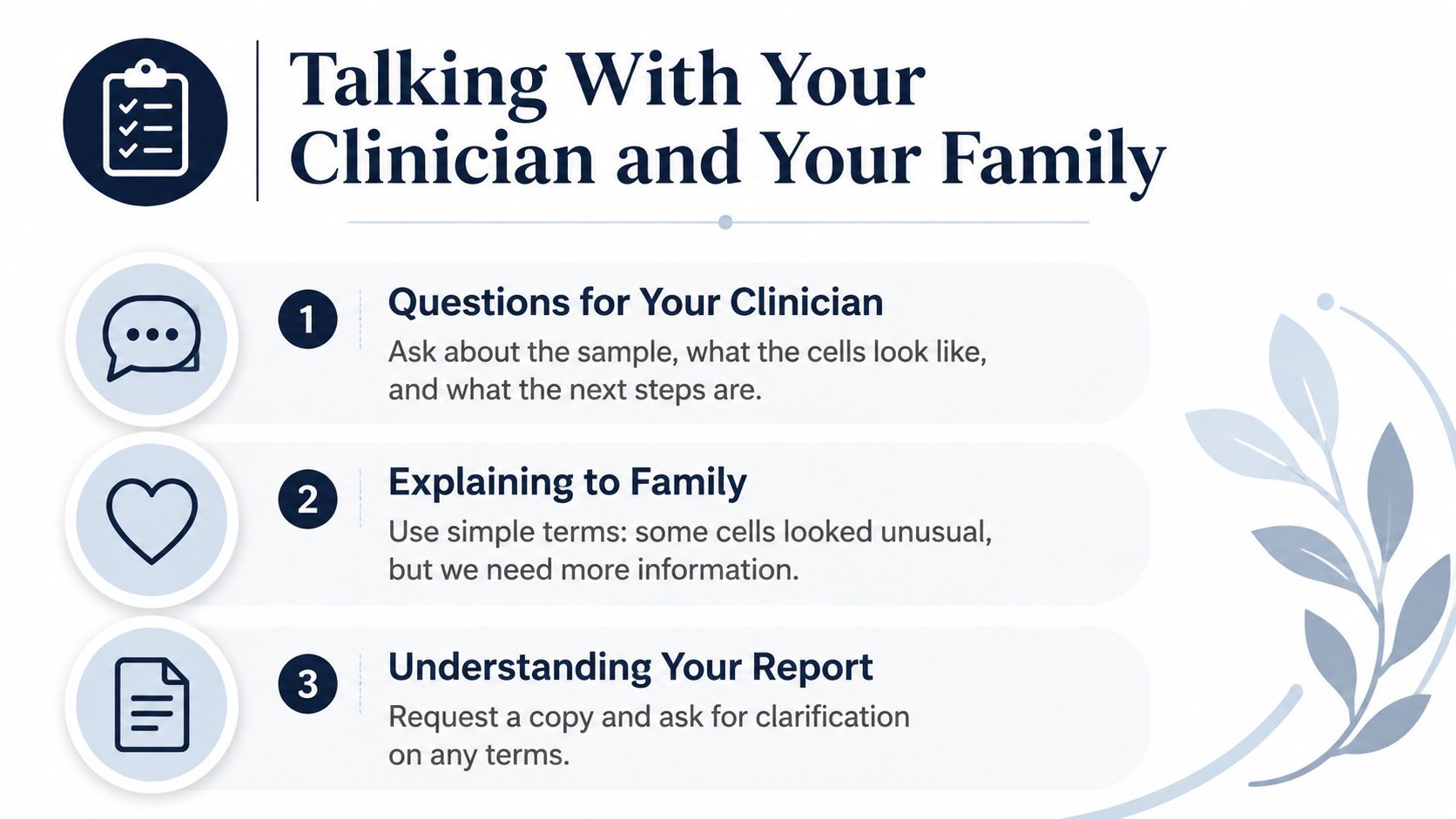
A short list of focused questions can make a difficult appointment more useful. Ask how the sample was obtained, whether the lesion was fully sampled, and whether the imaging matches the pathology. If the report uses a category like AUS/FLUS or another indeterminate term, ask what that means for the next step rather than assuming it already answers the diagnosis.
Questions that help keep the conversation clear
- How was the tissue collected? The method affects how much of the lesion the pathologist could see.
- Was the sample adequate? If not, the report may be describing only part of the problem.
- What do the imaging findings show? Pathology and imaging should be read together.
- What does the follow-up interval mean? A planned recheck isn't the same as a diagnosis.
- Can the slides and report be shared for another review? That matters if a second opinion is being considered.
When you talk with family, keep the language simple. You can say, some cells looked unusual, but we need more information, instead of reciting the report verbatim. That keeps the facts intact without creating confusion around technical terms.
If the situation involves death investigation, cause of death means the disease or injury that directly led to death, while manner of death describes the medicolegal category, such as natural, accident, homicide, suicide, or undetermined. Those terms belong to forensic review, not routine biopsy, but families often hear them in the same broader conversation about records and tissue handling.
Communication tip: Ask for a copy of the report in plain language before the conversation ends. People remember the explanation better when they can read it again later.
If a private second-opinion review is being considered, ask whether the slides can be released promptly and whether all related records are included. The goal isn't to challenge care, it's to make sure everyone is working from the same information.
Key Takeaways and How to Reach Our Team
Atypical cells are a classification, not a cancer diagnosis. Their meaning depends on the organ, the imaging, and the quality of the sample. In some settings, atypia is a high-alert warning. In others, it's a long-term risk marker or an indeterminate category that needs repeat sampling.
For families, attorneys, and county officials who need an independent review, Texas Autopsy Services provides forensic pathology work across all 254 Texas counties. Every examination is performed by a board-certified forensic pathologist certified by the American Board of Pathology, with full chain of custody, transparent flat-rate pricing, and direct communication with our team. Our work is carried out within the Texas Funeral Service Commission framework and Texas Health & Safety Code Chapters 711, 716, and 651.
If you still have questions after reading a biopsy report, it's reasonable to ask for a second set of eyes before making decisions. A clear review can help separate a sampling issue from a true disease process and can give families a steadier path forward.
If you need an independent review, a second-opinion autopsy, or guidance on how biopsy findings fit into a broader cause of death investigation, contact Texas Autopsy Services for a direct conversation with our team. We'll help you understand the records, explain the options, and decide what level of review makes sense for your situation.