April 13, 2026 · Texas Autopsy Services
What Percentage of Skin Biopsies Are Cancer
Discover what percentage of skin biopsies are cancer based on clinical data. We explain the factors affecting rates and how to interpret results.

On this page
- The Search for Certainty in a Biopsy Result
- The Baseline Numbers for Skin Biopsies and Cancer
- Factors That Influence Malignancy Rates
- How Different Biopsy Methods Affect Cancer Detection
- A Pathologists View on Interpreting Your Biopsy Report
- After the Diagnosis Implications and Next Steps
- Common Questions About Skin Biopsies and Pathological Review
- A frequently cited benchmark is that 44.5% of skin biopsies were diagnosed as malignant in one study of dermatologists’ biopsy practices, with important variation by subspecialty and case mix (PubMed).
- Most biopsied moles are not melanoma. In an analysis of 80,368 skin biopsies, 23% were melanocytic lesions, and most of those were benign or low risk (University of Washington Newsroom).
- The number changes because the biopsy method, lesion type, patient age, and the clinician’s level of suspicion all affect yield.
- A biopsy report is an interpretation of tissue, not a simple yes-or-no machine output. Borderline lesions can be difficult, which is why second opinions matter.
- That same search for certainty drives forensic pathology. Whether I’m reviewing a skin specimen or performing a postmortem examination, the work is about reaching defensible medical conclusions.
Waiting for a skin biopsy result can feel longer than it is. A patient is told a spot looked irregular, a sample is taken, and then life narrows to one question. Is it cancer or not?
That moment is familiar to anyone who works in pathology. In my field, families and attorneys come to me for a different kind of answer, but the need is similar. They want a clear, medically sound conclusion when the facts matter most.
A skin biopsy and a postmortem examination are different procedures, but they share the same principle. Tissue is examined because appearance alone is not enough. The eye raises suspicion. The microscope settles it, or at least narrows the uncertainty in a disciplined way.
If you’re in the stage of evaluating a suspicious lesion, a careful clinical exam still matters before any tissue is taken. For readers looking for a patient-facing example of that front-end assessment, a thorough mole check can help explain what clinicians are trying to sort out before a biopsy is chosen.
The Search for Certainty in a Biopsy Result
In pathology, certainty is built step by step. A clinician identifies a lesion that doesn’t fit the usual pattern. Tissue is sampled. The specimen is processed, embedded, sliced thinly, stained, and reviewed under the microscope. The report then translates cellular findings into terms the treating physician can act on.
That process sounds straightforward. It often is. But it is never casual.
What a biopsy actually answers
A biopsy does not answer every question. It answers a focused one. What is this tissue?
That distinction matters because patients sometimes hear “biopsy” and assume the procedure itself creates the diagnosis. It does not. The biopsy creates the opportunity for diagnosis by providing tissue for review.
In forensic pathology, I work with the same logic. An external appearance may suggest one story, but histology, toxicology, and full examination often refine or correct that first impression.
A medical conclusion is only as strong as the specimen, the context, and the interpretation behind it.
Why uncertainty feels so heavy
Uncertainty is difficult because people tend to think in extremes. They expect either benign reassurance or a firm cancer diagnosis. Real pathology includes a middle ground.
Some lesions are plainly benign. Some are plainly malignant. Others sit in a gray zone of atypia, limited sample size, or borderline features.
That doesn’t mean the process is unreliable. It means the process is honest. A careful pathologist does not overstate what the slide can prove.
The connection to postmortem investigation
Families often ask me why an autopsy is needed if there was already a hospital course, imaging, or a death certificate. The answer is the same reason a skin biopsy is done after a visual exam. Clinical impressions are important, but tissue can confirm, challenge, or sharpen them.
That’s the central overlap between dermatopathology and forensic pathology. Both fields exist because medicine sometimes needs final proof, not assumption.
The Baseline Numbers for Skin Biopsies and Cancer
A dermatologist removes a spot because it looks irregular, bleeds, or refuses to heal. The patient hears “biopsy” and often translates that to “possible melanoma.” The actual numbers are more specific than that.
One published analysis found a mean malignancy rate of 44.5% across dermatologists’ skin biopsy practices (PubMed). In practice, that figure is high for a simple reason. Biopsies are usually performed on lesions that have already cleared a threshold for concern.

The main benchmark
That same analysis showed meaningful differences by subspecialty:
- General dermatologists: 41.7% malignant
- Mohs micrographic surgeons: 57.4% malignant
- Pediatric dermatologists: 4.1% malignant
Those gaps reflect referral patterns and patient selection, not a disagreement about what cancer looks like. Mohs surgeons often biopsy lesions that are already strongly suspected to be skin cancer. Pediatric dermatologists work in a population where skin cancer is much less common.
Forensic pathology works the same way. A case selected for full postmortem examination is not a random death from the community. It is a case where the history, scene, or examination raises enough concern to justify tissue review and a closer search for the truth.
Which cancers appeared most often
The diagnostic breakdown reported in that study is worth reading carefully:
| Diagnosis | Share of biopsies |
|---|---|
| Basal cell carcinoma | 22.7% |
| Squamous cell carcinoma | 12.0% |
| Invasive melanoma | 1.4% |
The same report also listed melanoma in situ at 1.5%, squamous cell carcinoma in situ at 6.1%, nevi at 10.0%, benign neoplasms at 10.2%, actinic keratosis at 8.0%, and seborrheic keratosis at 7.6%.
The practical point is straightforward. Most malignant skin biopsy results are not melanoma. Basal cell carcinoma and squamous cell carcinoma account for a much larger share of positive cases in routine practice.
That distinction matters at the microscope. In dermatopathology, as in an autopsy, the public often focuses on the most feared diagnosis, while the pathologist has to sort through the diagnoses that are most prevalent.
For readers who want to connect these percentages to the wording they may see on pathology paperwork, this explanation of skin biopsy results gives useful context.
Laboratory certainty also depends on handling the specimen correctly from the start. Even a small container choice can affect sample integrity in other testing environments, which is why basic tools like a PCR reaction tube matter so much in bench science.
Clinical takeaway: A biopsy series can show a high malignancy rate because clinicians are sampling lesions that already look suspicious, not because half of all skin spots are cancer.
Factors That Influence Malignancy Rates
The number attached to a biopsy report does not come from chance alone. It comes from selection. A dermatologist chooses to biopsy because something about the lesion, the patient, or the history raises concern.
That’s why one practice can have a very different malignancy yield from another without either one doing poor medicine.
The lesion itself drives the first decision
A lesion’s visual pattern matters before any tissue is sampled. Irregular borders, asymmetry, unusual pigmentation, surface change, bleeding, or persistent nonhealing behavior can all increase concern.
Some lesions also stand out because they do not match a patient’s other moles. Others are biopsied because they recur, ulcerate, or continue changing despite time and treatment.
The point is simple. The biopsy population is already enriched for suspicious findings.
Patient context changes the probability
A biopsy result is never read in a vacuum. Clinicians weigh:
- Age: Older patients are more likely to have certain skin cancers than children.
- Sun exposure history: Long-term ultraviolet exposure shifts the likelihood toward sun-related lesions.
- Family history: A strong family history can change how aggressively a lesion is investigated.
- Body site: Sun-exposed locations often carry a different level of concern than protected areas.
These are not abstract details. They are the background that makes one lesion worth watching and another worth sampling now.
Site and specialty matter
The verified data also notes that lesion location affects malignant yield, particularly on sun-exposed areas, and that subspecialty patterns differ. That is exactly what pathologists expect. A highly selected specimen from a high-risk setting will not produce the same results as a broadly sampled low-risk population.
In forensic pathology, this is similar to case context. The same autopsy finding means different things depending on history, scene information, medical records, and timing. Tissue findings gain meaning when they are interpreted within the right frame.
If you’ve been told a result was “abnormal,” that term needs context before it becomes a conclusion. This discussion of skin biopsy results abnormal reflects that distinction well.
Practical rule: A biopsy is usually the end of a clinical suspicion process, not the beginning of one.
How Different Biopsy Methods Affect Cancer Detection
Not all biopsies collect tissue in the same way. That affects what the pathologist can see and how likely the sampled lesion is to prove malignant.
The three methods most patients hear about are shave, punch, and excisional biopsy.

Shave, punch, and excisional biopsies
A shave biopsy removes a more superficial portion of skin. It is often used when the lesion appears raised or more surface-based.
A punch biopsy removes a cylindrical core of tissue and captures more depth. It can be helpful when architecture below the surface matters.
An excisional biopsy removes the whole lesion, usually with a surrounding margin. When the clinical concern is high, this approach often gives the pathologist the best overall view.
The yields differ
Verified data reports different malignancy yields by biopsy type: 15% for shave biopsies, 25% for punch biopsies, and 40% for excisional biopsies (Liv Hospital summary).
That doesn’t mean excisional biopsy is always the best method for every lesion. It means clinicians tend to use excision when the lesion already looks more concerning or when complete architecture is important.
A pathologist also cares about handling and specimen integrity. In laboratory medicine, even the container matters because sample quality starts before the slide is ever cut. For readers interested in how specimen handling tools fit into molecular work, this explanation of a PCR reaction tube gives a useful example from another part of the lab world.
More biopsies can find more early melanoma
The same verified data notes that for every 1,000 additional biopsies performed in a population, about 6.9 extra melanoma cases were diagnosed, mostly in early stages (Liv Hospital summary).
That is an important trade-off. More tissue sampling can improve early detection, but it can also increase the number of very early lesions found without changing the burden of advanced disease in the same way.
In practice, what works is matching the biopsy method to the clinical question. What does not work is treating every lesion as though one sampling approach fits all.
A Pathologists View on Interpreting Your Biopsy Report
A patient checks the portal at 10:30 at night and sees words like “atypical melanocytic proliferation” or “cannot exclude early melanoma.” The specimen is only a few millimeters wide, but the implications feel much larger. I see the same search for certainty in forensic casework. A family wants one clear answer from limited tissue, limited context, and language that has to be precise enough to guide the next decision.
What the pathologist is deciding
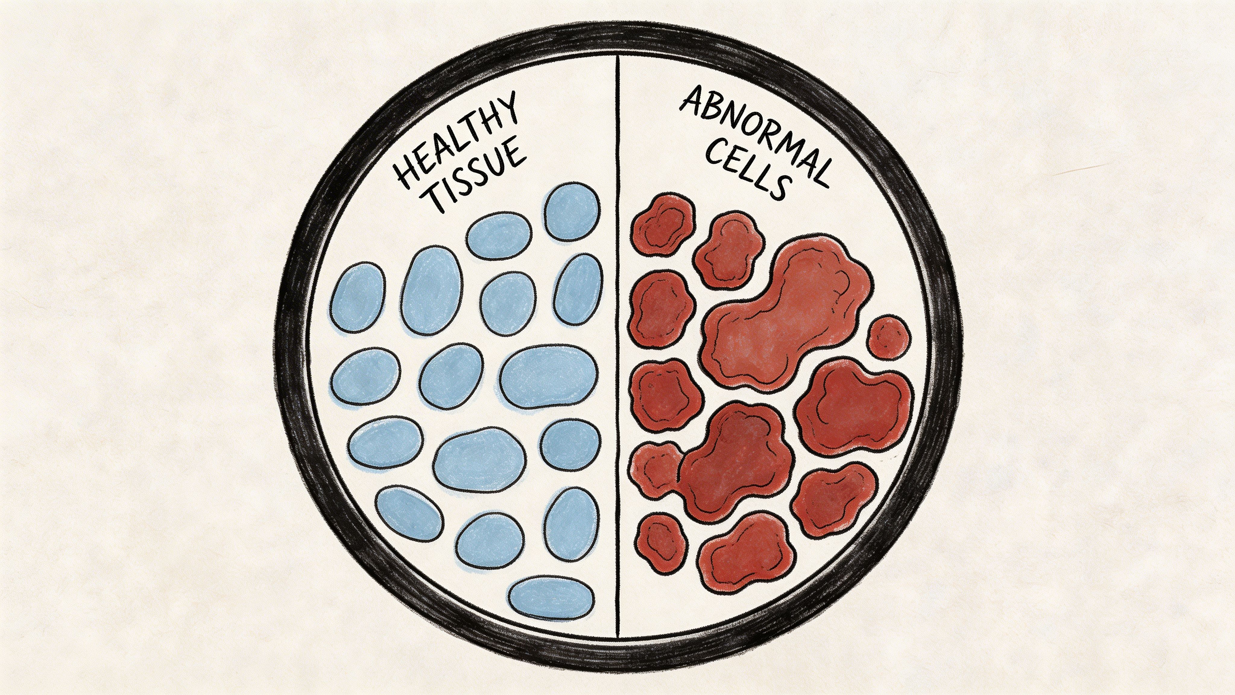
A skin biopsy report is an interpretation of patterns, not a transcript of what the eye sees on a single glance. The pathologist examines architecture, cytology, maturation, symmetry, depth, mitotic activity, inflammation, and whether abnormal cells cross the boundaries that separate a confined process from an invasive one.
Common report terms include:
- Benign for a lesion that lacks malignant features
- Atypical for changes that are real but fall short of a clean malignant diagnosis
- Dysplastic for abnormal structure or cytology, often in melanocytic lesions
- In situ for malignant cells confined to the expected compartment
- Invasive for malignant cells that extend beyond that boundary
Those distinctions affect excision margins, follow-up intervals, and whether the clinician treats the biopsy as the final procedure or the first step.
Why some reports use guarded language
Melanocytic lesions are a good example. Many are straightforward. Some are not. Borderline lesions can show overlapping features, and pathology reports reflect that uncertainty with careful wording rather than false certainty.
In my field, that restraint matters. In a postmortem investigation, overstating a finding can misdirect an entire case. Dermatopathology has the same discipline. Phrases such as “favor,” “compatible with,” or “cannot rule out” are not evasive. They tell the clinician how confident the pathologist is, where the limits are, and whether more tissue or correlation is needed.
For readers who want a plain-language guide to the report itself, this explanation of a biopsy pathology report breaks down the terminology patients often see in the portal.
What a second review actually looks like
The value of second review here is procedural, not philosophical. If the diagnosis will change management, the treating physician can request the original glass slides, tissue blocks, or both for outside review by a dermatopathologist. The consulting pathologist then issues a separate written report, often noting whether the outside interpretation agrees, refines the diagnosis, or recommends additional levels, special stains, or complete excision.
That process is familiar to forensic pathologists. Independent review is often requested when the first opinion is incomplete, disputed, or carrying legal significance. The parallel is direct. In both settings, the point is not to collect opinions until one feels comfortable. The point is to test whether the diagnosis holds up when another specialist examines the same evidence.
A second review is especially reasonable when:
- The report contains borderline language such as atypical, uncertain malignant potential, or suspicious
- The pathology result will change treatment such as wider excision, surveillance, or referral
- The clinical appearance and the microscopic diagnosis do not fit well together
- The biopsy is partial, making it hard to judge the full architecture of the lesion
Turnaround times vary by laboratory and shipping, but the practical sequence is usually straightforward. Request records. Authorize slide release. Confirm whether the consultant wants recuts or the original material. Then compare the addendum or consult report with the first diagnosis line by line.
This video gives a brief visual introduction to how a dermatopathologist evaluates a skin lesion under the microscope and why subtle pattern differences can change the final wording.
The best pathology reports are precise about what they know, careful about what they do not know, and clear about what should happen next.
After the Diagnosis Implications and Next Steps
Once the report is final, the practical consequences begin. For some people, the next step is reassurance. For others, it is treatment planning, margin evaluation, follow-up, or referral.
The report also has legal and evidentiary value in some settings. Timing, classification, and wording can matter far beyond the clinic.

For patients and families
If the diagnosis is benign, the main issue is often whether the lesion needs no further care, routine observation, or complete removal for comfort or appearance.
If the diagnosis is malignant, the treating team has to decide whether the biopsy was both diagnostic and therapeutic, or whether more tissue needs to be removed.
A pathology report should never be read as a stand-alone life sentence. It is one part of a larger medical decision.
For attorneys and case review
A skin biopsy result can become important evidence in disputes involving delay in diagnosis, adequacy of follow-up, or the sequence of medical events.
In that setting, the exact wording matters. “Suspicious for,” “consistent with,” and “diagnostic of” are not interchangeable phrases.
That is one reason independent review can be valuable. In death investigation, the same logic applies when official findings need closer scrutiny. Objective review is often what resolves conflict between assumption and evidence, much as public agencies rely on formal findings in County Forensic Autopsies.
What tends to work and what doesn’t
Some responses help immediately:
- Ask for the actual pathology report. A portal summary is often too brief.
- Review the result with the treating physician. Clinical context belongs beside the slide interpretation.
- Request slide review when the language is uncertain. This is especially important for borderline melanocytic lesions.
Other approaches usually create confusion:
- Relying on internet image comparisons alone
- Treating every abnormal result as proof of aggressive cancer
- Assuming every disagreement between doctors means negligence
Independent review is not a sign of distrust. It is a standard way to strengthen medical certainty when the consequences are significant.
Common Questions About Skin Biopsies and Pathological Review
Can a skin biopsy be wrong
A biopsy can be limited by sampling, interpretation, or the biology of a borderline lesion. That is why pathology uses careful terminology and why second opinions are sometimes appropriate.
A “wrong” result is not always a careless result. Sometimes the specimen captures only part of the lesion, or the features fall in a difficult category.
What is the difference between a pathologist and a dermatopathologist
A pathologist is a physician trained to diagnose disease by examining tissue, cells, and laboratory data. A dermatopathologist is a pathologist with focused expertise in diseases of the skin.
Both work within pathology. The difference is the degree of specialty concentration on skin lesions.
Why might one doctor say a lesion is suspicious and another say it is benign
Different specialists are answering different questions at different stages. A clinician decides whether a lesion deserves sampling. A pathologist decides what the tissue shows microscopically.
Even among pathologists, some lesions are difficult. Borderline melanocytic proliferations are a classic example of why expertise and full context matter.
Does a malignant biopsy always mean a severe cancer
No. “Malignant” includes a range of disease. Some skin cancers are common and often managed locally. Others carry more concern because of invasion pattern, subtype, or stage.
That distinction is why the exact diagnosis matters more than the single word cancer.
When should someone seek a second pathology opinion
A second opinion is reasonable when the report contains borderline language, when treatment would be substantial, when the clinical appearance and pathology do not seem to fit, or when the case may have legal implications.
That is not overreacting. It is a measured response to an important medical record.
How does this relate to forensic pathology
The relationship is in method, not procedure. Both fields depend on tissue interpretation, disciplined language, chain of custody, and defensible conclusions.
In plain terms, cause of death is the medical reason a person died, such as a disease or injury. Manner of death is the classification of how the death occurred, such as natural, accident, suicide, homicide, or undetermined. Chain of custody means documenting who handled evidence or specimens and when, so the integrity of the material can be defended.
That same respect for documentation is one reason pathology findings hold weight in both medicine and law.
If you need clear, independent answers after a death, Texas Autopsy Services provides private autopsy services Texas families, attorneys, and agencies can use when they need an experienced forensic pathologist to review the facts carefully and objectively. I approach each cause of death investigation, independent autopsy, and second opinion autopsy with the same standard that good pathology requires: accurate records, careful examination, and conclusions that stay within the evidence. If speaking directly would help, you can learn more through Texas Autopsy Services.