September 18, 2026 · Texas Autopsy Services
Digital Imaging Examples in Forensic Pathology
See real digital imaging examples used in forensic pathology, from postmortem CT to autopsy photography, with privacy and chain-of-custody guidance.
On this page
- What Digital Imaging Means in a Forensic Autopsy
- Postmortem CT and X-Ray Imaging Examples
- Autopsy Photography and Microscopic Digital Slides
- How Images Appear in the Final Autopsy Report
- Chain of Custody and Privacy for Forensic Images
- Choosing the Right Imaging Approach for Your Case
- Working With an Independent Pathology Practice
An unexpected death often leaves a family with two immediate concerns: what happened, and whether the available evidence can answer that question. During an initial call, families and attorneys commonly ask what digital images a private autopsy produces, whether those files can be reviewed, and how the images will be protected.
At Texas Autopsy Services, we treat imaging as part of a careful medical and legal record. Every examination is performed by a forensic pathologist certified by the American Board of Pathology, and our team explains findings in plain language while preserving the technical detail needed for attorneys, healthcare professionals, agencies, and Texas County officials.
- Digital imaging supplements the hands-on examination. It helps document findings, preserve details for later review, and support the final report.
- Four core modalities serve different purposes: postmortem CT, conventional X-ray, high-resolution examination photography, and whole-slide digital microscopy.
- The report gives meaning to the images. A picture alone isn't a diagnosis. The pathologist's observations, interpretation, and conclusions remain essential.
- Every file requires custody and privacy controls. Case identifiers, access records, secure storage, and authorized release help protect the integrity of the evidence.
- Texas law distinguishes autopsy reports from other investigative materials. The distinction can affect what families, attorneys, agencies, and courts receive.
A digital image can preserve a detail that isn't visible during an external examination, show the position of an implanted device, or allow a consulting pathologist to review tissue from another location. It can also raise difficult questions about privacy, authorization, and whether an image has been altered. Those questions deserve direct answers, especially when a family is considering private autopsy services in Texas.
What Digital Imaging Means in a Forensic Autopsy
The first digital image was created in 1957, when Russell Kirsch at the U.S. National Bureau of Standards used the SEAC computer to scan a photograph of his three-month-old son. The image measured 5 centimeters by 5 centimeters, yet NIST describes the work as the event that kicked off the field of image processing and a foundation for later technologies such as satellite imaging, CT scans, bar codes, desktop publishing, and digital photography. NIST's history of the first digital image shows why converting a physical image into computer-readable data still matters across medicine, documentation, and forensic work.
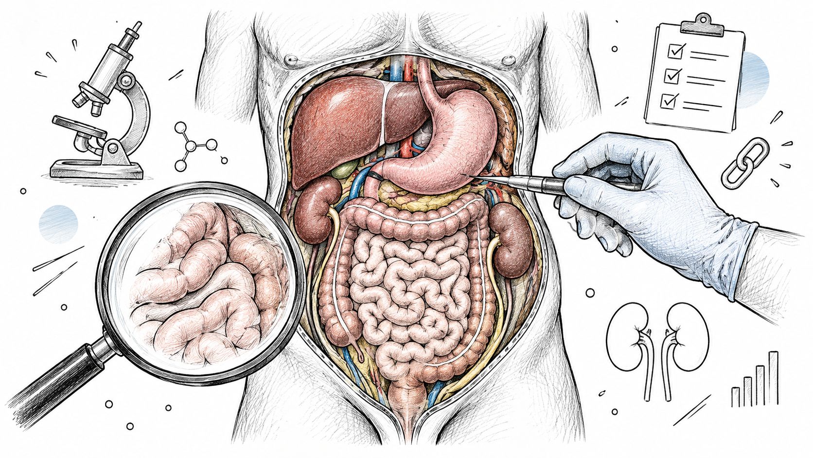
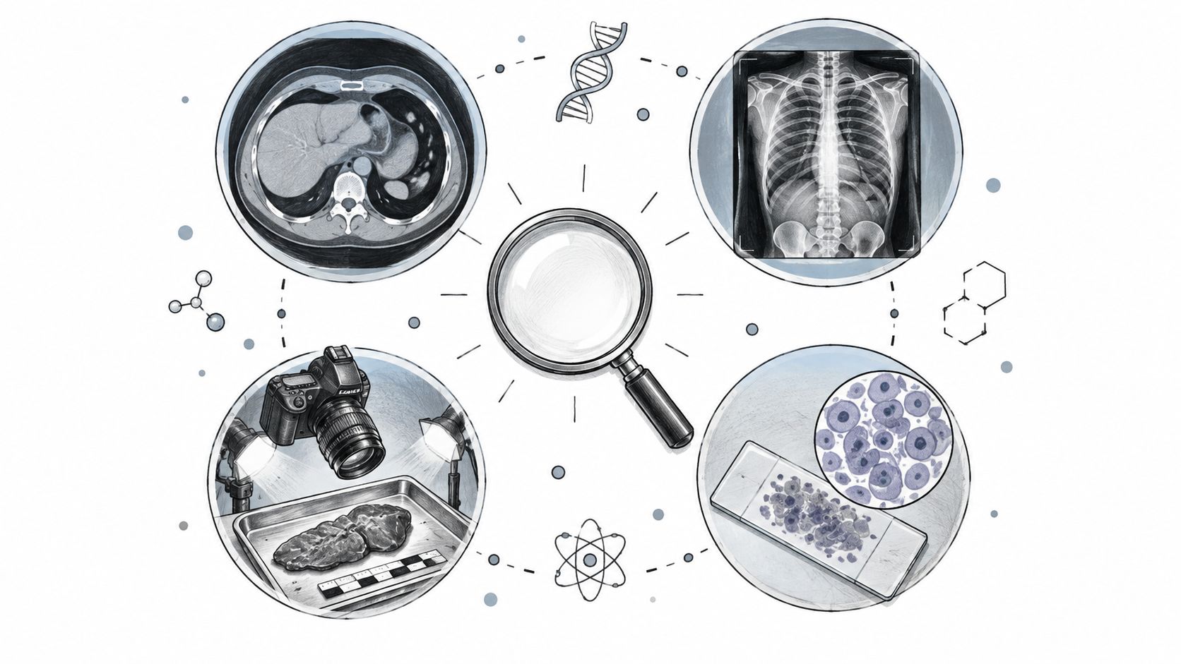
In a private autopsy, digital imaging means capturing, storing, reviewing, and linking visual information to the postmortem examination. Four modalities form the practical foundation:
- Postmortem computed tomography, or PMCT, creates cross-sectional views that can reveal skeletal injury, foreign objects, air or fluid patterns, and other internal features before dissection.
- Conventional radiography, or X-ray, produces flat views that help answer focused questions about fractures, metallic objects, dental structures, or medical devices.
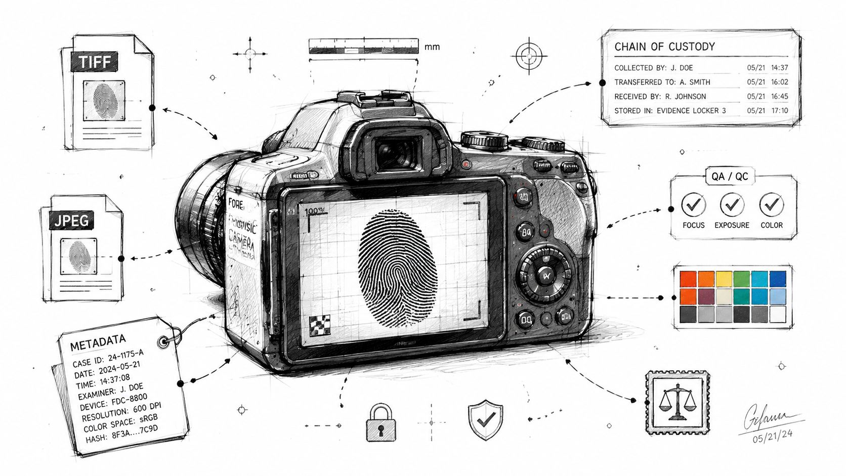
- High-resolution examination photography documents external findings, surgical sites, organ surfaces, and areas of interest with orientation, scale, and case identification.
- Whole-slide digital microscopy scans prepared tissue slides so pathologists can examine cellular findings at high magnification and share them with authorized consultants.
These methods don't replace a hands-on postmortem examination. They supplement it by preserving findings the forensic pathologist can revisit, compare with laboratory results, discuss with consulting experts, and reference while preparing the final report. Imaging can document what was present, but it still requires trained interpretation in the context of medical history, scene information, toxicology, and physical findings.
The scale of digital imaging is especially clear in healthcare. One estimate places worldwide diagnostic medical examinations at about 3.6 billion each year, or roughly 10 million imaging procedures each day. The medical imaging volume estimate illustrates how digital images have become part of regulated workflows for storage, consultation, and review.
Families who want broader context about technology in healthcare may also find this healthcare digital transformation guide from Nutmeg Technologies useful. In forensic pathology, the same basic principle applies: a physical finding becomes structured information that can be examined again, provided the file remains properly identified and protected.
Postmortem CT and X-Ray Imaging Examples
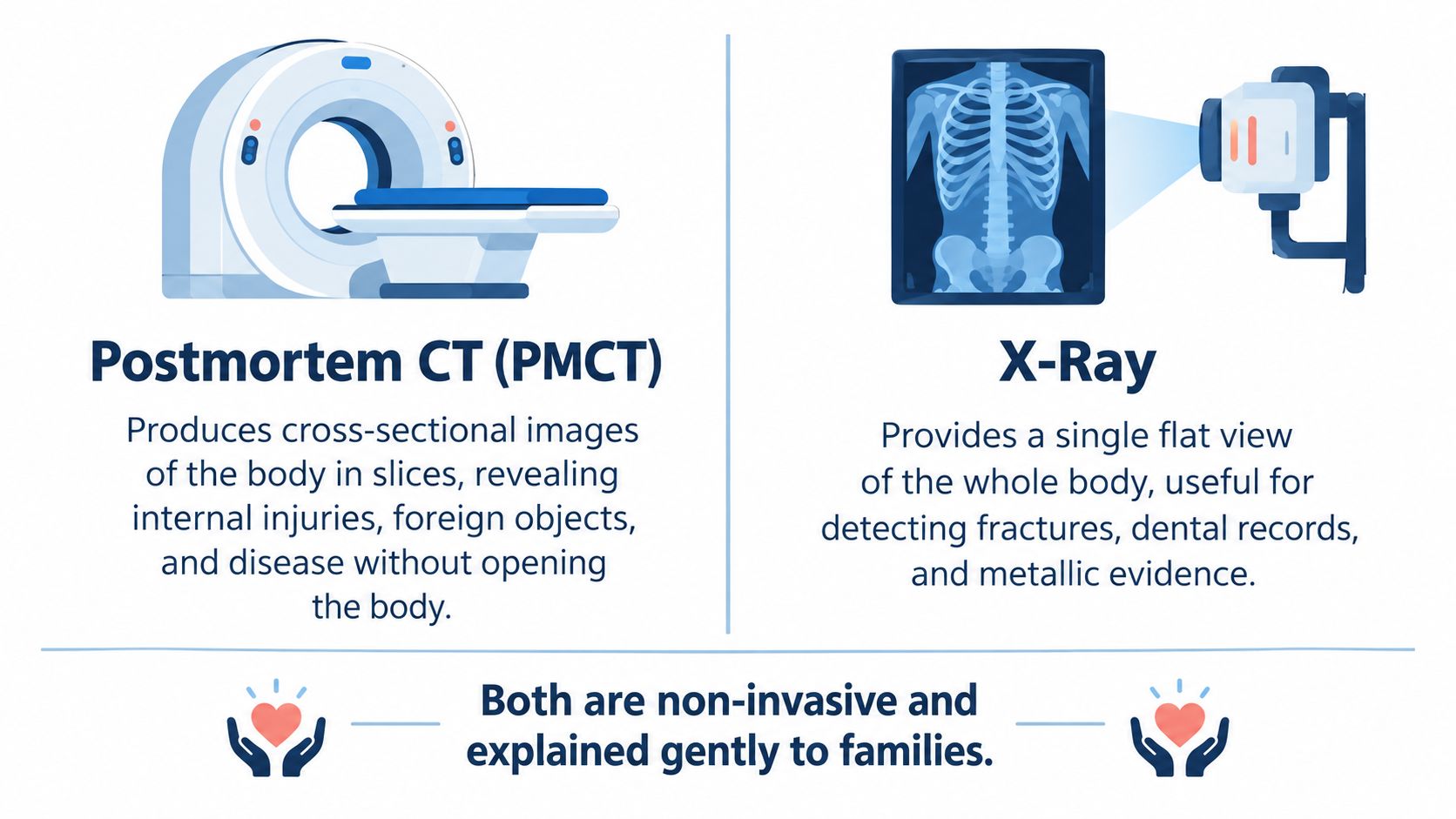
PMCT and X-ray answer related questions, but they don't produce the same kind of evidence. PMCT creates slices through the body, allowing the pathologist to inspect anatomy in multiple planes. X-ray provides a quicker, flat projection that can be valuable when the question is narrower.
Consider a case in which an external examination shows no obvious injury, but the history raises concern about a fall or other trauma. A PMCT review might identify a subtle cervical fracture that isn't visible from the outside. The scan gives the pathologist a precise location to evaluate during the examination and creates a record that can be reviewed later by an authorized expert.
A different situation may involve a hospital death where the position of a central line matters. Plain films can map the device's location and help document whether it remains in an expected position. X-ray may also assist with screening for fractures, metallic evidence, dental structures, or retained medical material.
What each scan can and cannot show
PMCT is particularly useful for bone, gas, foreign objects, and spatial relationships. It can show the path of a projectile or the location of an implanted device without requiring immediate dissection. It doesn't provide the same level of soft-tissue contrast as every other imaging method, so a normal scan doesn't rule out microscopic disease or all forms of injury.
X-ray is useful when the team needs a focused view quickly. It can reveal a fracture or metallic object, but it provides less layered information than CT. The forensic pathologist therefore chooses the modality based on the case question rather than ordering every scan automatically.
Practical rule: The scan is selected to answer a defined question, not simply to create more files.
The image files are archived under controlled access. The final report describes the relevant findings in narrative form, while the underlying studies remain available for authorized review. A family or attorney should ask which image files are included, which are retained as supporting records, and what authorization is required for release.
For readers who want to see how a non-invasive scan is presented, the following video provides visual context for medical imaging technology:
Digital imaging must be interpreted cautiously. It can show a fracture, device, or structural pattern, but cause of death means the disease, injury, or condition that started the fatal sequence. Imaging is one part of that determination, not an independent substitute for pathology.
Autopsy Photography and Microscopic Digital Slides
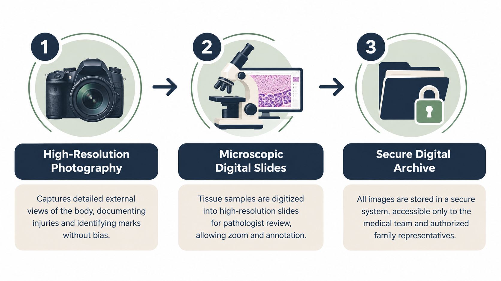
High-resolution examination photography creates the visual record of what the pathologist observes during the autopsy. The purpose isn't to dramatize the examination. It's to document external features, surgical sites, organ surfaces, and specific findings in a way that another qualified reviewer can understand.
Photographs are standardized, oriented, and labeled. A close-up of a contusion pattern, vascular anomaly, or implant should be paired with a ruler and case identifier so the image shows both what was observed and how large or where it was. Wider views establish location, while closer views preserve detail. The sequence matters because an isolated photograph can be misunderstood if the viewer doesn't know the anatomical orientation or examination stage.
From tissue sample to digital slide
Microscopic examination begins with tissue sampling, processing, and preparation on glass slides. Those slides can then be scanned at high magnification, creating a digital file that allows the forensic pathologist to zoom into cellular structures and annotate areas for discussion.
Digital slides support remote consultation without sending the original glass slide to every reviewer. An authorized subspecialist can examine the same scanned material, compare annotations, and offer a focused opinion while the practice retains the original specimen and the digital record.
The result is a reproducible visual record that supports three connected purposes:
- Report preparation: The pathologist can revisit tissue findings while correlating microscopy with the gross examination and medical history.
- Second-opinion review: An authorized forensic pathology expert can review the digital slide alongside the report and other evidence.
- Legal proceedings: Properly identified images can help explain a finding to attorneys, agencies, or a court without relying on memory alone.
Families should expect respectful documentation. Images may include sensitive identifying features, and not every photograph is appropriate for unrestricted circulation. The practice discusses the purpose and scope of requested images before release.
Our explanation of forensic photography provides additional detail about orientation, scale, labeling, and the role of photographs in a medicolegal examination. The central principle remains simple: the image documents a finding, while the report explains its significance.
How Images Appear in the Final Autopsy Report
A final autopsy report isn't a photo album. It's a structured medical-legal document in which the narrative, observations, laboratory findings, and conclusions are connected. Digital images may appear as inline figures, supporting plates, or referenced source files, depending on what best communicates the finding.
A properly identified image commonly includes:
- Case identifier: Connects the file to the correct decedent and examination.
- Anatomical orientation: Shows the body region and direction of view.
- Magnification: Matters for microscopic images and close-up documentation.
- Scale bar or ruler: Helps the reader understand size.
- Capture or scan information: Supports recordkeeping and later review.
The sequence usually follows the logic of the examination. Gross findings may appear first, followed by supporting radiographs or CT still frames, with microscopic plates later when tissue interpretation is relevant. Some reports place selected images beside the related finding, while others place a larger collection in an appendix.
Texas Health and Safety Code section 671 defines an autopsy report broadly. It includes the postmortem examination report, X-rays, photographs taken during the examination, toxicology reports, and other internal-organ examination reports, while excluding investigative reports and other documents reviewed to help determine cause of death. The Texas statute's definition of an autopsy report is important because it distinguishes the formal autopsy record from background material.
Captions do not replace interpretation
A caption may identify an image as a radiograph showing a fracture or a microscopic slide showing a particular tissue feature. It doesn't establish the full medical conclusion by itself. An image without interpretation isn't a diagnosis.
Still frames from a CT volume may be printed when they clearly demonstrate a relevant finding. The complete study may instead be referenced by its DICOM identifier, preserving access to the broader dataset. Microscopic slides are commonly referenced by block and slide number, rather than reproducing every scanned field in the report.
Families and attorneys should ask whether they're receiving the signed report, selected figures, the complete image set, or a combination. The answer depends on the requesting party, authorization, the case status, privacy concerns, and whether an agency or court controls the underlying record.
Chain of Custody and Privacy for Forensic Images
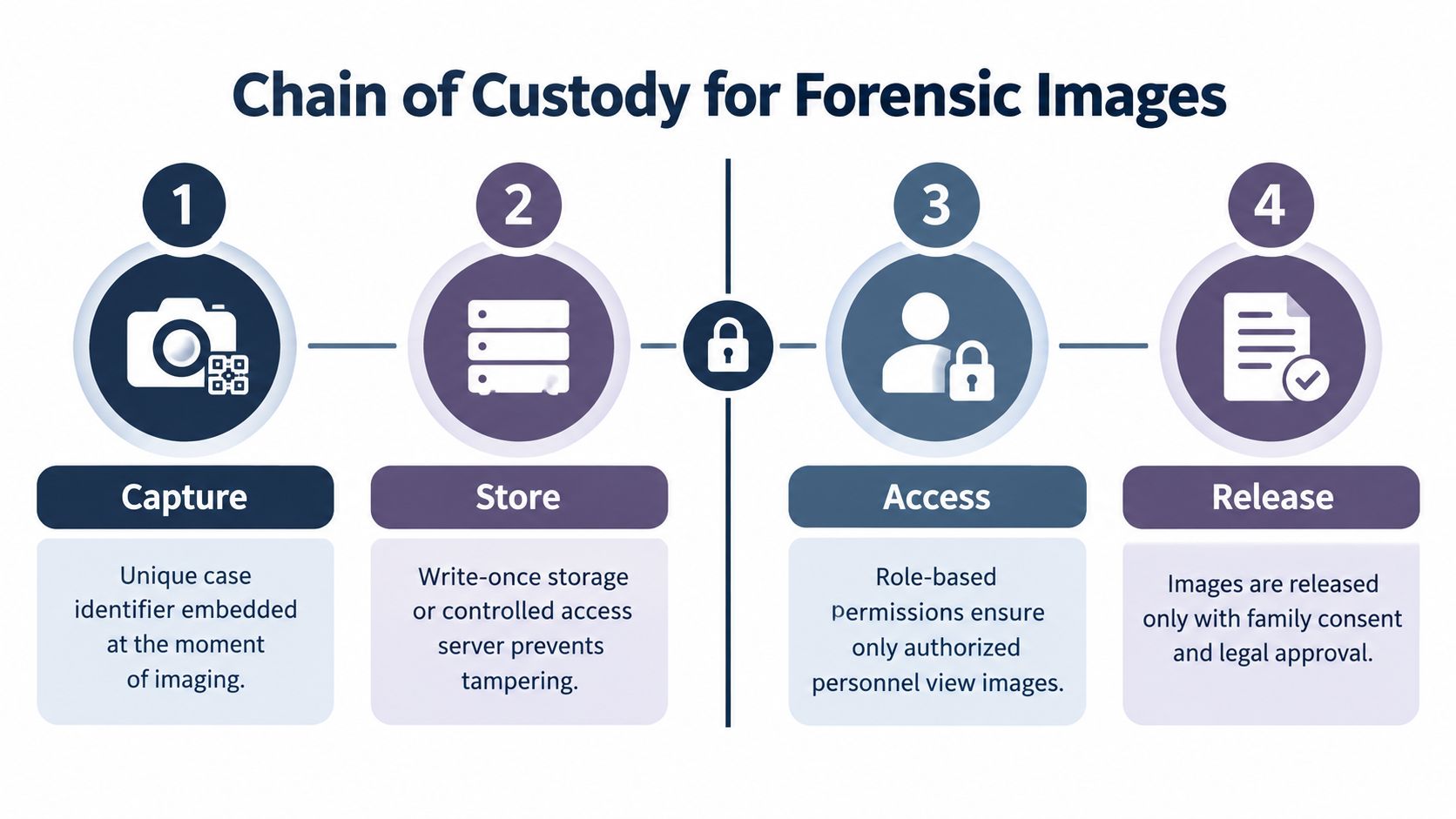
Chain of custody is the documented history of evidence from collection through storage, review, transfer, and release. For digital images, that history must show which case the file belongs to, who could access it, whether it was transferred, and when an authorized copy was provided.
Our team applies custody controls throughout the imaging workflow. The exact technical system can vary, but the purpose remains consistent:
- Capture: A unique case identifier is associated with the image when it's created.
- Store: Original files are placed in controlled storage designed to prevent unauthorized modification.
- Access: Role-based permissions limit viewing to the pathologist and authorized staff whose work requires it.
- Transfer: Images leaving the facility are sent through protected methods, with the transfer recorded.
- Release: Copies provided to a family, attorney, agency, or court are documented in a release log.
HIPAA and Texas privacy obligations may apply to health information and identifying details. A family's request for the autopsy report isn't always identical to an attorney's request for underlying DICOM files, original photographs, metadata, or access logs. Law enforcement or a court may require separate legal authority, particularly when the case is under investigation or subject to litigation.
Access controls matter
A password alone identifies a user but may not adequately limit which files that user can view. An allowlist can restrict access to approved people, systems, or destinations. This comparison of password and allowlist controls offers useful general background on why authorization design matters for sensitive digital records.
Retention and destruction must follow the practice's records policies and applicable legal requirements. At the end of an authorized retention period, secure destruction should prevent routine recovery. If an image includes identifying features of an unrelated person, the team evaluates whether redaction is possible and whether redaction would impair the evidentiary value of the file.
A request should identify the case, the requesting party, the desired file types, and the purpose of the request. Our explanation of the chain of custody form can help families and attorneys understand why each transfer needs a clear record.
Choosing the Right Imaging Approach for Your Case
The right imaging approach depends on the question being asked. A family concerned about an unexplained fracture needs a different record from an attorney evaluating a possible device complication or a pathologist reviewing suspected microscopic disease.
| Modality | Best for | Report artifact | Key limitation |
|---|---|---|---|
| Postmortem CT | Skeletal injury, projectile or foreign-object location, airway and dental detail | DICOM study, selected still frames, radiology or pathology narrative | Bone and air are well depicted, while soft-tissue contrast has limits |
| X-ray | Focused fracture screening, medical devices, metallic objects | Radiographic views and written findings | Flat projections provide less layered information than CT |
| High-resolution examination photography | External documentation, injury mapping, organ-surface findings | Labeled photographs with orientation and scale | Photography documents appearance but cannot show cellular disease |
| Whole-slide digital microscopy | Cellular disease, tissue changes relevant to toxicology, and injury assessment | Scanned slide referenced by block and slide number, with microscopic interpretation | It examines sampled tissue, not the entire body |
PMCT may help locate a foreign object or show a skeletal injury before dissection. X-ray can answer a focused question about a fracture or device without requiring a full CT examination. Examination photography preserves the appearance and location of visible findings, while digital microscopy addresses changes that can only be evaluated at the cellular level.
Decision rule: Match the primary question to the modality. Ask about CT for internal structure, X-ray for focused projection, photography for visible findings, and microscopy for tissue-level disease or injury.
This approach avoids treating digital imaging as a checklist. More images don't automatically produce a clearer conclusion. The forensic pathologist considers the medical history, circumstances, examination findings, and testing plan before deciding which studies will add meaningful information.
Our discussion of digital imaging standards covers the importance of image quality, calibration, labeling, and controlled review. For forensic work, technical quality must serve interpretation and documentation, not replace them.
Working With an Independent Pathology Practice
An independent pathology practice can coordinate the imaging record from intake through final archive. The process begins with the family's or attorney's question, not with a predetermined collection of images.
Our team reviews the circumstances, available medical records, existing autopsy materials, and the purpose of the examination. A board-certified forensic pathologist then determines which modalities are appropriate. PMCT or X-ray may be scheduled through a partner facility, while standardized examination photographs and tissue sampling are completed as part of the autopsy workflow.
The practice's operational responsibilities include:
- Case planning: Selecting imaging based on the question the examination must address.
- Evidence handling: Maintaining identification and custody records for images, specimens, and related documents.
- Report preparation: Connecting visual findings with examination observations, laboratory testing, and the cause-of-death analysis.
- Communication: Explaining technical terms and clarifying what the images do and don't establish.
- Authorized release: Providing reports and image files to the appropriate requesting party under applicable privacy and legal requirements.
For families, a single point of contact can make a difficult process easier to follow. Attorneys may need a complete image set, metadata, or documentation of transfers for litigation. County officials may need a forensic autopsy service that can coordinate transport, examination, reporting, and communication across Texas.
The practice serves all 254 Texas counties and operates from Elgin, Texas, with in-house licensed transport. Texas law can require an inquest when a person dies without having been attended by a licensed physician or when the attending physician can't certify the cause of death with certainty. The Texas Code of Criminal Procedure provision on these inquest triggers explains why private services must coordinate carefully with the appropriate local authority rather than treating every death as a purely private matter.
Texas law also addresses public-health autopsies. With survivor consent, an attending physician or health authority may request an autopsy when more information is needed to protect public health. If there are no survivors or consent is withheld, the health authority shall order the autopsy to determine cause of death, and the results must be reported to the department. Texas Health and Safety Code section 81.045 describes that formal reporting endpoint.
Frequently asked questions
Will every private autopsy include a CT scan?
No. The forensic pathologist selects imaging based on the case circumstances and the question being investigated. CT may be appropriate for internal structure or skeletal injury, while X-ray, photography, or microscopy may provide more useful information in another case.
Can a family receive the original digital images?
A family may request the report and related images, but release depends on authorization, privacy obligations, case status, and whether an agency or court controls the record. The practice can explain which files exist and what process applies to the request.
Does a normal scan rule out a cause of death?
No. Imaging has limitations, particularly for conditions that require microscopic examination, toxicology, medical-record review, or a full postmortem examination. The final opinion depends on the complete evidence.
What do cause of death and manner of death mean?
Cause of death is the disease, injury, or condition that began the fatal sequence. Manner of death describes the classification of how the death occurred, such as natural, accident, suicide, homicide, or undetermined, when the available evidence supports that classification.
How quickly will the report be available?
Timing depends on the examination, testing, medical records, consultations, and legal circumstances. Some cases may be completed quickly, while others require additional work. Families should ask the practice for a case-specific explanation rather than rely on a guaranteed timeline.
An independent autopsy can provide a second opinion when a family has unresolved medical questions, when an attorney needs objective documentation, or when a county needs forensic pathology support. Texas Autopsy Services provides private autopsy services, second-opinion reviews, secure digital imaging, licensed transport, and court-oriented reporting with direct communication from the team.
Texas Autopsy Services can review your case question, explain which digital imaging examples may be relevant, and coordinate an independent autopsy or second-opinion review across Texas. Please visit Texas Autopsy Services to contact our team and discuss the next appropriate step with dignity and clarity.