August 22, 2026 · Texas Autopsy Services
Forensic Digital Imaging Standards: A Guide
Learn forensic digital imaging standards for private autopsies in Texas. Ensure court-admissible evidence with our guide on resolution, formats, and chain
On this page
- The Critical Role of Digital Imaging in Forensic Autopsies
- Understanding Foundational Digital Imaging Standards
- Key Aspects of Forensic Digital Imaging Standards
- Preservation, Chain of Custody, and QA/QC Integration
- Court-Admissibility Considerations Under Texas Law
- Verifying Standards in Private Autopsy Reports
- Why Standards Matter for Trust and Outcomes
A family may receive photographs, scans, or other digital records after a death and assume that the files speak for themselves. They don't. Their value depends on what the images show, how they were created, whether the original files remain protected, and whether another qualified reviewer can understand their context and history.
At Texas Autopsy Services, we treat digital imaging as part of the evidentiary record, not as decoration for a report. Every examination is performed by a forensic pathologist certified by the American Board of Pathology, with documentation designed to support clear cause-of-death analysis, responsible communication, and legal review across Texas.
- Digital images can influence interpretation: Image quality, context, and preservation affect how findings are reviewed.
- DICOM supports medical interoperability: The standard covers communication, storage, retrieval, display, and related information management, not only file transfer. ISO describes DICOM as ISO 12052:2017.
- Original files matter: Forensic guidance emphasizes transparency, security, integrity, and availability throughout the evidence lifecycle. NIST and OSAC forensic digital image guidance addresses those safeguards.
- Chain of custody connects technology to court review: A reviewer should be able to trace a digital image from capture through processing, storage, reporting, and presentation.
- Standards don't replace careful implementation: Consistent procedures, conformance checks, secure archiving, and complete documentation remain necessary.
- Families and attorneys can ask informed questions: A report should explain what was imaged, how files were preserved, and how the images support the written findings.
A private autopsy often begins when a family, attorney, healthcare professional, or county official needs an independent examination or second opinion. The medical findings may be important, but the digital record must also withstand scrutiny from people who weren't present during the examination.
The Critical Role of Digital Imaging in Forensic Autopsies
A Texas family may receive an autopsy report months after an examination, then learn that an attorney or independent expert needs to review the underlying images. The report might describe an injury, surgical site, or other finding relevant to the cause of death. If the images lack context, omit useful metadata, or exist only as exported copies, a later reviewer may have difficulty determining exactly what was documented.
That difficulty does not prove the examination was wrong. It shows that the supporting record may be harder to verify. A court, opposing attorney, or reviewing expert must be able to separate the original observation from later editing, labeling, compression, or presentation choices.
Why image quality affects forensic interpretation
Digital images can preserve details that words alone cannot fully convey. A photograph may show the location and appearance of an external finding. A radiographic image may document a fracture or retained material. Microscopic or specialized images may support documentation of tissue findings. An image does not determine the medical conclusion by itself, but it can support, clarify, or challenge the interpretation in the written report.
Cause of death means the disease, injury, or condition that began the events leading to death. Manner of death describes how the death occurred, such as natural, accident, suicide, homicide, or undetermined, when that classification applies under the investigation. Digital images may inform these determinations, while the pathologist's complete postmortem analysis supplies the medical interpretation.
Practical rule: An image should remain understandable outside the room where it was captured. Its identity, connection to the examination, and processing history should be documented.
Texas Autopsy Services documents forensic photography and related imaging with attention to original files, examination context, and later review. Families and attorneys can also read the practice's explanation of forensic photography in autopsy work to understand how photographic documentation supports an autopsy record.
What can go wrong without standards
A copied image may lose information that helps establish when, where, and how it was created. A derivative file may work well in a report but remain unsuitable as the only preserved version. An export in an unfamiliar format may open on one system while becoming difficult to inspect or compare on another.
Those failures can create avoidable disputes. They may make it harder to reproduce a view, identify an alteration, connect an image with a specimen or body location, or explain the difference between an original file and an enhanced presentation. Standards cannot settle every disagreement about medical meaning. They give families, attorneys, and courts a clearer record to authenticate and evaluate.
Understanding Foundational Digital Imaging Standards
A forensic case may involve CT images from a hospital, photographs created during a private autopsy, and files prepared for a court exhibit. Those records must remain identifiable and reviewable as they move between systems. The history of medical imaging standards explains why that requirement became practical rather than optional. In 1983, the American College of Radiology and the National Electrical Manufacturers Association formed a working group to address image exchange and interoperability problems. Their first standard, ACR-NEMA 300, appeared in 1985, was revised in 1988, and was substantially replaced by DICOM in 1993. A historical review in the medical literature describes how DICOM expanded image transfer from point-to-point connections to networked communication using TCP/IP.
DICOM became a European formal standard in 1995. By 1996, it supported at least seven major modalities, including x-ray angiography, computed radiography, CT, MRI, nuclear medicine, radiofluoroscopy, and ultrasonography. The development reflects a direct operational need: equipment and information systems required a common method for communicating while retaining image and workflow information.
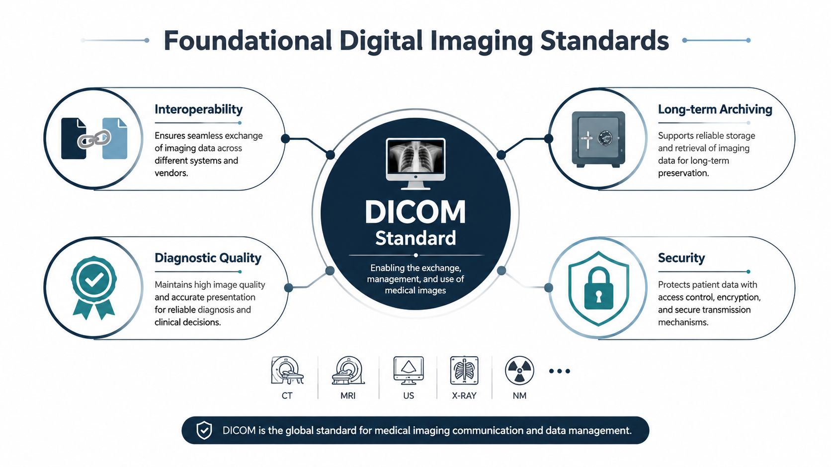
DICOM is more than a file extension
DICOM specifies network communication protocols, command syntax and semantics, and media storage and file-format rules. For forensic pathology, that scope matters because an image needs its surrounding information to remain useful. The record may identify the subject, examination, body region, modality, acquisition details, and connection to a report or finding.
DICOM is particularly relevant when a private autopsy practice reviews CT or other medical images received from a hospital or clinical imaging system. The standard can help retain the connection between an image and its clinical context. It also supports viewing, archiving, printing, processing, and retrieval, as described in ISO's DICOM standard description. In a legal proceeding, that context helps a pathologist explain what the file represents and how it relates to the examination.
JPEG, TIFF, and the trade-off between access and preservation
JPEG was standardized in the late 1980s and became widely used for digital images across industries. Compression can make files easier to transmit and review, but a convenient email attachment or report image may not be suitable as the archival original. The practice should identify which file is the source and which is a working or presentation copy.
TIFF was first published in 1986. TIFF/IT was later standardized in ANSI IT8.8-1993 and ISO 12639:1998, reflecting the role of formal image standards in publishing and healthcare during the 1990s. This historical review of digital image standards places JPEG, TIFF, DICOM, and PACS within the broader development of standardized image exchange.
For forensic work, the goals should remain distinct. Interoperability lets authorized reviewers open and exchange records. Preservation protects the evidentiary original. Presentation produces a clear derivative for a report, hearing, or testimony. One format may not serve all three purposes equally well.
Key Aspects of Forensic Digital Imaging Standards
In a Texas private autopsy, a digital image may later be reviewed by a family, attorney, opposing expert, or court. The record should answer four questions: what was captured, how it was captured, what happened to the file, and whether another qualified reviewer can understand it without guessing. Resolution, file format, metadata, and color management each support that record.
Resolution should match the finding
Resolution should serve the forensic question. An overview photograph establishes location and relationships. A close image may document surface detail, margins, a pattern, or another feature that requires closer inspection. Both views can be necessary when the finding's appearance and position matter.
Higher resolution cannot correct weak technique. Focus, lighting, scale, orientation, and an unobstructed view still determine whether the image is useful. A large file that is blurred, poorly labeled, or separated from its examination context remains unreliable documentation.
File formats should preserve purpose
The practice should retain the original capture in the format produced by the imaging system when practical. A report may use a converted copy because common software displays it more easily. That copy must be identified as a derivative, not presented as the source file.
DICOM can preserve medical image information with metadata and communication details. JPEG may support access and presentation, while TIFF may fit particular archival or publishing needs. The appropriate choice depends on the source, intended use, and whether conversion could remove information.
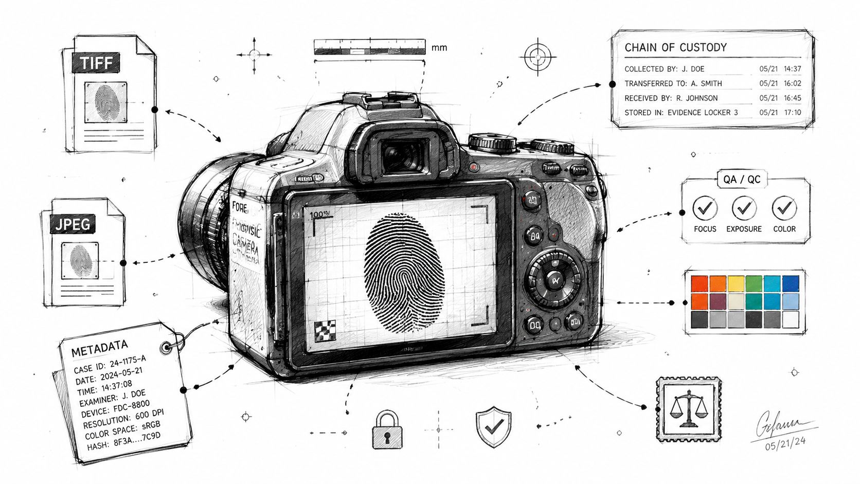
- Original file: Preserve the source image without alteration.
- Working derivative: Use a clearly identified copy for necessary adjustments or report placement.
- Presentation copy: Label the image so readers know it was prepared for viewing rather than retained as the untouched source.
- Export record: Document conversions, transfers, or other processing that affects the file.
These distinctions matter in legal review because an attorney or expert should be able to separate evidence from a convenience copy.
Metadata provides context
Metadata can identify the examination, source, acquisition details, and file history. It may also connect an image to a specific specimen, body region, imaging study, or report entry. Metadata does not replace a written chain-of-custody record. The two records should agree, and discrepancies should be investigated rather than ignored.
A photograph without context may show a real finding while leaving the reviewer uncertain about its identity and place in the examination.
Color management requires discipline
Color can affect how a viewer perceives tissue, bruising, staining, or other findings. The forensic team should use consistent lighting and equipment practices, avoid unnecessary changes, and record material processing. If an image is adjusted for brightness, contrast, cropping, or annotation, the original must remain available for comparison.
Forensic image interpretation is part of a postmortem examination. It should be documented with the same care as the written findings and should not be treated as medical advice for a living person.
Preservation, Chain of Custody, and QA/QC Integration
Digital imaging becomes legally useful when technical controls work together. Preservation protects the original. Chain of custody records who possessed or handled the evidence, when the transfer occurred, and what happened during that transfer. Quality assurance and quality control, often shortened to QA/QC, provide checks that the files and documentation remain complete, readable, and consistent.
The NIST and OSAC guidance emphasizes transparency, security, integrity, and availability across the digital image lifecycle. It identifies hash-verified originals and documented processing chains as central safeguards. A hash is a calculated value used to help detect whether a file changed after it was recorded. It doesn't explain the medical meaning of an image, but it can support the claim that the preserved file is the same file later reviewed.
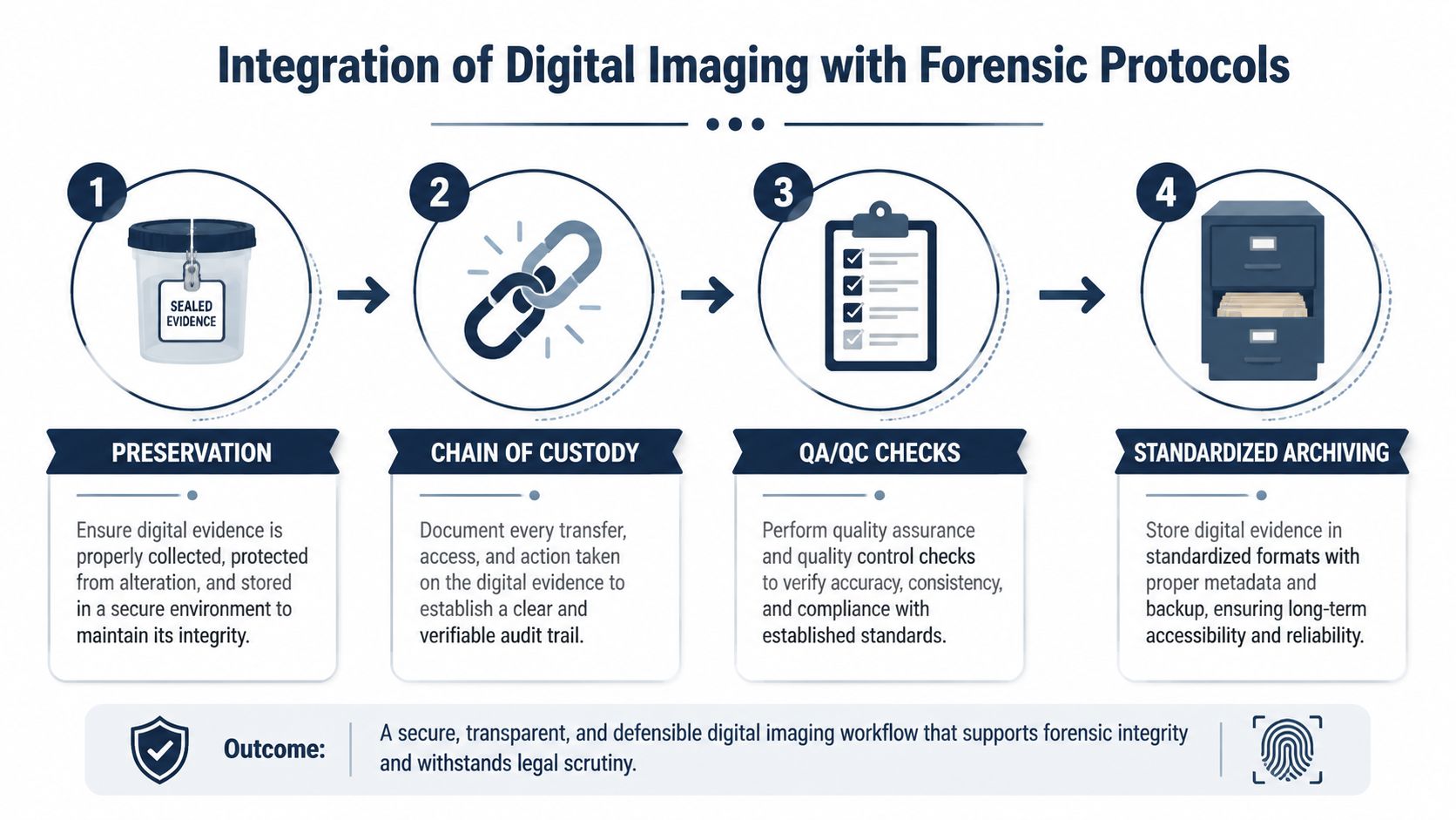
Four controls that should operate together
Preservation: The team identifies and protects the original digital files. Originals shouldn't be overwritten by edits, annotations, resizing, or report preparation.
Chain of custody: Each transfer or access event should be documented according to the case workflow. The record should connect the image to the examination and identify the people or systems responsible for handling it.
QA/QC checks: Staff verify that images are present, associated information is consistent, files can be opened, and the report references the intended images. These checks can identify missing files or mismatched labels before production.
Standardized archiving: The practice stores records in a controlled system that supports retrieval, access management, and long-term review. Archiving should include the original and clearly identified derivatives, not just the image inserted into a PDF.
Texas Autopsy Services maintains digital imaging on site as part of its examination and laboratory workflow. The practice also uses licensed in-house transport, secure temperature-controlled storage, digital imaging, testing coordination, and HIPAA-compliant reporting with chain-of-custody documentation. Families and attorneys can review the related chain-of-custody procedures when they need a plain-language explanation.
Why quality checks matter at the handoff
The greatest weakness may appear after the examination, when records move between a pathology practice, attorney, expert, agency, or court. A file can be renamed, compressed, detached from its metadata, or copied without the documentation that explains its origin.
Evidence principle: The courtroom record should make it possible to distinguish an original, a processed derivative, and a presentation copy.
A complete system doesn't promise that every dispute will disappear. It gives each reviewer a clearer basis for evaluating authenticity, processing, and interpretation.
Court-Admissibility Considerations Under Texas Law
Texas courts evaluate evidence under legal rules that address relevance, reliability, authentication, and other requirements. The precise decision depends on the case, the judge, the offered evidence, and the testimony or records used to establish it. Digital imaging standards don't independently make an image admissible, but they can strengthen the foundation for showing what the image is and how it was handled.
Texas-specific work also requires attention to the agency responsible for the death investigation and to applicable provisions of the Texas Health & Safety Code, including Chapters 711, 716, and 651, as well as the role of the Texas Funeral Service Commission. Those authorities don't all govern the same issue, so families and attorneys should identify which agency, statute, or proceeding applies to their situation. A private autopsy doesn't automatically replace a justice of the peace, medical examiner, law enforcement, or other authorized investigation.
Comparing stronger and weaker practices
| Aspect | Compliant Practice | Non-Compliant Risk |
|---|---|---|
| Original files | The source files are retained separately from edited or report-ready copies. | The only available image may be a resized, cropped, or altered copy. |
| Metadata | Available identifying information is preserved and connected to the examination record. | The reviewer may be unable to establish context or source. |
| Processing | Adjustments, annotations, and exports are documented, with the original retained. | The opposing side may challenge whether the image accurately reflects the original. |
| Chain of custody | Transfers, access, storage, and handling are recorded. | Gaps may create uncertainty about who possessed or changed the file. |
| File access | The image can be reviewed using an appropriate system or documented export. | A proprietary or incomplete export may prevent independent review. |
| Report connection | The written report identifies the finding and the corresponding image. | The court may have difficulty linking the image to the stated conclusion. |
Attorneys often need more than a printed photograph. They may need the underlying file, processing history, report references, and testimony from the person who created or maintained the record. Our broader discussion of forensic science and law explains why technical documentation and legal foundations must be considered together.
Verifying Standards in Private Autopsy Reports
Families and attorneys don't need to become imaging specialists to ask effective questions. A transparent private autopsy practice should be able to explain the imaging workflow in ordinary language and identify which files are originals, derivatives, and report illustrations.
Questions to ask before relying on an image
- What was captured: Ask what findings, views, studies, or specimens were imaged and how the images relate to the report.
- What is original: Confirm whether the practice retained the source files and whether the report images are copies prepared for presentation.
- What metadata exists: Ask whether available acquisition and identification information remains associated with the file.
- What processing occurred: Request a plain explanation of cropping, contrast changes, annotations, conversions, or other adjustments.
- How custody was recorded: Confirm that transfers, access, storage, and delivery are documented.
- How the files can be reviewed: Ask whether an attorney or qualified expert can receive a usable production set with supporting documentation.
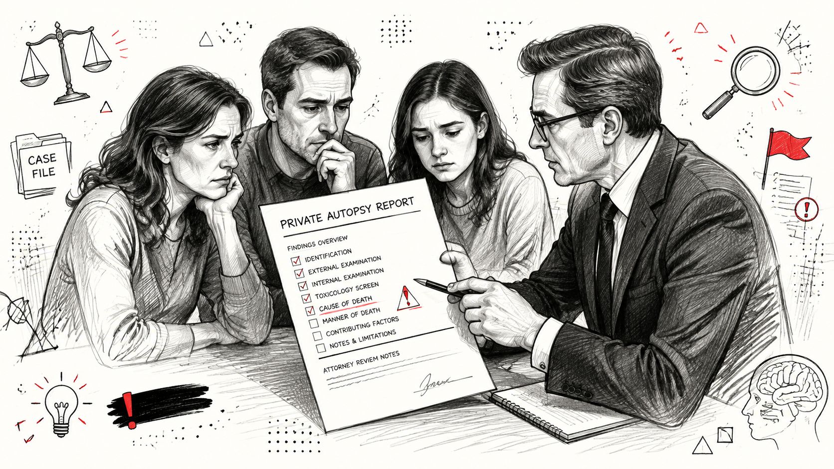
Red flags that deserve clarification
A report that contains only low-quality embedded images may still be useful, but the family should ask whether higher-quality originals exist. Unexplained file names, missing dates, inconsistent body-location descriptions, or a lack of processing notes can make later review more difficult.
Another concern is an image that appears to have been enhanced without a corresponding original. Enhancement may improve visibility for a reader, but it shouldn't erase the source or obscure what changed. A court or expert should be able to compare the presentation image with the preserved original.
What a complete production may include
Depending on the examination and the request, a record may include the written autopsy report, relevant medical imaging, forensic photographs, laboratory findings, chain-of-custody records, and documentation of processing or export. The exact contents depend on the case, authorization, privacy requirements, and the responsible agency.
We explain these issues directly because families often receive records during grief and attorneys may be working under procedural deadlines. A request for a second opinion autopsy or review of prior records should address both the medical conclusions and the quality of the supporting digital evidence.
Why Standards Matter for Trust and Outcomes
Digital imaging standards connect observation to accountability. They help preserve the difference between an original file and a later presentation, maintain context across systems, and give attorneys, families, agencies, and courts a clearer record to examine.
DICOM's development shows that interoperability requires more than moving a file from one computer to another. In forensic pathology, the same principle applies to photographs, radiology studies, pathology images, annotations, reports, and related records. DICOM has also been extended for digital pathology, including static and whole-slide images, and pathology implementations can encode annotations and quantitative measurements derived from images. A review of DICOM in digital pathology and research on pathology image annotations and measurements describe that broader role. DICOM Working Group 26 was established in 2005 to extend the standard for pathology and whole-slide imaging, as documented by the Digital Pathology Association.
Our board-certified forensic pathologists use imaging as one part of an independent cause-of-death investigation. The practice serves families, attorneys, healthcare professionals, and county officials throughout all 254 Texas counties, with direct communication and documentation suited to medical and legal review.
Texas Autopsy Services provides independent private autopsy services, second-opinion reviews, digital imaging, forensic pathology reporting, and county forensic autopsy support across Texas. Contact our team to discuss how original files, metadata, processing records, and chain-of-custody documentation can be handled in your case, then visit Texas Autopsy Services to request guidance with dignity and clarity.