August 26, 2026 · Texas Autopsy Services
Abdominal Autopsy: What the Procedure Reveals
Learn what an abdominal autopsy reveals, how the procedure works, and what families and attorneys in Texas can expect from findings and reporting.
On this page
- What an Abdominal Autopsy Is and When It Is Performed
- Why Abdominal Autopsy Findings Matter More Today
- Inside the Abdominal Examination Procedure
- Common Findings and What They Mean for Families
- Distinguishing True Disease From Postmortem Change
- When a Standard Abdominal Exam Is Not Enough
- Texas Law, Chain of Custody, and What to Expect From the Report
- Key Takeaways and How to Reach Our Team
A family member dies after abdominal pain, vomiting, a fall, or a surgical complication. The medical examiner's office may decline the case, or the official examination may not address the question troubling the family. In that moment, many people ask what an abdominal autopsy can show and whether it can distinguish a real disease from changes that naturally occur after death.
At Texas Autopsy Services, we explain the process in plain language. An independent examination can document abdominal disease, trauma, surgical complications, and tissue findings that help clarify the cause of death, while respecting the authority of the medical examiner or justice of the peace when that jurisdiction applies.
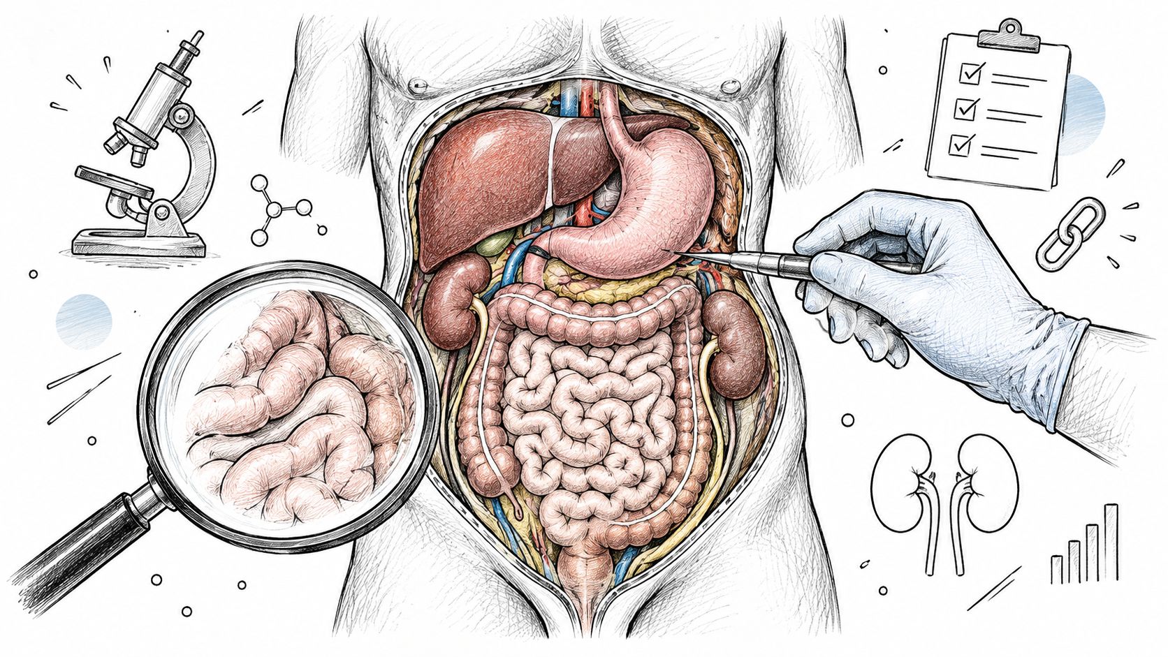
- An abdominal autopsy examines the organs and structures inside the peritoneal cavity, including the stomach, bowel, liver, pancreas, spleen, kidneys, and major vessels.
- The examiner documents fluid, organ position, injuries, disease, and tissue changes before removing organs.
- Histology, toxicology, microbiology, imaging, and medical records may be needed to answer the actual forensic question.
- Postmortem gas, tissue softening, fluid redistribution, and discoloration can mimic disease, so interpretation requires experience and context.
- Texas law governs authorization, jurisdiction, and custody of the body and specimens.
What an Abdominal Autopsy Is and When It Is Performed
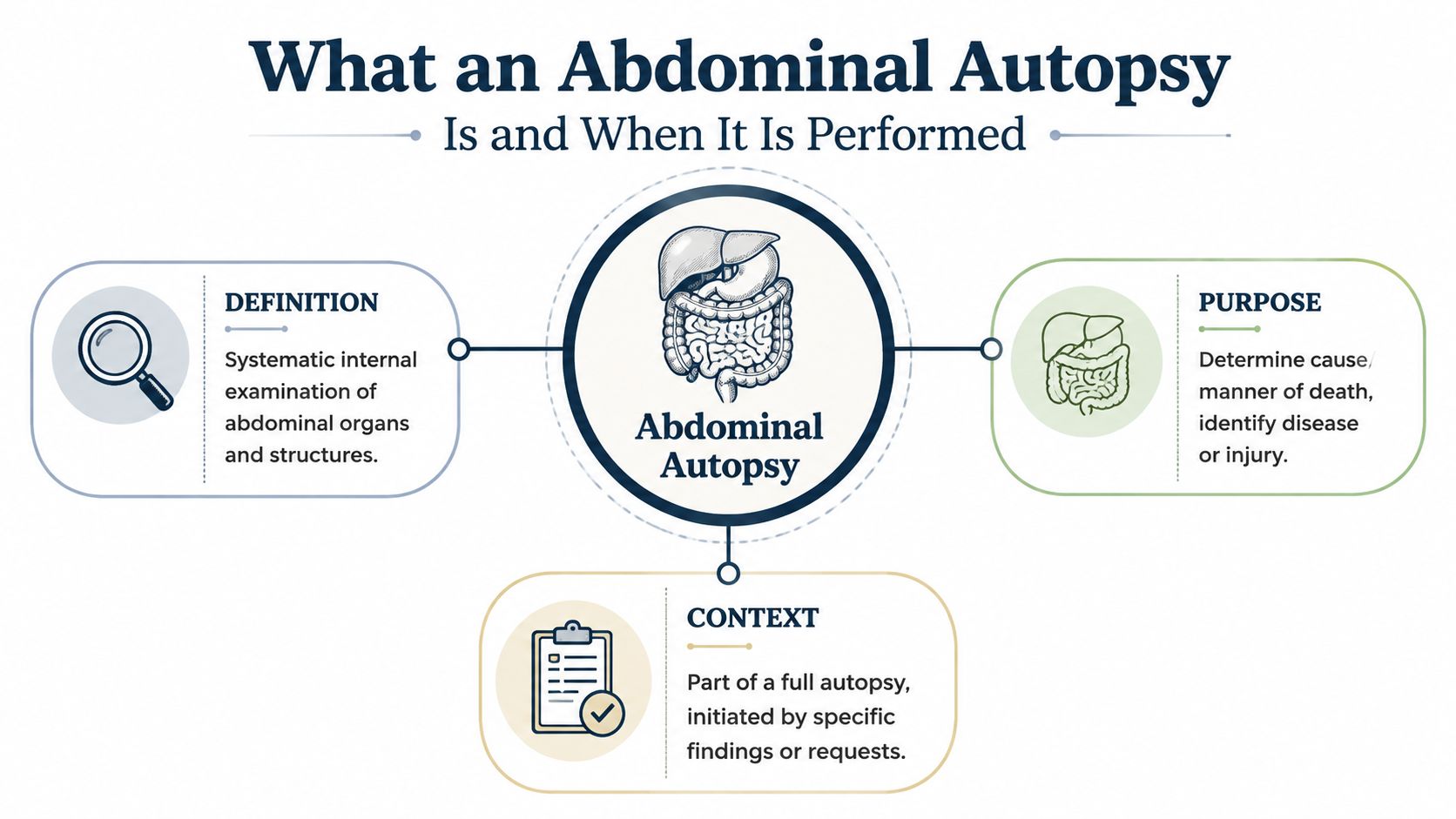
A grieving family member or retained attorney often starts with a simple concern: what happened inside the abdomen? An abdominal autopsy is the systematic dissection, inspection, and sampling of the peritoneal cavity, the internal space that contains many abdominal organs. It may be part of a complete postmortem examination or a focused private study directed at a particular medical or legal question.
The examination can include the stomach, liver, gallbladder, pancreas, small and large bowel, kidneys, adrenal glands, spleen, aorta, pelvic organs, and related blood vessels. The examiner evaluates both the organs themselves and their relationships to one another. A tear in a major vessel, blood around a damaged organ, or leakage from the bowel may matter as much as an abnormal organ.
The terms families commonly hear
Evisceration means removing internal organs for detailed examination. The organs can be removed individually, in connected groups, or together as an anatomical unit. In-situ inspection means examining organs while they remain in their original positions. This first view can preserve relationships that might be lost after removal.
The retroperitoneum is the space behind the main abdominal lining. It contains the kidneys, adrenal glands, portions of the pancreas, ureters, and major vessels. A superficial examination can miss injuries or disease in this area, so the retroperitoneum must be opened deliberately.
A private abdominal autopsy may be considered when a medical examiner declines jurisdiction, when the family believes a diagnosis was missed, or when an attorney needs documented findings for a potential claim. Texas law still controls whether a private examination may proceed, and a medical examiner's or justice of the peace's authority can override a family's preference.
The procedure can reveal whether a suspected disease was present, whether trauma caused internal bleeding, whether surgery altered or injured an organ, and whether an apparent abnormality is only a postmortem artifact. It can also show when the available evidence is insufficient to reach a more specific conclusion.
Why Abdominal Autopsy Findings Matter More Today
Routine hospital autopsies have declined sharply. In the United States, overall autopsy rates were about 25% to 35% of hospital deaths in the mid-1960s, compared with only 7% to 9% today, according to a review of autopsy practice and its decline (review of the medical autopsy tradition). Some historical sources noted that hospital autopsies were performed in 50% of hospital deaths during the 1940s and 1950s, while a separate review reported a 58% decline from 1972 to 2007 in the same source.
That change affects how medical knowledge is checked. When fewer postmortem examinations occur, fewer missed diagnoses are discovered after death. Abdominal disease can be especially important because gastrointestinal, hepatic, pancreatic, vascular, and adrenal conditions may be difficult to recognize before death or may be obscured by several competing diagnoses.
| Era | Approximate hospital autopsy rate | Typical missed abdominal diagnoses |
|---|---|---|
| 1940s and 1950s | 50% of hospital deaths | Gastrointestinal, hepatic, pancreatic, and vascular disease |
| Mid-1960s | About 25% to 35% of hospital deaths | Disease not fully recognized during life |
| Today | About 7% to 9% of hospital deaths | Abdominal disease or trauma clarified only after death |
The figures describe historical patterns, not the probability that a particular person had a missed diagnosis. They do show why each carefully performed abdominal autopsy carries greater diagnostic importance than it did when hospital autopsies were routine. The Joint Commission also no longer requires the earlier 20% to 25% in-hospital autopsy benchmark, as described in the cited review.
Why direct examination still matters
Imaging and laboratory results can be valuable, but they don't answer every postmortem question. A scan may show fluid, bowel gas, organ enlargement, or suspected ischemia, yet the examiner may need to open the organ, inspect blood vessels, and submit tissue for microscopy to determine whether a process occurred during life.
Autopsy findings remain relevant in fatal surgical disease and trauma. A Swedish forensic series reported fatal surgical diseases at 67.3 per 1,000 autopsies from 1970 to 1982 and 83.4 per 1,000 from 2000 to 2004, while fatal acute abdomen rose from 16.5 to 39.0 per 1,000 autopsies over those periods (PubMed summary of abdominal autopsy findings). These data support a practical conclusion: an abdominal autopsy isn't a narrow add-on. It can be the examination that establishes what clinical records couldn't.
Inside the Abdominal Examination Procedure
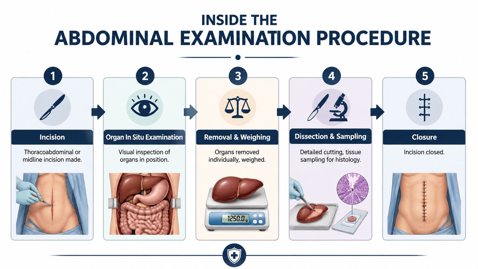
The procedure begins with an external examination and review of the available history. The incision may be a midline or thoracoabdominal incision, depending on the questions under investigation. A midline approach provides access from the chest or upper abdomen toward the pelvis, while a thoracoabdominal approach can help when the suspected disease or injury crosses the chest and abdominal compartments.
Opening and documenting the cavity
Once the peritoneal cavity is opened, our pathologists inspect it before moving organs. They document the presence, amount, and character of free fluid. Blood, bile, gastric contents, intestinal material, or ascites can each suggest a different source, but early collection is important because moving organs can spread material and contaminate a wound track.
Standard forensic technique recognizes four main evisceration approaches:
- Virchow method: Organs are removed and examined individually, which can provide detailed attention to routine natural disease.
- Rokitansky method: Organs are dissected largely in position, preserving relationships during the initial examination.
- Ghon method: Organs are removed in separate blocks that maintain selected anatomical connections.
- Letulle method: The organs are removed en masse, meaning as one connected group, when continuity is important.
The appropriate method depends on the suspected mechanism of death. Penetrating trauma and complex abdominal injury may require preservation of connected structures, while an uncomplicated natural death may benefit from individual-organ examination (forensic autopsy techniques and evisceration methods).
Organ examination and specimen collection
The liver, gallbladder, biliary tree, pancreas, ampulla, stomach, duodenum, small bowel, mesentery, colon, kidneys, adrenal glands, spleen, aorta, iliac branches, and pelvic organs are assessed according to the case history. The ampulla is the opening where bile and pancreatic secretions enter the duodenum. The mesentery is the tissue fold that supports the intestines and carries vessels, nerves, and lymphatic channels.
The team may collect peritoneal fluid for chemistry, swabs for microbiology, and tissue for histology, the microscopic examination of preserved tissue. Organs can be weighed, opened along appropriate anatomical planes, and examined for hemorrhage, obstruction, ulceration, perforation, tumor, infarction, or infection.
Before final reporting, the findings are compared with medical records, imaging, operative notes, toxicology, and witness or agency information. Families can review our specimen collection procedures to understand how samples support the final opinion.
Common Findings and What They Mean for Families
Consider a non-identifiable situation involving a middle-aged adult who develops worsening abdominal pain and vomiting, collapses at home, and dies before a complete clinical explanation is established. The medical examiner declines jurisdiction, and the family authorizes a private abdominal autopsy. The examination isn't designed to promise a particular answer. It is designed to test plausible explanations against physical evidence.
Mesenteric ischemia means that blood flow to the intestine has been blocked or severely reduced. The bowel may show injury from inadequate circulation, and examination of the mesenteric vessels can help determine whether a clot, embolus, or other vascular event was involved. The finding can address whether the death followed a sudden catastrophic event or a process that had been developing.
A perforated peptic ulcer is a hole through the wall of the stomach or duodenum, allowing digestive contents to escape into the abdominal cavity. The examination looks for the perforation, contamination, inflammation, and evidence of peritonitis, which is inflammation or infection of the abdominal lining.
Findings that can change the interpretation
A ruptured splenic artery aneurysm is a tear in an abnormal widening of the artery supplying the spleen. It can cause internal bleeding that isn't externally visible. A ruptured abdominal aortic aneurysm is a tear in a weakened section of the body's main abdominal artery and can likewise produce fatal internal hemorrhage.
Hemorrhagic pancreatitis means severe inflammation of the pancreas accompanied by bleeding into or around the organ. The examiner evaluates the pancreas, nearby tissues, blood, and microscopic changes to determine whether the condition contributed to death.
Practical rule: The autopsy finding must be matched to the medical history, timing, treatment, and scene evidence. A disease can be present without being the cause of death.
Histology may help determine whether an injury or disease process developed during life and whether it appears relatively recent or more established. Toxicology can identify contributing substances, including anticoagulant medications or alcohol, but a detected substance isn't automatically the cause of death. The final opinion depends on the complete record, not one isolated result.
Distinguishing True Disease From Postmortem Change
Families sometimes hesitate to ask whether an abdominal finding is real or just what happens after death. That concern is reasonable. Once circulation stops, tissues soften, fluids redistribute, bacteria produce gas, and organs change color. Some of these effects can resemble disease on imaging or during gross examination.
Greenish staining near the gallbladder bed can reflect bile movement rather than a living inflammatory process. Postmortem gastric autolysis, or self-digestion of the stomach lining, can produce dark discoloration that resembles injury. Livor mortis, the settling of blood after death, can appear in dependent bowel loops. Bacterial gas can create diffuse peritoneal haziness that may be mistaken for infection or another abdominal process.
| Finding | What it looks like | What it actually means |
|---|---|---|
| Green discoloration near the gallbladder | Possible tissue injury or inflammation | May reflect postmortem bile staining |
| Dark stomach lining | Possible hemorrhage or ulceration | May reflect postmortem gastric autolysis |
| Gas in bowel or portal veins | Possible ischemia or infection | Can occur during decomposition |
| Dependent dark areas in bowel | Possible bruising or bleeding | May reflect livor mortis |
| Diffuse abdominal haziness | Possible peritonitis or fluid | Can result from postmortem bacterial change |
The distinction depends on tissue planes, distribution, associated injury, and microscopic findings. A vital reaction is a body response that occurs during life, such as inflammation or tissue repair. Histology can help distinguish coagulation necrosis, tissue death caused by loss of blood supply during life, from cellular breakdown that occurs after death.
Postmortem CT can also show bowel gas, portal venous gas, liver softening, gallbladder distension, and fluid redistribution. The findings need context before anyone calls them ischemia, hemoperitoneum, ascites, or cholecystitis (review of postmortem abdominal changes). Our team also provides a detailed explanation of decomposition analysis for families and attorneys evaluating these difficult findings.
A defensible diagnosis explains not only what was seen, but why the finding represents disease rather than expected postmortem change.
When a Standard Abdominal Exam Is Not Enough
A routine abdominal examination may not answer a case-specific question after surgery. The dissection must change when anatomy has been altered, when bleeding is suspected, or when an operative complication requires precise documentation.
After bariatric surgery or gastric bypass, the stomach and intestine may no longer follow their original paths. The examiner may need to trace the gastric pouch, staple lines, bypassed segments, and anastomoses, which are surgical connections between organs or bowel segments. A complete postmortem examination is preferred after bariatric surgery, while an exam limited only to the surgical site is considered suboptimal (Pathology Outlines review questions).
Three situations that call for modification
- Bariatric or gastric bypass surgery: The examination follows altered anatomy and inspects staple lines and anastomoses for leakage, obstruction, bleeding, or other complications.
- Suspected upper gastrointestinal hemorrhage: The esophagus, stomach, and duodenum may require careful in-situ opening before removal so the bleeding source and blood distribution remain interpretable.
- Possible operative complications: The team documents the surgical site, anastomotic integrity, retained sponges, drains, mesh, and other foreign material with photographs and separate sampling when appropriate.
These cases may take additional time and require careful handling of foreign objects. Drains, mesh, surgical material, tissue blocks, photographs, and other evidence must remain identifiable and traceable. The team may also need operative records or consultation with the operating surgeon or an independent surgical pathologist.
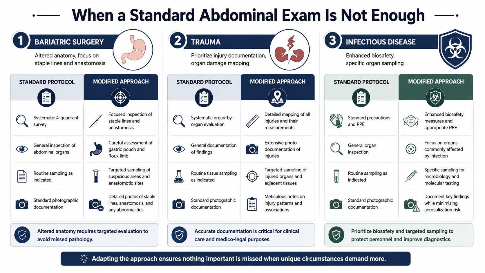
Recognizing that a standard protocol isn't enough is part of sound forensic practice. The examination should follow the clinical question rather than force every case into the same sequence.
Texas Law, Chain of Custody, and What to Expect From the Report
Texas authorization depends on the governing authority and the circumstances of the death. Texas law provides that when a commissioners court establishes a medical examiner's office, powers and duties related to death investigations and inquests transfer from justices of the peace to that office. In a county without such an office, a justice of the peace may order an autopsy in connection with an inquest (Texas criminal procedure provisions concerning death investigations and autopsies).
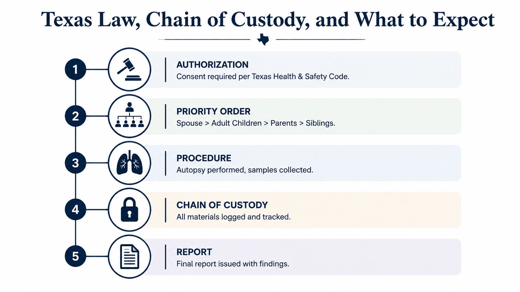
A physician may not perform or assist with a postmortem examination or autopsy without written informed consent unless the examination is authorized by the appropriate legal authority. Families should therefore confirm jurisdiction before arranging a private examination. Family consent can't override an active medicolegal hold or an order from the responsible authority.
Consent, custody, and documentation
The applicable Texas Health & Safety Code provisions address who may authorize certain postmortem services and how the body is handled. The precise priority can depend on the circumstances and applicable statute, so our team verifies the authority and documentation rather than relying on an informal verbal request.
Chain of custody means the documented history of who controlled or possessed the body, specimen, or item of evidence at each stage. Texas Health and Safety Code requires each person with control or possession of a body or anatomical specimen to complete the required chain-of-custody form, retain a copy, and transfer the form to the next person receiving control or possession (Texas Health and Safety Code custody requirements).
In practice, documentation can include:
- Intake records: The team records receipt, identification information, condition, and authorized release.
- Specimen tracking: Tissues, slides, blocks, fluids, photographs, and retained materials receive identifying documentation.
- Transfer records: Each handoff is recorded so the next custodian and the time of transfer remain clear.
- Report correlation: Laboratory results are matched to the correct case and specimen.
The final report
A report generally addresses external findings, internal findings, microscopic results, toxicology, microbiology when performed, cause of death, and manner of death. Cause of death is the disease, injury, or combination of conditions that led to death. Manner of death is the broader classification of how the death occurred, such as natural, accident, suicide, homicide, or undetermined, when the available evidence supports a classification.
Turnaround depends on the case, the need for special testing, the condition of the remains, record availability, and legal review. Most cases are completed within 24 to 48 hours for the examination phase, while the final report may require additional laboratory and interpretive work. Families and attorneys should request a case-specific estimate rather than rely on a fixed promise.
Every examination is performed by a forensic pathologist certified by the American Board of Pathology. The board's standard requires applicants to hold prior certification in AP/CP or AP and complete 12 months of training in an ACGME-accredited forensic pathology program (American Board of Pathology standards).
Families can also review our explanation of chain-of-custody procedures when a case may involve an attorney, agency, or court.
Key Takeaways and How to Reach Our Team
An abdominal autopsy answers more than the question of whether an organ looks abnormal. It evaluates the abdominal cavity in context, preserves fluid and anatomical relationships, tests tissue under the microscope, and compares the findings with medical records, imaging, medications, surgery, and circumstances of death.
The central questions often include:
- Disease: Was there bowel ischemia, ulcer perforation, pancreatitis, vascular rupture, infection, or another abdominal condition?
- Timing: Does the tissue show a process that occurred during life rather than after death?
- Causation: Did the finding cause death, contribute to it, or merely coexist with another fatal condition?
- Procedure: Does altered anatomy or a prior operation require a modified examination?
- Evidence: Were specimens, photographs, and records handled in a way that supports independent review?
A private examination may be appropriate when a medical examiner declines jurisdiction, when the family seeks a second opinion, or when an attorney needs an independent evaluation of abdominal findings. It won't replace the authority of a medical examiner or justice of the peace, and it can't guarantee that every unanswered question will have a definitive answer.
Our team serves families, attorneys, healthcare professionals, and Texas county officials across all 254 Texas counties. Texas Autopsy Services is based in Elgin, provides in-house licensed transport, and communicates directly with the people responsible for the case. To begin a consultation, gather the decedent's identifying information, available medical and surgical records, any agency correspondence, and documents showing authorization.
Texas Autopsy Services provides independent abdominal autopsies, second-opinion reviews, specimen handling, and forensic pathology reporting for families and attorneys seeking clearer answers after a death. Visit Texas Autopsy Services to contact our team, discuss the circumstances, and learn what documentation may be needed before an examination can proceed.