August 8, 2026 · Texas Autopsy Services
What Is Microscopic Examination? a Complete Guide
What is microscopic examination - Learn what microscopic examination reveals in forensic and clinical pathology, from sample prep to analysis. Clear answers
On this page
A family member often first hears the word microscopic examination after a death report is already underway. A lawyer may see it in a pathology summary and want to know whether it supports the cause of death, while a grieving family just wants to know what a slide can show that the naked eye cannot.
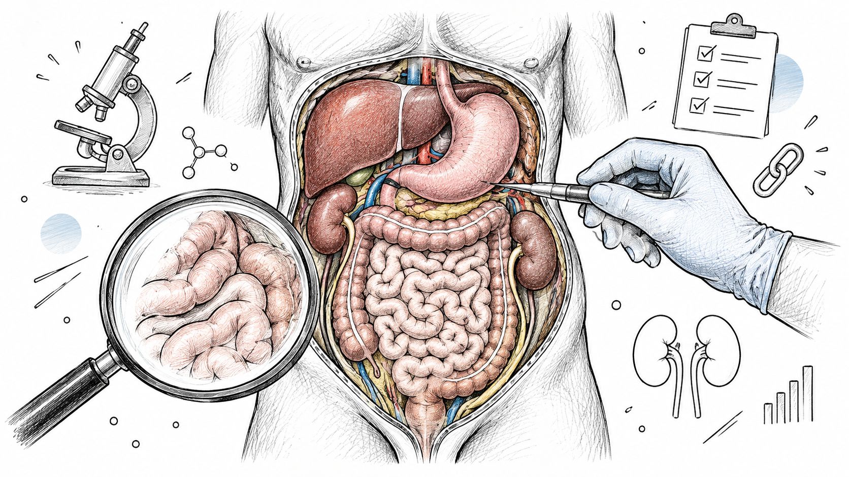
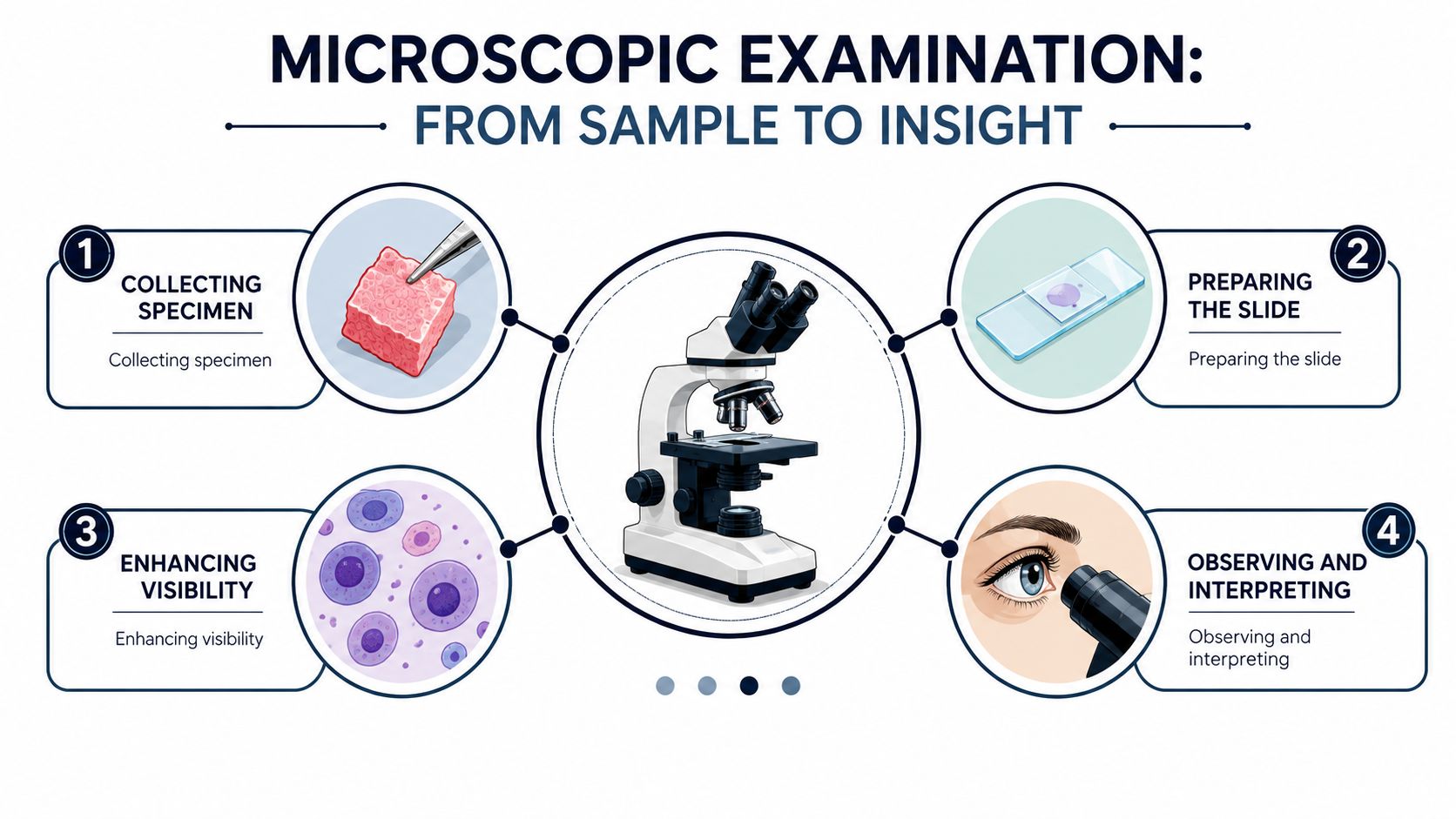
In our work, the answer starts with a simple idea. Microscopic examination is the process of turning a sample into a prepared slide, then studying it under a microscope so we can see cells, microorganisms, and tissue patterns that are too small to judge by eye alone. It is one of the main ways a forensic pathologist separates a broad impression from a defensible medical finding, especially when the questions are about cause of death, hidden disease, or whether an injury or illness was really present at the tissue level.
Microscopic Examination in Plain Language
A useful way to think about what is microscopic examination is to picture a single slide on a pathologist's desk. The tissue or fluid has already been collected, fixed, and prepared. Now the question is whether the slide shows a pattern that explains illness, injury, infection, or another cause of death.
What the term actually covers
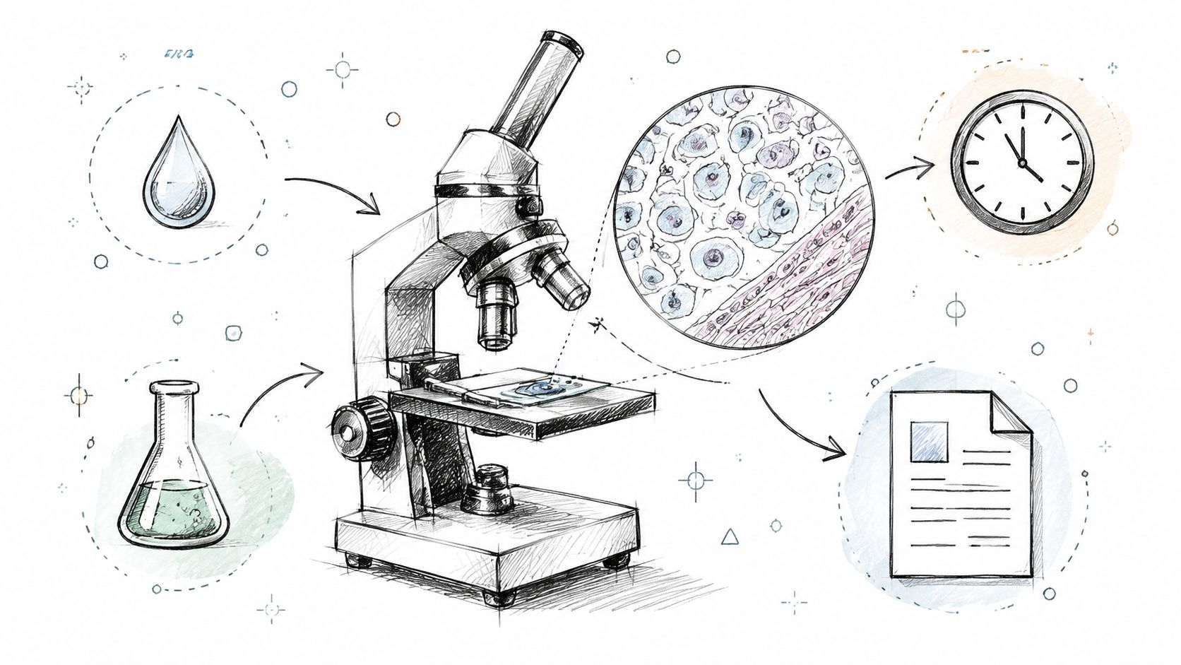
Microscopic examination is not one single action. It is a workflow that starts with a specimen and ends with a medical interpretation. In histology, that often means a tissue sample is fixed, processed, embedded, sliced thin enough for light to pass through, stained, and mounted on a slide.
That preparation matters because the microscope does two jobs at once. Resolution tells us what a lens can separate, and contrast tells us what the eye can distinguish once the slide is stained. Without both, the same tissue may look flat, blurry, or incomplete.
Practical rule: a good slide does not just magnify, it makes the right structures visible in a way that can be reviewed and explained.
Why families and attorneys keep hearing it
Families usually want to know whether the microscope confirmed something the gross exam hinted at, or whether it ruled something out. Attorneys often need the same answer in different language, whether the finding supports a cause of death opinion or shows that the tissue did not contain enough evidence to be definitive.
A microscope is useful because it reaches below naked-eye resolution, but it is still only one part of the record. The meaning comes from the slide, the stain, the sampling method, and the pathologist's interpretation together. That is why microscopic examination can be central to a death investigation without being the whole story.
From Specimen to Slide
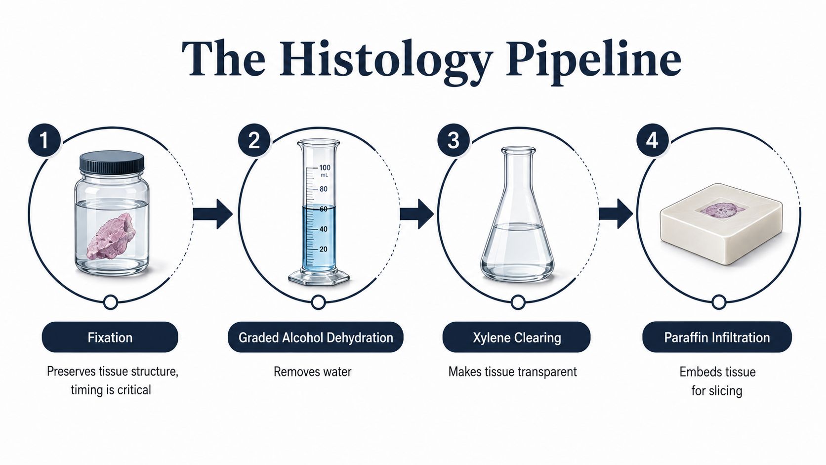
The slide tells the truth only if the preparation is disciplined. In routine histology, tissue is fixed first, then dehydrated through graded alcohols, cleared in xylene, infiltrated with paraffin, embedded, and cut into thin sections. Those steps preserve structure and make the sample thin enough for light microscopy to work cleanly, which is why section thickness is commonly around 5 µm, and sometimes as thin as 3 µm for especially careful visualization microscopy workflow and section thickness.
Why each step exists
Fixation preserves tissue before decay distorts the architecture. Dehydration removes water so the sample can accept wax. Clearing replaces alcohol with a medium that paraffin can penetrate, and embedding holds everything in a firm block that can be sliced.
Fluid and sediment specimens follow a different path. Urine, aspirates, and some trace-evidence washes are concentrated, placed as a thin film, and scanned rather than embedded. That matters because the goal is to spread sparse material out so a pathologist or technologist can find rare cells, crystals, casts, or microorganisms without burying them in excess debris.
The common failure points usually show up later in the report, not in the lab room. Chatter, knife marks, folds, under-dehydration, and stain artifacts all reduce confidence because they interfere with the tissue pattern the pathologist needs to read.
Why preparation affects the final answer
A slide can only support a reliable opinion if the underlying sample is intact. Poor fixation can blur cell detail, excess thickness can create overlap, and weak processing can hide the very structure the review is meant to reveal. The point is not to make the specimen look perfect, it is to make it readable in a way that another qualified reviewer could follow.
For a broader specimen-preservation context, we also explain the handling side of the process in a guide to forensic specimen preservation.
Tools Pathologists Actually Use
A microscope is not one tool, it is a family of tools, each answering a different question. Routine pathology relies most often on a compound light microscope, because it shows tissue architecture, cell shape, inflammation, necrosis, and many common organisms within the limits of visible light. That is the workhorse instrument in most slide reviews.
Matching the tool to the question
The boundary matters. Visible-light microscopy is limited to resolving structures down to about 200 nm according to the optical work that followed Ernst Abbe's findings, which is why some questions need a different instrument altogether history of microscopes. When a structure sits below that range, an electron microscope may be used to see ultrastructural detail.
Other tools answer different problems. Polarized light helps separate crystals and certain fibers, which is useful in trace evidence and some tissue reviews. Phase contrast can help with unstained wet material. Fluorescence microscopy is valuable when tagged antibodies or microorganisms need to stand out against the background.
A tool is only as useful as the question behind it. Light microscopy handles most routine tissue diagnosis. Electron microscopy is reserved for smaller structures. Polarized and fluorescence methods are chosen when the slide itself needs help revealing what's present.
Where digital imaging fits
Many readers now encounter microscopic work through digital capture, not just a person looking into eyepieces. That does not replace the pathologist's judgment, but it can help with documentation, consultation, and later review. For a plain-language discussion of digital image handling in medical settings, how AI upscaling improves medical scans is a useful adjacent resource because it explains why image clarity matters before interpretation begins.
In our own practice, the choice of instrument follows the evidentiary question, not the other way around. Texas Autopsy Services uses microscopic review as part of a broader forensic pathology process when tissue-level findings are needed in a private autopsy or second-opinion case.
Stains, Objectives, and Field Counts
Magnification alone does not make a diagnosis. A slide has to be stained correctly, scanned at the right power, and read with enough discipline to avoid missing sparse findings. Microscopic examination is a method, not just a look. It is closer to following a recipe than glancing at a photograph, because each step affects what can be seen and what can be missed.
How staining changes what you can see
Gram staining is a useful example because the sequence itself creates diagnostic contrast. The slide is exposed to crystal violet, then iodine, then a decolorizer, then a counterstain. That order lets organisms retain different colors based on cell-wall structure, which helps separate likely gram-positive from gram-negative bacteria in one review microscopy guidance.
Parasitology works differently, but the logic stays the same. A stool specimen review starts with the 10× objective for broad screening, then moves to 100× oil immersion when detail matters. Guidance also recommends examining at least 200 to 300 oil-immersion fields when low-burden protozoa are a concern, because a few organisms can be scattered across a slide and easy to miss in a quick pass microscopy guidance.
Why the scan has to be systematic
A quick glance misses sparse organisms. So does using the wrong objective. The pathologist has to match the field count and stain to the specimen type, especially when the diagnostic target is small, scattered, or partly obscured by debris. In practice, that means the slide is not read like a single snapshot. It is worked through field by field, with attention to where the finding is most likely to hide.
For lab quality and reagent discipline, we also point readers to Herbilabs reagent testing protocols, because consistent staining depends on controlled materials, not just good eyesight.
| Stain | Targets | Typical Use Case |
|---|---|---|
| Hematoxylin and eosin | General tissue structure | Routine tissue review in autopsy and surgical pathology |
| Gram stain | Bacteria with different cell-wall structures | Suspected bacterial infection |
| Special organism stains | Microorganisms and selected tissue components | When organisms are hard to see on routine stain |
| Polarized light method | Crystals and some fibers | Trace evidence and material identification |
| Fluorescent labeling | Tagged antigens or organisms | Targeted pathogen or immune-marker review |
The same scanning logic applies in forensic pathology when we look for subtle myocardial fibrosis, aspirated material, or other sparse findings. The instrument changes, but the discipline does not. A stain may make a pattern visible, but it cannot rescue a poor section, a weak stain, or a rushed scan.
For readers who want to connect these slide findings to the final write-up, what a histology report includes explains how microscopic observations are organized into a report that families, attorneys, and county officials can read.
Reading the Pathology Report
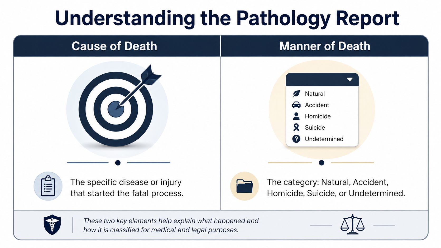
A pathology report only helps if the reader knows which part answers which question. Cause of death is the disease or injury that started the fatal process. Manner of death is the category assigned to that death, such as natural, accident, suicide, homicide, or undetermined.
What a complete report usually contains
A solid report normally separates the gross findings, the microscopic findings, and any ancillary studies such as toxicology, microbiology, or molecular testing. That structure matters because one section may support another, or explain why a conclusion stays cautious.
Families sometimes receive a summary that feels compressed. Attorneys may receive a broader packet that includes slides, blocks, and supplemental material. For readers who want a simpler framework for medical paperwork, simplified medical document formats can help with the layout of a report, even though the medical content still has to be judged on its own terms.
Why chain of custody matters
Slides and tissue blocks are not just lab byproducts. They are part of the evidence record, and they have to be retained under documented conditions if they may be reviewed again later. That is what supports a second-opinion review, a court question, or a county file review without changing the underlying material.
For a more detailed pathology format discussion, see what is a histology report.
Most preliminary cause-of-death findings are reached within 24 to 48 hours, with the final report following after ancillary testing returns. That estimate depends on the case, not on a promise, because some questions need extra testing before the opinion is complete.
Limits of the Microscope
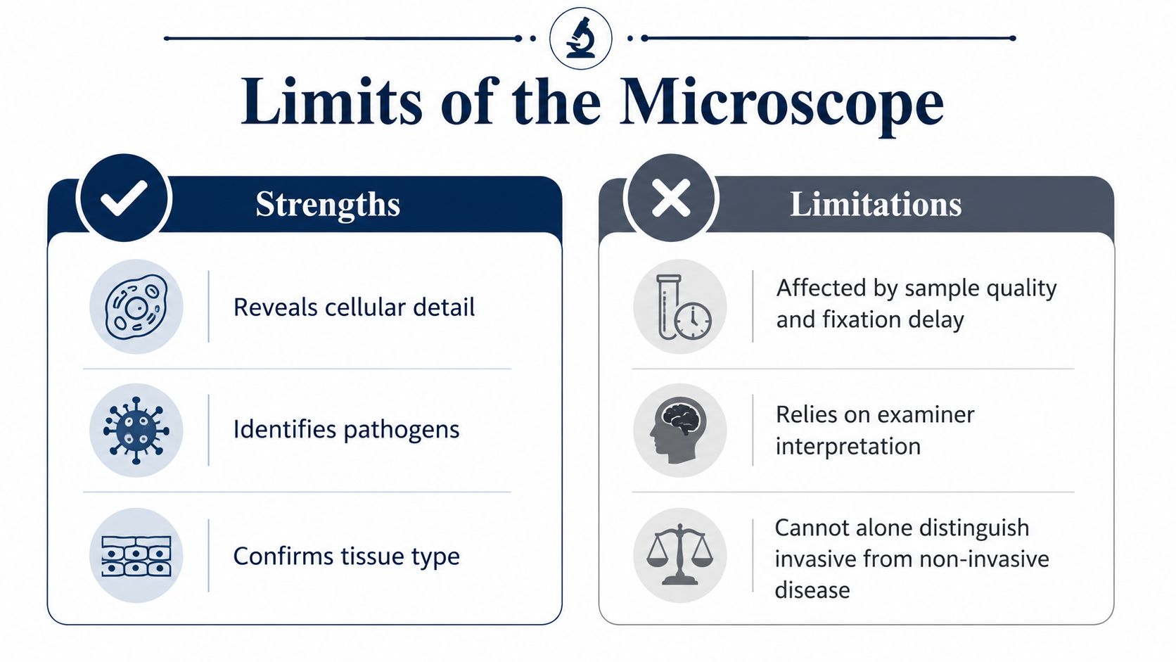
Microscopy is powerful, but it's not self-interpreting. The result depends on sample quality, fixation delay, stain choice, and the pathologist's reading of what's on the slide. A clean-looking field can still be misleading if the tissue was poorly sampled or if the stain failed to show the structure that mattered.
What microscopy can't settle by itself
Microscopy often helps distinguish a real tissue process from a guess, but it can't always tell invasive disease from surface colonization on its own. That distinction may need culture, molecular testing, or correlation with the gross exam and medical history. A stain can be non-diagnostic even when the disease is real, especially if organism burden is low or the sampled area missed the lesion.
In plain terms, a slide can support an answer, narrow a differential, or show that the evidence is too thin to conclude more. It can't fix a limited sample.
When another review is worth considering
An independent review becomes more useful when the prior autopsy report conflicts with the clinical history, when the original sampling was narrow, or when an attorney needs a separate analysis of the same slides. Families also ask whether a new set of eyes can change the answer. The honest answer is that it sometimes can, especially when the original material was incomplete.
For readers who want the testing side of that decision, molecular genetic testing explains why some questions move beyond the microscope and into targeted laboratory methods.
Microscopy is strongest when it's part of a larger evidentiary chain. It is weakest when people expect it to do everything alone.
Microscopy in Texas Death Investigations
Texas families and attorneys need the medical answer, but they also need to know which legal pathway applies. A private autopsy is authorized by a family or attorney, while a medicolegal autopsy may be directed by a justice of the peace under Texas Health and Safety Code Chapter 716. Cremation and related disposition issues are governed through the Texas Funeral Service Commission framework and Texas Health & Safety Code Chapters 651 and 711.
How the microscope fits each pathway
In a private autopsy, microscopic review is part of the tissue-level work when the cause warrants histology. In a county-directed case, the same slides may later support an independent review if questions remain. The microscope does not change the legal process, but it often determines how complete the medical answer can be.
What to look for in a provider
A careful autopsy provider should be able to show how the slides were handled, who kept custody of the material, and whether the report can be reviewed again later.
Look for board certification by the American Board of Pathology, documented chain of custody, transparent flat-rate pricing, and the ability to retain slides and blocks for later review. Those features matter in a private autopsy, a second opinion autopsy, or a county forensic case because the evidence has to remain readable long after the first report is finished.
Texas Autopsy Services provides independent forensic pathology services across all 254 Texas counties, with in-house licensed transport and direct communication with our team. The practice approach is built around medical precision, documented handling, and plainspoken reporting.
Questions Families and Attorneys Ask
Does microscopic review always take extra time
It can, because tissue has to be processed, stained, examined, and sometimes re-cut if the first slide isn't readable. Some cases move quickly, while others need extra stains or ancillary testing before the opinion is complete.
Can a family request slides or tissue blocks
Yes, in many cases the slides and blocks can be retained and reviewed later under documented conditions. That's especially important when a family wants a second opinion autopsy or when counsel needs the underlying material for independent review.
When is a second opinion worth pursuing
A second opinion is most useful when the original report is thin, the history doesn't fit the conclusion, or the family needs a clearer explanation of how the answer was reached. It's less about disagreement for its own sake and more about whether the sample, the stains, and the interpretation were complete.
How does microscopy relate to toxicology or molecular testing
Microscopy answers tissue questions. Toxicology answers chemical exposure questions. Molecular testing can resolve cases where organisms or genetic changes need a different method than the microscope can provide.
For a case-specific question, direct conversation matters more than guesswork. If you're comparing reports, waiting on ancillary results, or trying to understand whether slides should be reviewed again, contact us for a careful look at the facts in your case.
Texas Autopsy Services offers independent, court-conscious forensic pathology across Texas, with microscopic review used when tissue-level detail matters to the cause of death investigation. If you need a second opinion autopsy, a county forensic autopsy service, or a clearer explanation of a report, visit Texas Autopsy Services and contact our team directly for guidance on your specific case.