August 28, 2026 · Texas Autopsy Services
Immunohistochemistry Testing in Autopsy Work Explained
Learn how immunohistochemistry testing supports cause-of-death investigations, common markers, sample handling, and what families and attorneys can expect.
On this page
- What Immunohistochemistry Testing Is and Why Autopsies Use It
- How the Stain Actually Works Step by Step
- Sample Collection and Fixation Before the Stain
- Common Markers and Panels Used in Autopsy Cases
- Reading the Slide and Interpreting Results
- Limits, False Negatives, and When IHC Is Not Enough
- Turnaround, Cost Drivers, and How to Request IHC
- Questions Families and Attorneys Ask Most Often
Immunohistochemistry testing is a staining technique that uses antibodies to make specific proteins visible in tissue under a microscope. That visibility can help clarify an unclear cause of death, but it cannot by itself prove that a detected protein caused death.
A family may receive an autopsy report stating that routine microscopy did not fully explain a sudden death. An attorney may see a reference to an “IHC panel” and need to know whether it adds meaningful evidence or merely confirms a possibility. In both situations, the important question is not whether a stain is positive in isolation. It's what the stain shows, whether the tissue was preserved adequately, and how the result fits with the complete medicolegal investigation.
At Texas Autopsy Services, we view immunohistochemistry through that practical lens. Every examination is performed by a forensic pathologist certified by the American Board of Pathology, with attention to tissue quality, documentation, chain of custody, and the limits of postmortem evidence.
- IHC makes selected proteins visible within preserved tissue sections.
- The result is interpreted with morphology, toxicology, microbiology, and case circumstances, not alone.
- Fixation and postmortem tissue changes matter because they can weaken or eliminate staining.
- A negative stain isn't proof that a protein was absent, especially when controls or tissue quality are inadequate.
- IHC can support a cause-of-death investigation, but it can't independently establish causation.
- A clear report should explain the stain, its limitations, and its significance in plain language.
What Immunohistochemistry Testing Is and Why Autopsies Use It
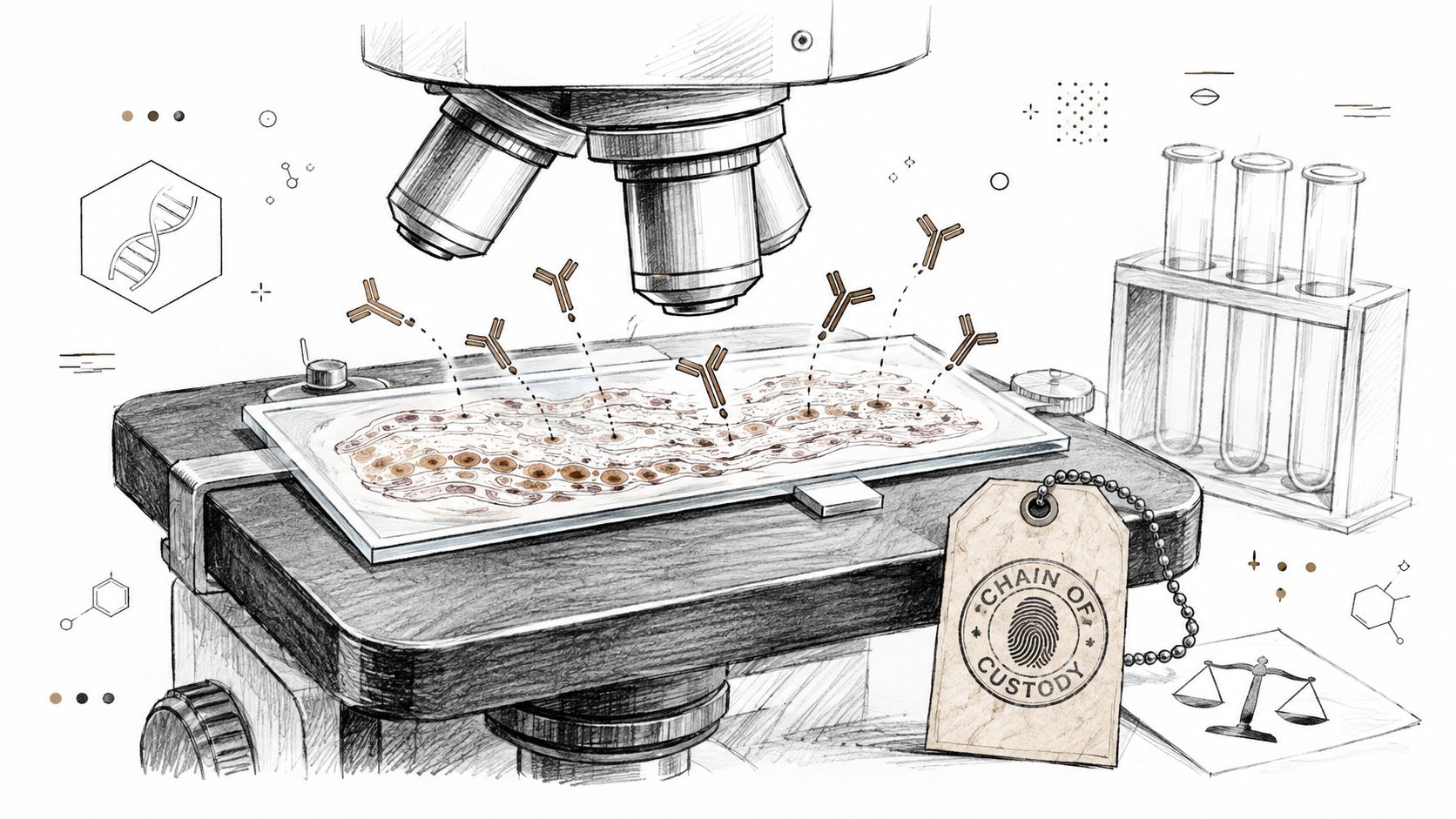
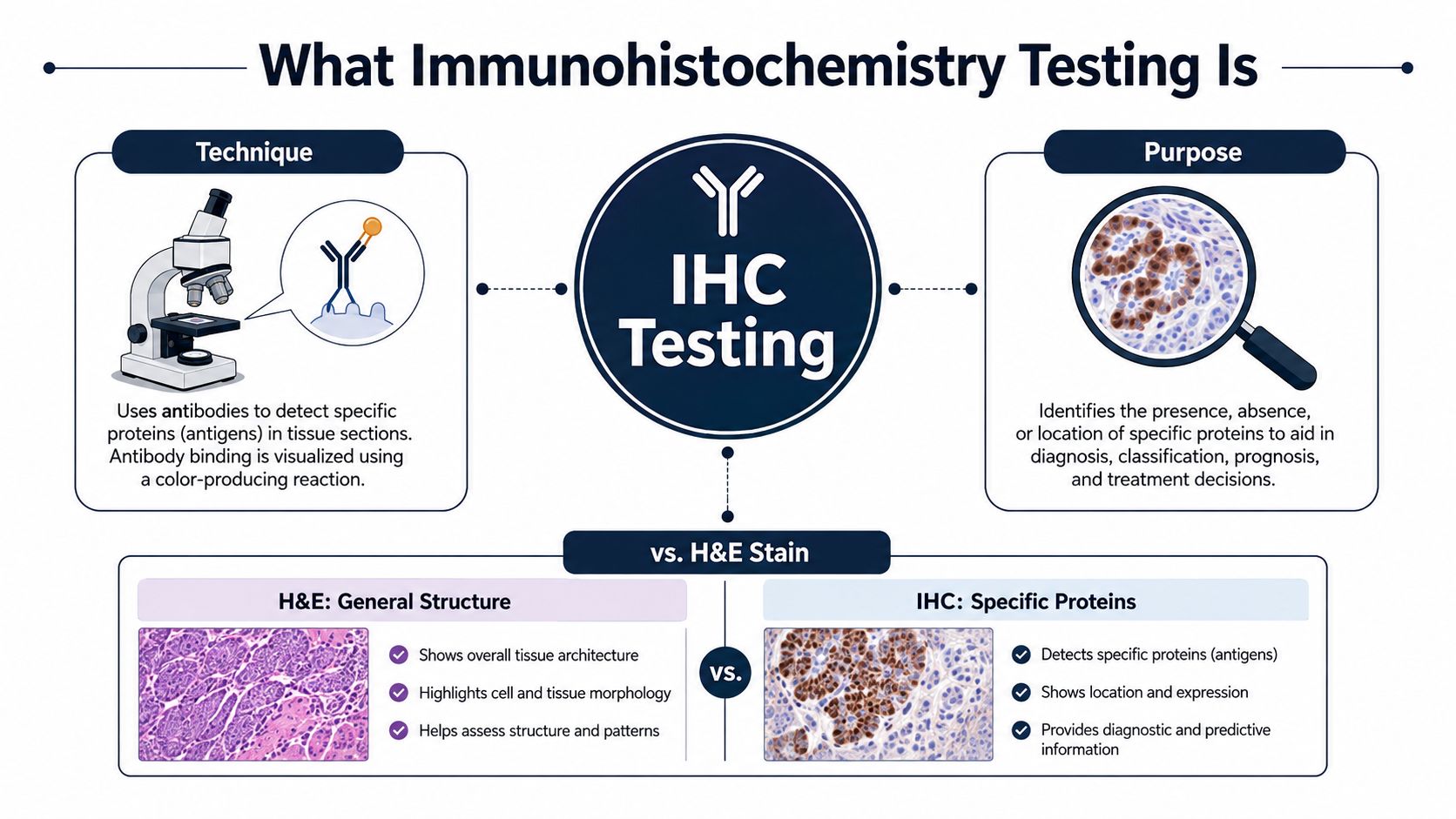
Immunohistochemistry testing, often shortened to IHC, uses labeled antibodies to locate a particular protein inside a tissue section. The antibody binds to its target, and a detection system produces a visible signal, commonly a brown deposit, where that binding occurs. A forensic pathologist then examines the signal in relation to the tissue's structure.
Routine hematoxylin and eosin, or H&E, staining answers a different question. H&E shows general architecture, cell injury, inflammation, bleeding, and other structural features. It usually can't identify with certainty which cell type is producing a particular protein or whether a selected molecular signal is present in a specific compartment.
Why the distinction matters after death
A gross autopsy can show no obvious explanation for a sudden death. Histology may reveal changes, but those changes can be subtle, incomplete, or difficult to separate from postmortem artifact. IHC may then help investigate questions involving hypoxic injury, myocardial injury, sepsis-related lung damage, anaphylaxis, drug-related liver injury, or tumor classification.
The method has a long foundation in pathology. The first reported use of antibodies to detect antigens by IHC occurred in 1942, enzyme-labeled antibodies were described in 1966, and the demonstration of antigens in formalin-fixed, paraffin-embedded tissue in 1974 helped make IHC practical for routine diagnostic work. These milestones explain why archived autopsy tissue can still be useful for targeted review (pathology history and development of IHC).
IHC is often ordered after the pathologist has reviewed routine histology and considered toxicology, microbiology, medical records, and scene information. It's not a replacement for those sources. It adds a more specific view of selected tissue features.
Practical rule: A stain is evidence about a target protein in a particular tissue location. It isn't, by itself, a complete explanation for a death.
The same antibody-based methods used in cancer diagnosis can be adapted for forensic questions. Readers who want to understand the physical setting in which tissue processing and staining occur can browse pathology lab casework, including the controlled work areas that support specimen handling.
How the Stain Actually Works Step by Step
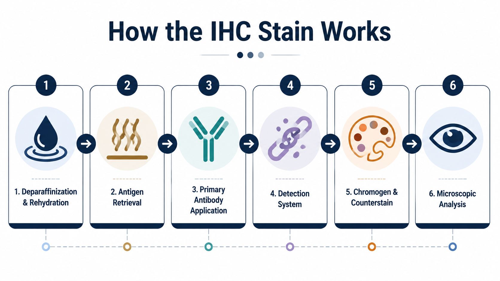
An IHC slide begins with a thin section of tissue, commonly cut from a paraffin block and mounted on glass. The laboratory first removes the paraffin and rehydrates the section so water-based solutions can reach the tissue. This opening stage prepares the specimen for the antibody reactions that follow.
From tissue section to visible signal
The laboratory then performs antigen retrieval. Formalin can create chemical cross-links that hide the protein sites an antibody needs to recognize. Heat-based or enzyme-based retrieval helps expose those sites, although the appropriate method depends on the tissue and target.
The next steps control unwanted background. The laboratory blocks endogenous peroxidase and other nonspecific binding sites so the final color reaction is more likely to represent the intended antibody binding rather than unrelated tissue chemistry.
A primary antibody is applied next. It is selected for a particular protein, not for “death” as a general condition. After washing, a detection system binds to the primary antibody. This may use a polymer-based reagent or a secondary antibody system linked to an enzyme such as horseradish peroxidase or alkaline phosphatase.
The enzyme reacts with a chromogen. With DAB, the reaction commonly creates a brown precipitate at the sites where the target protein is detected. Hematoxylin is then used as a counterstain to show the surrounding nuclei and tissue structure. Finally, the slide is dehydrated, covered, and reviewed under the microscope.
The pathologist doesn't interpret the brown color alone. The review considers whether the signal appears in the expected cells, whether it occupies the expected location, whether the intensity is plausible, and whether the controls worked.
A positive control contains tissue expected to express the target. A negative control helps identify nonspecific or reagent-related staining. If the controls fail, the patient or decedent's slide may not be interpretable, even if some color is visible.
The following video provides a visual overview of the laboratory sequence:
Sample Collection and Fixation Before the Stain
The quality of IHC is often determined before the first antibody reaches the slide. Tissue must be collected, labeled, sampled, fixed, processed, and stored in a way that preserves both morphology and antigenicity. In a postmortem case, the postmortem interval, temperature exposure, autolysis, and the condition of each organ can affect what remains detectable.
For most tissues, a published IHC protocol recommends 10% neutral buffered formalin fixation for 24 hours at room temperature, with a tissue-to-fixative ratio ranging from 1:1 to 1:20 (published IHC protocol). Actual handling depends on tissue thickness, organ type, suspected disease, and laboratory requirements.
Formalin fixation protects structure, but it can also mask epitopes through cross-linking. A pathology review reports that approximately 85% of antigens fixed in formalin require some form of antigen retrieval to optimize immunoreactivity (formalin fixation and antigen retrieval review). Delayed fixation, excessive fixation, inadequate penetration, or autolysis may produce weak or false-negative staining.
Variables that deserve documentation
Tissue is usually sliced to allow fixative to penetrate. The laboratory records the block site, cassette identifier, slide number, and transfer history. This documentation matters when an attorney later needs to establish that a particular slide came from a particular specimen.
| Variable | Ideal practice | Risk if ignored |
|---|---|---|
| Tissue collection | Sample representative areas promptly and document the site | Autolysis or missed focal injury can limit interpretation |
| Tissue thickness | Use sections that allow fixative penetration | The center may remain poorly fixed |
| Fixative | Use an appropriate formalin-based method and adequate volume | Antigen masking or uneven preservation may occur |
| Fixation duration | Record the start and end of fixation | Retrieval conditions may be chosen incorrectly |
| Block identity | Link the tissue, cassette, block, and slide | The result becomes harder to trace or defend |
| Custody record | Document every transfer and storage step | A later challenge may focus on specimen integrity |
The specific collection method is part of the broader forensic record. Our team also considers forensic specimen collection procedures when selecting and documenting tissue for later laboratory examination.
Fresh or frozen tissue may be retained in selected cases when a target is especially sensitive to fixation. That decision must be made early because it affects specimen allocation and later testing options.
Common Markers and Panels Used in Autopsy Cases
Forensic IHC is most useful when the panel is built around a clear investigative question. Ordering many stains without a defined question can create confusing findings, consume limited tissue, and make the final interpretation less focused.
A hypoxic-ischemic injury panel may include HIF-1alpha, HSP70, and selected neuronal markers. These stains can help assess cellular responses associated with reduced oxygen, but they still must be considered alongside scene information, medical history, microscopic findings, and the timing of death.
A myocardial injury panel may include troponin, C5b-9, CD68-positive macrophages, and fibronectin. The purpose is to investigate whether a suspected myocardial infarction has tissue support when gross findings and routine histology don't align.
Other panels address different problems:
| Question | Typical markers | What a positive result supports |
|---|---|---|
| Is there evidence of hypoxic or ischemic injury? | HIF-1alpha, HSP70, selected neuronal markers | Supports a tissue response associated with oxygen deprivation, subject to timing and preservation limits |
| Could myocardial injury be present despite subtle routine findings? | Troponin, C5b-9, CD68, fibronectin | Supports investigation of myocardial injury or infarct-related change |
| Is amniotic material present in pulmonary vessels? | Cytokeratin and mucin stains | Supports identification of squames or mucin in the pulmonary vasculature, with clinical and autopsy correlation |
| Could a tumor be classified more precisely? | Lineage and tumor-associated markers | Supports a tumor type or possible origin when morphology alone is insufficient |
| Could cardiac amyloid be present? | Kappa, lambda, transthyretin, AA | Supports an amyloid pattern and helps direct further typing |
| Could an infectious process be involved? | Organism-specific or host-response markers | Supports the presence of selected infectious material or tissue response |
Mutation-specific and companion-diagnostic IHC is also moving beyond simple protein detection. Recent literature describes multiplex immunohistochemistry and immunofluorescence for spatial immune-cell analysis, while mutation-specific IHC may serve as a surrogate for molecular testing in selected settings. An IHC companion diagnostic for MET expression in non-squamous non-small-cell lung cancer received FDA approval in 2025, illustrating how the method can contribute to treatment-selection decisions (review of newer IHC developments).
A positive result usually changes the level of support for a specific hypothesis. It doesn't automatically change the cause-of-death conclusion. For example, a stain supporting myocardial injury still has to be reconciled with coronary anatomy, toxicology, medical history, and the distribution of injury.
Reading the Slide and Interpreting Results
The pathologist begins by checking whether the controls behaved as expected. A positive control should show the anticipated staining pattern, while a negative control should remain appropriately clean. Without valid controls, the laboratory can't confidently distinguish specific signal from technical background.
The slide is first scanned at low power. This shows distribution, symmetry, focality, and relationship to the tissue architecture. The pathologist then moves to higher power and asks whether the signal is in the correct cell type and compartment. Nuclear, cytoplasmic, membranous, vascular, and extracellular staining can have very different meanings.
What positive and negative actually mean
In surgical pathology, some markers use formal scoring systems, including Allred scores or H-scores, to translate semiquantitative observations into a standardized format. In postmortem work, reporting may instead use terms such as positive, focal, weak, or negative, depending on the target and the clinical question.
A negative result means the laboratory didn't detect the target under the conditions used. It doesn't necessarily mean the protein was absent from the body. Fixation, autolysis, antigen retrieval, antibody selection, and control performance all affect detectability.
The report should therefore record relevant pre-analytic facts. A weak stain from tissue with delayed fixation may carry a different evidentiary weight from a negative stain in well-preserved tissue with strong controls.
A negative IHC result is a laboratory observation, not a complete statement about biology.
Readers who need a broader explanation of how tissue findings are reviewed can consult our discussion of microscopic examination in forensic pathology. The same principle applies here: the slide must be interpreted within the tissue, the case, and the testing conditions.
For attorneys, the wording matters. “No staining identified” is not always equivalent to “the protein was absent,” and “positive staining” is not equivalent to “this finding caused death.” A defensible report explains those distinctions directly.
Limits, False Negatives, and When IHC Is Not Enough
IHC can provide highly specific spatial information, but it has hard limits. Formalin may mask the target epitope. Autolysis may damage the protein. The selected antibody clone may not recognize a postmortem-modified form of the antigen. Cross-reactivity can also create an unexpected signal.
Validation creates another important boundary. An antibody validated on fresh surgical tissue may not perform the same way on decomposed or partially autolyzed postmortem tissue. A forensic laboratory must understand the antibody, platform, retrieval method, controls, and tissue context before relying on the result in a high-stakes conclusion.
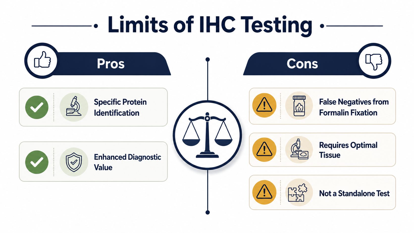
A published review notes that IHC quality failures more often involve weak or false-negative staining than false-positive staining. Updated validation expectations referenced by the College of American Pathologists emphasize at least 90% concordance for assay validation, with separate validation by clone and scoring combination when applicable (IHC validation review). A separate validation document recommends at least 90% overall concordance between a new assay and its comparator or expected result, with investigation required when concordance falls below that level (CAP-referenced validation guidance).
What other testing contributes
IHC can show that a protein is present in a place. It can't, by itself, prove that the protein caused death or distinguish a therapeutic drug effect from an overdose.
Other methods answer different questions:
- PCR and related molecular tests can investigate selected infectious organisms or genetic targets.
- Toxicology evaluates drugs, poisons, metabolites, and concentrations in appropriate specimens.
- Molecular sequencing may help investigate inherited or acquired variants associated with arrhythmia syndromes.
- Microbiology and culture can contribute evidence about organisms when viable material and proper sampling are available.
- Routine histology remains necessary because the cellular architecture gives the IHC signal its context.
The strongest medicolegal interpretation usually comes from layering these sources rather than asking one test to carry the entire conclusion.
Turnaround, Cost Drivers, and How to Request IHC
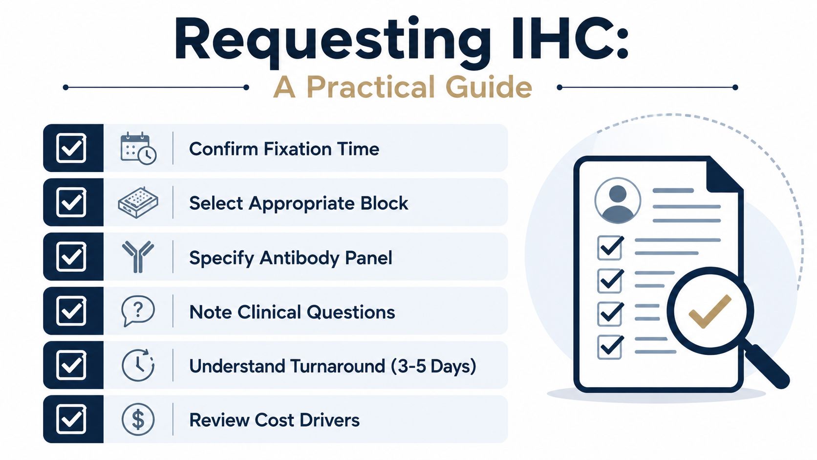
IHC turnaround depends on the condition of the tissue, the number of stains, the availability of controls, the need for retrieval optimization, and the pathologist's review. Most cases within 24–48 hours may still require additional time when tissue must be located, recut, sent to an outside laboratory, or tested with a broader panel. A final amended report is issued only after the results are reviewed in the context of the complete case.
Cost also varies by the work required. Common drivers include the number of antibodies, whether the request involves one stain or a panel, clone selection, control slides, repeat runs, specialized retrieval, slide preparation, and interpretation time.
Information to provide with a request
Families, attorneys, healthcare professionals, and agencies can help the laboratory by supplying:
- Case identification: The case number, decedent's identifying information, and agency or facility contact.
- Investigative question: The suspected diagnosis or unresolved cause-of-death issue.
- Tissue location: The organ, block identifier, or slide number when available.
- Existing findings: Prior H&E observations, autopsy conclusions, toxicology, microbiology, and relevant medical records.
- Custody information: Whether the tissue comes from a family-authorized private autopsy, a medical examiner investigation, or a court-ordered examination.
- Legal needs: Any deadline, subpoena, deposition, or anticipated testimony requirement.
The FDA recommends that primary antibodies be evaluated on panels of normal tissues, with testing on samples from three different persons for each listed tissue. It also recommends positive tissue controls from fresh autopsy, biopsy, or surgical specimens that were fixed, processed, and embedded as promptly and similarly as the patient material (FDA IHC guidance).
For an overview of related laboratory work, including how testing may fit into a larger postmortem investigation, review laboratory analysis services. The request should identify the question first. The antibody panel comes second.
Questions Families and Attorneys Ask Most Often
Does an IHC result appear in the cause-of-death statement?
It may, but the wording depends on the full evidence. The cause of death is the disease, injury, or condition that resulted in death. The manner of death describes the circumstances classification, such as natural, accident, suicide, homicide, or undetermined, when the responsible authority makes that determination.
An IHC result may support a disease or injury listed in the cause-of-death statement. It generally won't establish the manner of death by itself. A final opinion should explain whether the stain is supportive, contributory, inconclusive, or not relevant to the final conclusion.
Can a negative stain be repeated?
Sometimes. Retesting may be possible if paraffin blocks or unstained sections remain, the tissue is adequately preserved, and the requested antibody or retrieval method is appropriate. A second review should include the original report, controls, fixation history, block identifiers, and the reason the result is being challenged.
The second pathologist should distinguish a true independent interpretation from a technical repeat. If the original control failed, a repeat may address a laboratory problem. If the tissue is severely autolyzed, repeating the same stain may not solve the underlying limitation.
How are slides handled for a second opinion?
Slides and blocks should be packaged, identified, and transferred with documentation of who released them, who received them, and when. That record is the chain of custody, meaning the traceable history of evidence from collection through storage, testing, review, and presentation.
A second-opinion reviewer typically needs the case history, autopsy report, H&E slides, IHC slides, control information, toxicology, microbiology, medical records, and relevant photographs. The reviewing pathologist may also need access to the original block if the question involves recutting or repeating the stain.
Can IHC evidence be challenged in court?
An attorney may examine the antibody's validation, intended use, control results, tissue preservation, clone selection, retrieval method, scoring method, and the pathologist's reasoning. Depending on the court and jurisdiction, admissibility may involve standards associated with Daubert or Frye. The applicable standard is a legal question for the court, not a conclusion that a laboratory can guarantee.
Research-grade antibodies and validated clinical or forensic assays aren't interchangeable. A deposition should clarify how the reagent was validated, whether the tissue type matched the validation material, and whether the result was reproduced.
What if the original laboratory closes?
The responsible agency, facility, record custodian, or successor laboratory may hold the report, blocks, or slides. Families and attorneys should request the complete laboratory file, including specimen identifiers, custody records, control information, raw interpretations, and amended reports. If only a report remains, a reviewing pathologist can assess the language, but the absence of slides or blocks may limit independent verification.
Texas Autopsy Services provides independent autopsy examinations and second-opinion reviews for families, attorneys, healthcare professionals, and county officials across Texas. Our team can evaluate whether retained tissue, prior slides, and the investigative question support additional immunohistochemistry testing, with every examination performed by a board-certified forensic pathologist certified by the American Board of Pathology.
If an autopsy report leaves important questions unanswered, contact Texas Autopsy Services for a careful review of the available records, tissue, and laboratory findings. Visit Texas Autopsy Services to discuss an independent autopsy or second-opinion pathology evaluation with our team.