August 17, 2026 · Texas Autopsy Services
What Is Pulmonary Embolism and How Forensic Pathology Helps
Learn what is pulmonary embolism, its causes, symptoms, and treatments. Discover how independent autopsy services clarify cause of death in complex cases.
On this page
- Understanding Pulmonary Embolism in Plain Language
- How Blood Clots Form and Travel to the Lungs
- How Doctors Diagnose Pulmonary Embolism
- Recognizing Symptoms and Warning Signs
- Treatment Options and Long-Term Recovery
- Why Pulmonary Embolism Appears in Autopsy Findings
- When to Seek Emergency Care or Request an Autopsy
Pulmonary embolism is a blood clot that travels to the lungs and blocks blood flow, most often after forming in the deep veins of the legs. It can become fatal quickly when the blockage interferes with oxygen exchange or places severe strain on the heart.
A person may feel sudden shortness of breath, sharp chest pain, or an unexplained rapid heartbeat and assume the problem is anxiety, pneumonia, or a heart condition. In other cases, the first clear evidence appears after death, when medical records are incomplete or the symptoms were too subtle to prompt emergency evaluation. For families and attorneys, understanding what is pulmonary embolism can help clarify both clinical decisions and postmortem findings.
Understanding Pulmonary Embolism in Plain Language
A pulmonary embolism, commonly called PE, occurs when a clot blocks an artery in the lungs. The clot usually begins as a deep vein thrombosis, or DVT, in the leg. If part of the clot breaks away, the bloodstream carries it through the heart and into the pulmonary arteries, which transport blood into the lungs.
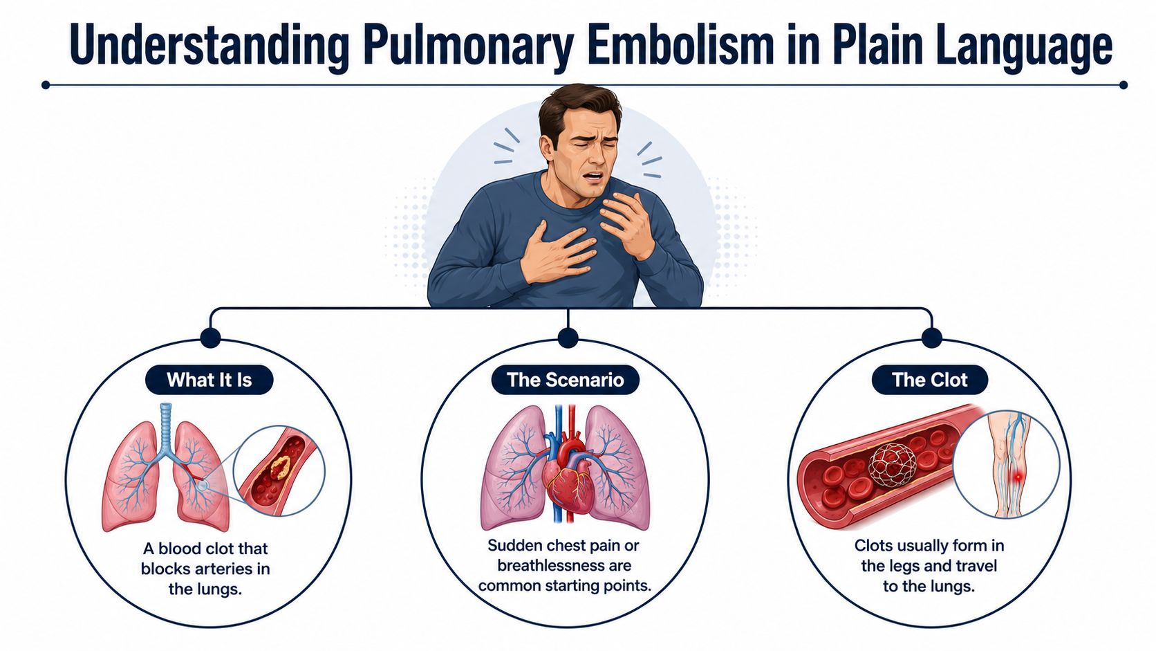
A reader may be searching for answers after a relative suddenly developed breathlessness while resting, walking, or recovering from an illness. PE symptoms can begin abruptly, but they aren't always dramatic. A smaller clot may cause limited symptoms, while a larger clot can obstruct enough pulmonary circulation to impair oxygen exchange and strain the right side of the heart.
Pulmonary embolism is a major global cardiovascular condition. Annual incidence estimates range from about 39 to 115 cases per 100,000 people, while some European guidance places venous thrombosis and PE together at roughly 0.5 to 1.0 per 1,000 inhabitants each year (clinical review of pulmonary embolism). In the United States, reported incidence increased from 37.3 per 100,000 person-years in 2000 to 83.5 per 100,000 person-years in 2020 (review of PE incidence and mortality).
Safety point: Sudden breathing difficulty, chest pain, fainting, or coughing up blood requires emergency medical attention. A postmortem explanation can't replace urgent evaluation during life.
PE matters in forensic pathology because it may cause death before a person reaches medical care or before clinicians can confirm the diagnosis. When the cause remains uncertain, an independent examination can help distinguish pulmonary embolism from heart disease, pneumonia, drug effects, trauma, or other possible causes.
How Blood Clots Form and Travel to the Lungs
The biological process behind many blood clots is described through Virchow's triad. It includes three conditions that make clot formation more likely: slowed venous blood flow, injury to the inner lining of a blood vessel, and an increased tendency for blood to clot.
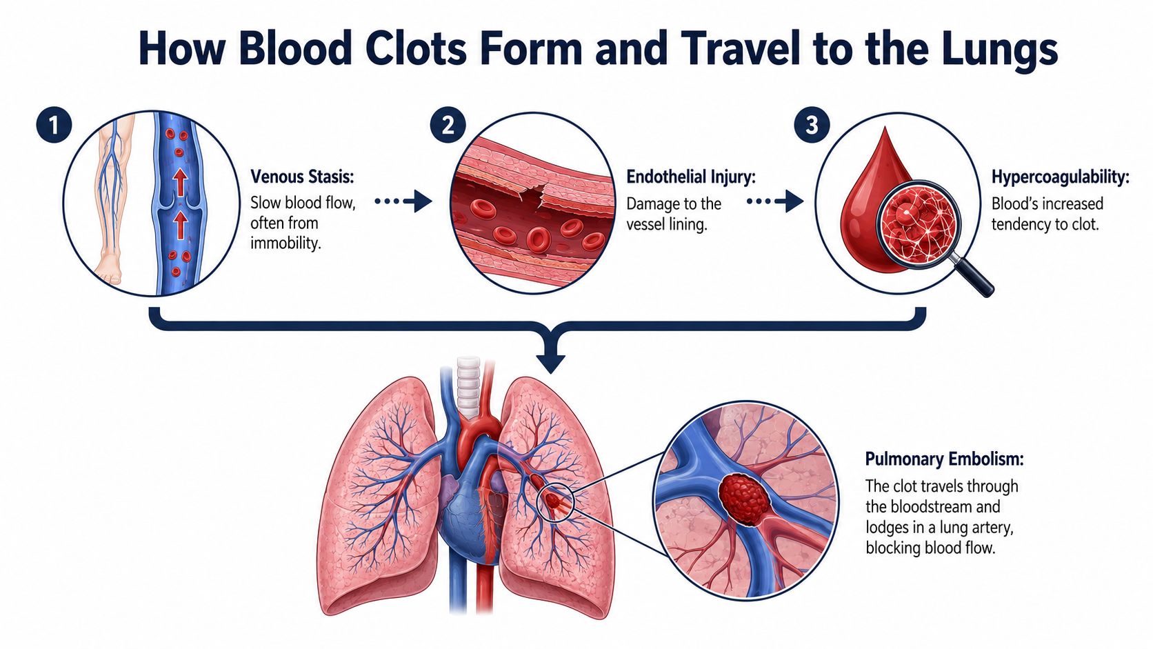
The three parts of Virchow's triad
Venous stasis means blood moves slowly or pools, especially in the deep veins of the legs. Immobilization, bed rest, and recovery after surgery can reduce the normal muscle movement that helps return blood toward the heart.
Endothelial injury involves damage to the vessel lining. Surgery, physical trauma, and the presence of an indwelling venous catheter can contribute to this condition.
Hypercoagulability means the blood has a greater tendency to clot. Active cancer, pregnancy or the postpartum period, estrogen exposure, thrombophilia, and some chronic illnesses can contribute. A prior venous thromboembolism also matters because recurrence risk can remain clinically important.
The clinical overview of PE causes and diagnosis identifies these mechanisms and risk factors as central to practical assessment. A clot may remain in a leg vein, or a portion may detach and travel through the venous circulation into the right side of the heart. From there, it enters the pulmonary arteries.
Severity depends on the clot's size, location, and the person's cardiopulmonary reserve. A large obstruction can abruptly reduce blood flow through the lungs and cause right-ventricular strain. Smaller emboli may produce less obvious symptoms, which can complicate both treatment decisions and later interpretation of medical records.
For people advised to use compression garments, practical instructions on using tools for compression stockings may help with application. Compression products should be used according to a clinician's recommendation, particularly when circulation problems or other medical conditions are present.
How Doctors Diagnose Pulmonary Embolism
A patient arrives with breathlessness and chest discomfort, but the same complaints can arise from pneumonia, heart disease, panic symptoms, or other conditions. Clinicians begin by estimating how likely PE is, then select laboratory tests and imaging that can support or challenge that assessment. This stepwise record also matters later when families or attorneys need to understand how a suspected PE was evaluated.
Clinical probability comes first
The Wells score helps estimate pretest probability from clinical findings and history. One interpretation classifies a score below 2 as low probability, 2 to 6 as intermediate, and 6 or higher as high probability. A modified approach describes PE as unlikely at 4 or below and likely above 4 (Wells score reference).
A D-dimer test often follows when probability is low or intermediate. In an appropriate patient, a negative result can help rule out PE. A positive result does not establish the diagnosis, because many conditions can raise D-dimer levels. Imaging may then be required.
Imaging confirms the suspected blockage
CT pulmonary angiography, or CTPA, is commonly used when clinical suspicion is high or D-dimer is elevated. It can display a clot inside the pulmonary arteries. If CTPA cannot be performed, a ventilation-perfusion scan may offer an alternative.
| Clinical probability | First test | Next step if positive | Next step if negative |
|---|---|---|---|
| Low | D-dimer | CTPA or further evaluation | PE becomes unlikely in the appropriate clinical setting |
| Intermediate | D-dimer | CTPA | PE may be ruled out when the result and clinical assessment support that conclusion |
| High | CTPA | Treat and risk-stratify confirmed PE | Reassess the diagnosis and consider additional evaluation |
Blood tests serve different purposes in emergency and hospital care. Reviewing what blood tests do can help families read medical records without treating every abnormal result as proof of PE.
The 2026 guideline summary supports risk-based evaluation, including D-dimer testing for low- or intermediate-probability cases, CTPA when indicated, and V/Q scanning when CTPA cannot be performed (2026 acute PE guideline summary). In forensic review, these clinical findings are compared with imaging, treatment records, and autopsy observations when the cause of death remains unclear.
Recognizing Symptoms and Warning Signs
After a long flight, a person may suddenly become short of breath, develop sharp chest pain, or feel an unusually rapid heartbeat. Those symptoms can reflect a pulmonary embolism, especially when they appear without an obvious explanation. Sudden shortness of breath, chest pain that worsens with deep breathing, a fast heart rate, and coughing up blood are recognized warning signs.
A smaller clot may cause fatigue, reduced ability to exercise, mild chest discomfort, or a vague sense that something is wrong. The same pattern can resemble anxiety, a respiratory infection, muscle strain, or a heart condition. Symptoms may therefore be overlooked, and a lack of dramatic symptoms does not rule out PE.
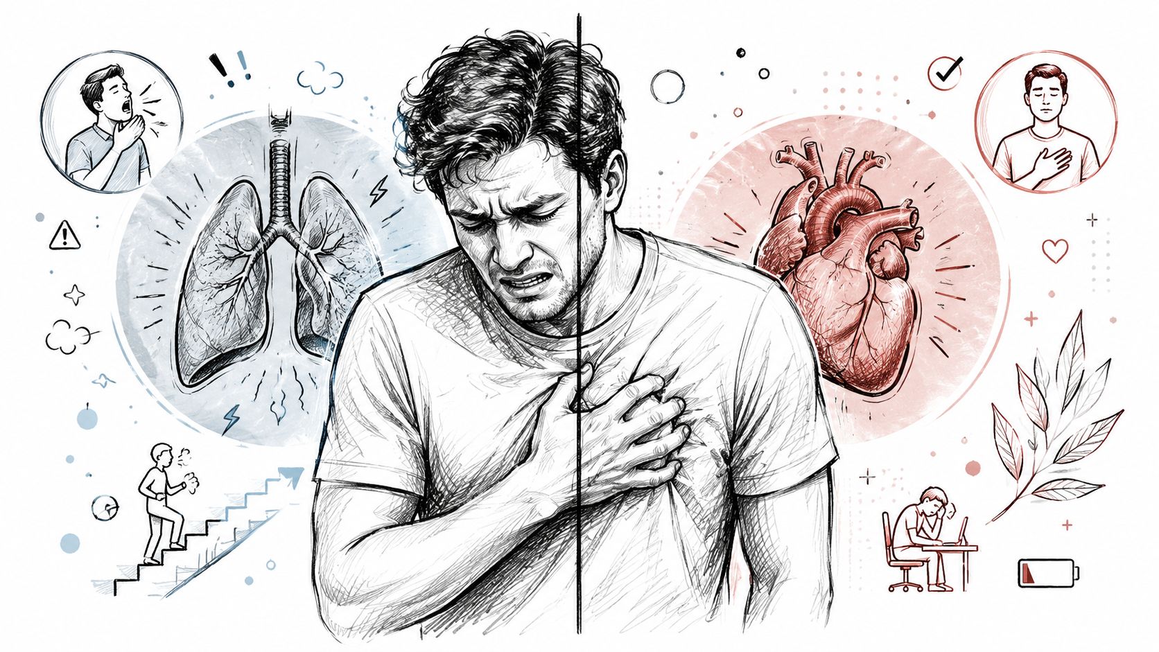
Symptoms that require emergency action
Seek emergency care for:
- Sudden breathing difficulty: Particularly when it has no clear cause.
- Chest pain: Especially sharp pain linked to breathing or paired with breathlessness.
- Fainting or severe weakness: These may indicate impaired circulation.
- Coughing up blood: This is an urgent warning sign when respiratory symptoms are also present.
- A rapid or irregular heartbeat: A fast pulse can reflect strain on the heart and lungs.
One-sided leg swelling, pain, warmth, or tenderness may provide additional context because these findings can occur with DVT. Some DVTs produce no noticeable leg symptoms.
For families reviewing a death, a record that mentions “anxiety,” “shortness of breath,” or “chest pain” does not establish PE alone. Those observations may matter when examined alongside risk factors, testing, medication history, and autopsy findings. A forensic pathologist assesses the complete medical and postmortem record, which can help families and attorneys evaluate an unclear cause of death without treating one symptom as conclusive.
Treatment Options and Long-Term Recovery
Treatment is chosen according to the PE's severity, bleeding risk, cause, and effect on the heart and circulation. Anticoagulation is the main treatment for most confirmed cases. It reduces the blood's ability to form new clots while the body gradually clears or organizes the existing obstruction.
Guidance recommends at least 3 months of therapeutic anticoagulation during the primary treatment phase (anticoagulation duration guidance). Many patients continue treatment for 3 to 6 months. Longer or indefinite therapy may be considered when recurrence risk remains high. The treating clinician balances that risk against the possibility of bleeding, taking the person's medical history and circumstances into account.
Severe PE may require hospital-based treatment beyond medication. Depending on the patient's condition, clinicians may consider clot-dissolving treatment, catheter procedures, surgical removal, or supportive measures for impaired circulation and breathing. These decisions address the living patient's immediate needs and are separate from any later autopsy evaluation.
Recovery continues after discharge
Guideline comparisons recommend follow-up at 3 to 6 months. Breathlessness or reduced exercise tolerance continuing beyond 3 months may lead clinicians to consider noninvasive testing (international PE guideline comparison).
Recovery can include taking medication as prescribed, reviewing persistent symptoms, deciding when work or physical activity is appropriate, and repeating tests when clinically indicated. The 2026 AHA/ACC guideline describes acute and early post-acute PE care and introduces five Acute PE Clinical Categories to support decisions about discharge, hospitalization, and follow-up (2026 AHA/ACC guideline announcement).
For families reading a discharge record: The hospital stay is only one part of PE care. Follow-up plans, medication instructions, symptom monitoring, and activity restrictions may help explain the treatment course and later medical review.
Why Pulmonary Embolism Appears in Autopsy Findings
Pulmonary embolism can be present without a confirmed diagnosis during life. A person may deteriorate too quickly for imaging, or symptoms may be attributed to another condition. In some deaths, the clot is discovered only during a complete forensic examination.
The mechanism is direct but complex. A clot blocks pulmonary blood flow, reducing circulation through the lungs and interfering with gas exchange. A large clot burden, low blood pressure, rapid heart rate, or right-ventricular dysfunction can indicate severe physiologic stress. A meta-analysis associated higher mortality with right-ventricular dysfunction, increased troponin, hypotension, tachycardia, malignancy, immobility, chronic lung disease, heart failure, and older age (pathophysiology and mortality review).
Untreated acute PE has been associated with hospital mortality as high as 30%, while treated cases were reported around 8% in classic clinical literature. Contemporary summaries describe death within one month in 12% to 28% of patients in some cohorts, and mortality reaching 30% by one year in higher-risk populations (clinical review of PE incidence and mortality).
What an independent examination can clarify
A forensic pathologist evaluates the pulmonary arteries, heart, other organs, medical history, toxicology when indicated, and the circumstances surrounding death. The pathologist must also consider competing explanations, because finding a clot doesn't automatically establish that it caused death.
Our explanation of how a coroner determines cause of death describes the broader reasoning involved. In a private autopsy or second-opinion autopsy, the written report can help families understand whether PE was the cause of death, a contributing condition, or an incidental finding.
The terms have distinct meanings. Cause of death is the disease or injury that produced death, such as pulmonary embolism. Manner of death describes the circumstances, typically classified as natural, accident, suicide, homicide, or undetermined. Chain of custody is the documented control and transfer of evidence from collection through examination and storage, which protects its integrity for legal review.
Pulmonary embolism's medical history is relatively recent. The first documented case is commonly dated to 1837, and historical accounts describe the condition as nearly universally fatal before about 1930. Heparin entered clinical trials around 1935, and anticoagulant therapy was shown in 1960 to reduce death and recurrent venous thromboembolism (history of pulmonary embolism).
When to Seek Emergency Care or Request an Autopsy
Sudden shortness of breath, chest pain, fainting, coughing up blood, or a rapid heartbeat may signal a pulmonary embolism. Call emergency services rather than waiting, particularly after recent immobility, surgery, trauma, cancer, pregnancy or postpartum status, estrogen exposure, or a previous clot.
After an unexplained death, families may seek an independent autopsy if official findings leave important questions unanswered, medical records are incomplete, or an attorney needs an objective forensic review. In Texas, a medical examiner, justice of the peace, or judge may order an autopsy when required for a death investigation under Texas criminal procedure statute.
Texas Autopsy Services provides private autopsy services that Texas families, attorneys, healthcare professionals, and county officials may consider. A forensic pathologist certified by the American Board of Pathology performs each examination, with licensed transport, documented chain of custody, and reporting for medical and legal review. Information about requesting an autopsy explains practical next steps.
Families may also review Private Autopsies, County Forensic Autopsies, or the Waterloo Mortuary Partnership for coordination details.
If records do not clearly explain a death that may involve pulmonary embolism, contact Texas Autopsy Services for guidance on independent autopsy, second-opinion review, transport, chain of custody, and forensic reporting. The team is available across Texas counties and explains the process with medical precision and compassion.