May 14, 2026 · Texas Autopsy Services
Cavitating Mass in Lung Explained by a Pathologist
A forensic pathologist explains a cavitating mass in lung, its causes (cancer, infection), and its meaning in a cause of death investigation or private autopsy.
On this page
- An Unexpected Finding on an Autopsy Report
- Understanding How a Lung Mass Becomes a Cavity
- Common Causes Behind a Cavitating Lung Mass
- The Forensic Investigation of a Lung Cavity
- Distinguishing Disease from Postmortem Changes
- What a Cavitating Mass Means for a Cause of Death Investigation
- Frequently Asked Questions About Lung Findings
When a family or attorney reads an autopsy report and sees the phrase cavitating mass in lung, it often lands with a shock. The words sound severe, but they don't explain themselves. My job as a forensic pathologist is to translate that finding into plain language and then determine what it meant in life and at death.
At Texas Autopsy Services, every examination is performed by a board-certified forensic pathologist experienced in medicolegal death investigations. Our work centers on one principle: follow the evidence carefully, explain it plainly, and treat the deceased and their family with respect.
TL;DR
- A cavitating mass in lung means there is a lung lesion with a hollow space inside it.
- That cavity can be caused by cancer, infection, inflammatory disease, or postmortem change.
- Wall appearance matters. In one classic study, 92% of cavities with walls ≤4 mm were benign, while 95% with walls >15 mm were malignant in solitary lung cavities, as summarized in a clinical review of cavitary lung lesions.
- In forensic pathology, the key question is not just what the cavity looks like, but whether it formed before death or after death.
- The most reliable answer usually comes from gross examination, tissue sampling from the cavity wall, microscopy, and sometimes microbiology.
An Unexpected Finding on an Autopsy Report
A common situation starts like this. A family receives an autopsy report, or an attorney reviews records for a case, and one line stands out: “cavitating mass in the lung.” The immediate concern is usually cancer. The next concern is whether it was missed, whether it caused death, and whether someone should have recognized it sooner.
That reaction is understandable. The phrase is technical, and it doesn't tell you whether the process was malignant, infectious, old, recent, or even truly present before death.
What the term actually means
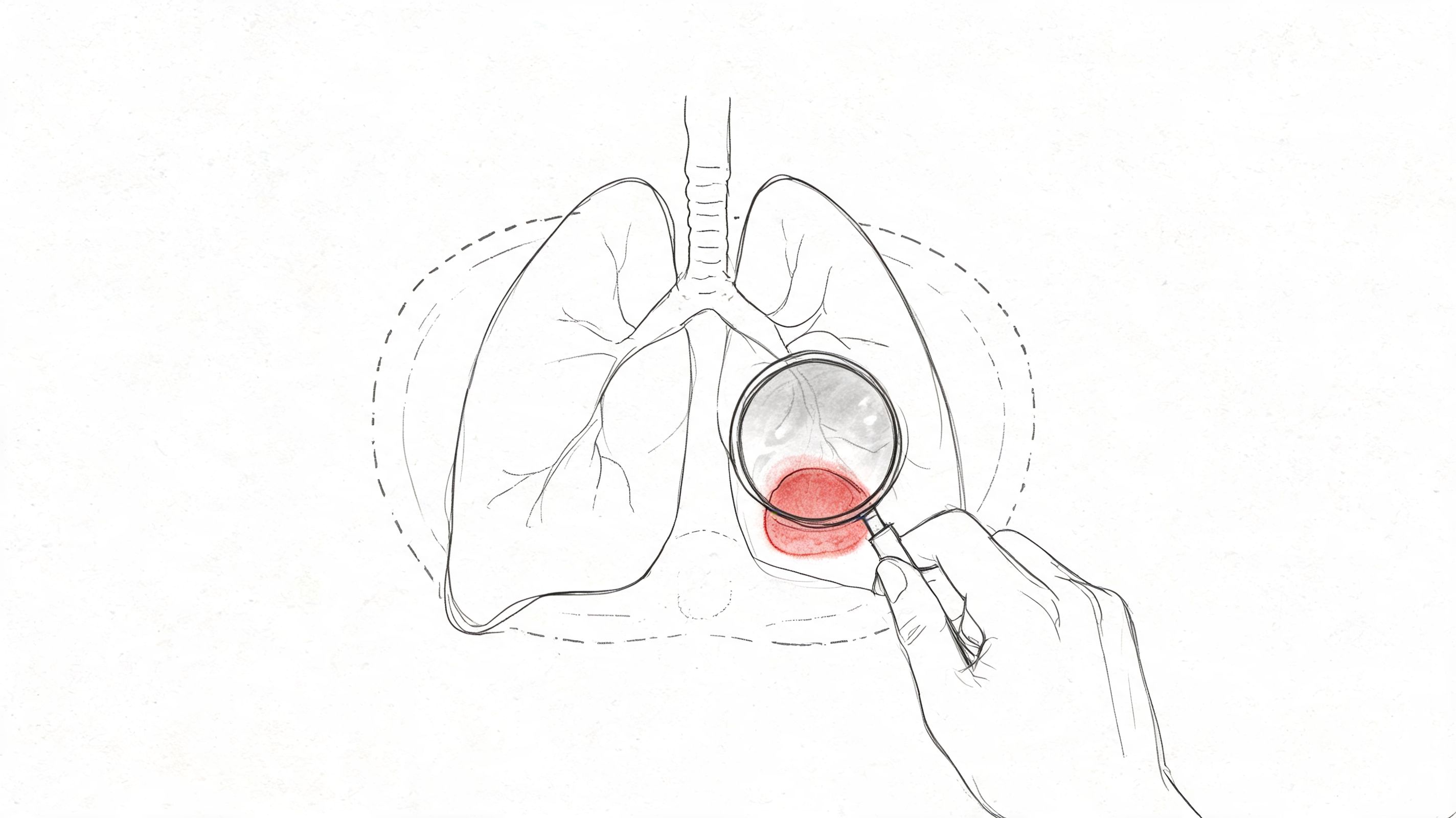
A mass is an abnormal area of tissue. Cavitating means that part of that abnormal tissue has broken down and left a hollowed-out space. In simple terms, it is a lung lesion with an interior void that may contain air, fluid, debris, or a mix of these.
What matters is that cavitation is a feature, not a diagnosis. It describes how a lesion looks. It doesn't by itself tell us the cause.
A cavity in the lung is a clue. It is not the final answer.
Why families and attorneys need more than the report wording
In clinical medicine, imaging often starts the conversation. In forensic pathology, imaging and report language are only part of the evidence. At autopsy, I can inspect the lesion directly, measure it, open it, examine its wall, and sample it under the microscope.
That matters because several very different diseases can produce a similar cavity. A lung cancer can do it. A bacterial abscess can do it. Tuberculosis can do it. A metastatic cancer can do it. After death, decomposition can also create appearances that confuse the picture if the case isn't interpreted carefully.
For families, this affects understanding and peace of mind. For attorneys, it affects causation, timing, standard-of-care questions, and whether a lesion was incidental or central to the death.
Understanding How a Lung Mass Becomes a Cavity
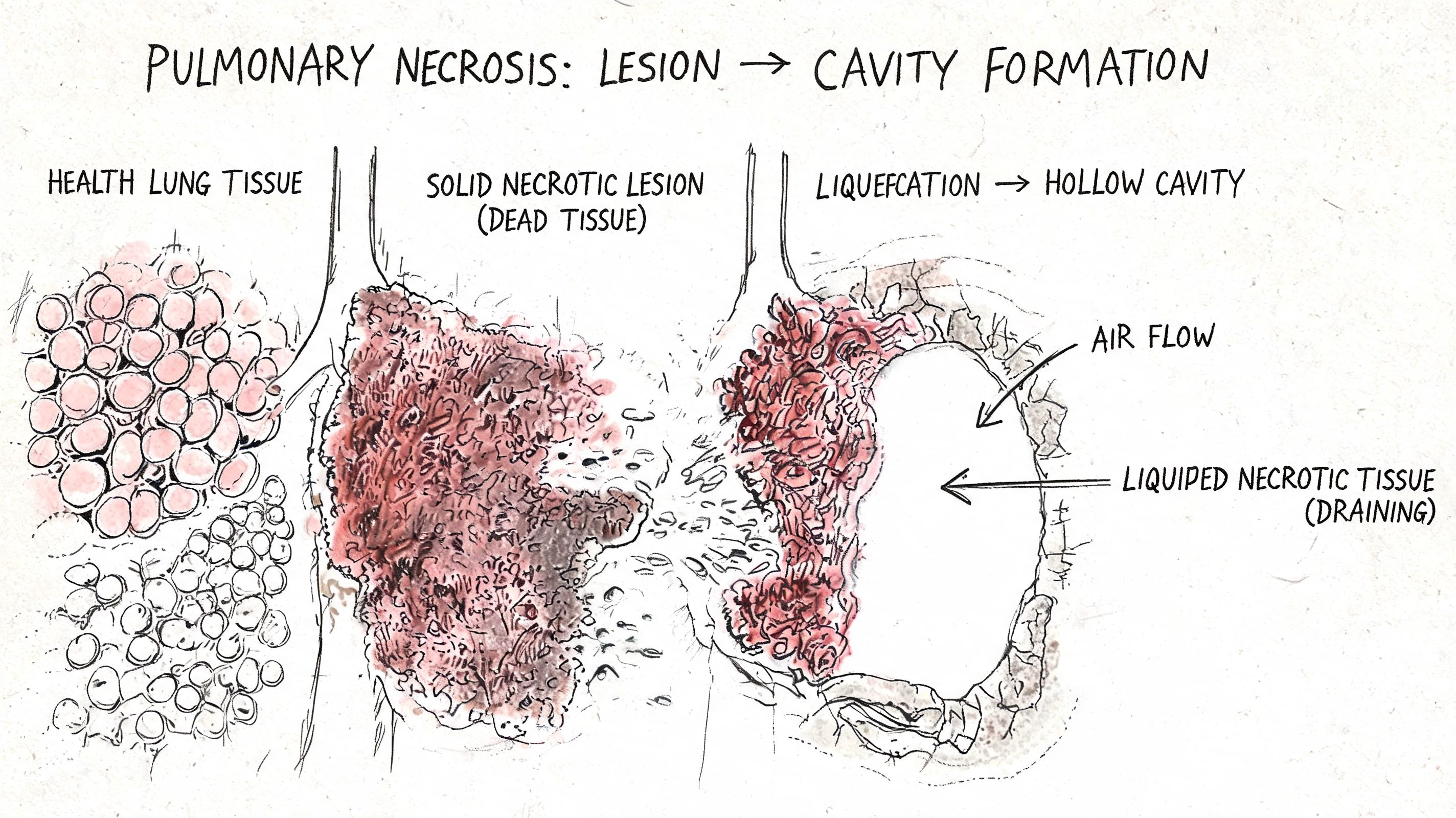
Most cavitating lesions form because tissue in the center dies. In pathology, we call that necrosis. Once the center breaks down, the damaged material may drain through the airways, remain as debris, or leave an air-filled space.
A simple way to picture it is a piece of fruit that begins to rot from the inside. The outer portion may still hold its shape for a time, but the center becomes soft, damaged, and hollow. Lung lesions can behave in a similar way.
The basic process
Several mechanisms can lead to cavitation:
- Tumor outgrows its blood supply: The center of the lesion loses adequate blood flow and dies.
- Infection destroys tissue: Bacteria or fungi can trigger intense inflammation and tissue breakdown.
- Airway connection forms: As the center empties into a bronchus, a hollow space can remain.
- Treatment effect appears: In metastatic disease, cavitation can emerge after chemotherapy because of tumor necrosis, as described in Radiopaedia's overview of cavitating pulmonary metastases.
Why the wall matters
When I explain a cavity to families, I often focus on the wall. The wall is the part that still contains living or once-living tissue, inflammation, scar, infection, or tumor. The center may contain debris, but the wall usually tells the story.
That's why pathologists pay close attention to wall thickness, texture, and lining. It's also why a detailed histology report is often the key document after the gross autopsy findings.
Practical rule: The center may be dramatic to look at, but the wall is usually where the diagnosis lives.
Common Causes Behind a Cavitating Lung Mass
A cavitating mass is a description, not a diagnosis. On an autopsy report, that phrase can alarm a family because it sounds specific, but several very different processes can leave behind the same hollowed-out appearance. In forensic work, the first question is not only what caused the cavity, but whether it formed during life from disease or appeared altered after death.
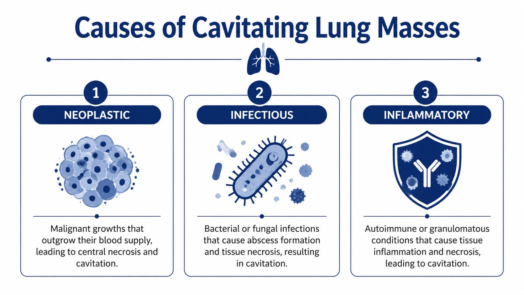
The main categories are neoplastic, infectious, and inflammatory. A helpful way to frame them is to ask what was eating away at the lung tissue. Cancer can outgrow its blood supply. Infection can digest and destroy tissue. Inflammatory disease can injure the lung through an abnormal immune response. Each route may create a cavity, but the clues around that cavity are different.
Neoplastic causes
Cancer remains one of the most concerning explanations, especially when the cavity has a thick or irregular wall and sits within an obvious destructive mass. Primary lung cancers, particularly squamous cell carcinoma, are well known for cavitation, as summarized in a clinical review of cavitary lung lesions. In practice, that means a cavity can represent a tumor whose center has died and broken down while the outer edge still contains viable cancer.
Wall appearance can help guide suspicion. Thin-walled cavities more often prove benign, while markedly thick walls raise concern for malignancy, though there is overlap and pathology still has the final word. A cavity does not diagnose cancer by itself.
If the question is whether a cavitary lesion found after death represents an undiagnosed primary tumor, the broader autopsy context matters. I look at the airways, hilar and mediastinal lymph nodes, pleura, and any spread beyond the lung. For that narrower issue, my discussion of a lung cancer autopsy may help families and attorneys understand what gets examined.
Here is a short clinical overview for readers who prefer visual explanation:
Infectious causes
Infection is another common explanation. A lung abscess can leave a cavity filled with pus, liquefied tissue, or foul-smelling debris. Tuberculosis is a classic cause of cavitary lung disease, often in the upper lobes, and fungal infections can produce a similar picture in the right clinical setting.
From a forensic standpoint, infection raises a different set of questions than cancer. Was there fever, weight loss, cough, aspiration risk, immune suppression, or a recent hospitalization? Was the cavity part of a localized abscess, or one piece of a wider pneumonia or disseminated infection? The answer may affect both the cause of death and whether the lesion was an incidental finding.
Good records matter here. Clear documentation and accurate transcription can make a real difference when reviewing prior admissions, radiology reports, or dictated pathology notes. That is one reason practices handling autopsy and hospital records pay attention to safely managing sensitive medical voice recordings.
Inflammatory and other causes
Some cavitating lesions are neither cancer nor infection. Autoimmune and inflammatory disorders, including granulomatosis with polyangiitis and rheumatoid nodules, can form nodules or masses that later cavitate. Septic emboli can also create multiple cavitary nodules, particularly when infection elsewhere in the body has seeded the lungs through the bloodstream.
These cases can be confusing because the gross appearance may overlap with tumor or abscess. The distinction often depends on the pattern across both lungs, the medical history, and what the microscope shows in the cavity wall and surrounding tissue.
A quick comparison
| Category | What often raises suspicion | What usually confirms it |
|---|---|---|
| Cancer | Thick or irregular wall, destructive mass, nodular inner margin, associated tumor pattern | Microscopy showing malignant cells |
| Infection | Pus, necrotic debris, surrounding pneumonia, abscess pattern, upper lobe cavitation in some infections | Histology, cultures, and clinical history |
| Inflammatory disease | Multiple nodules, vasculitic pattern, granulomatous inflammation, history of autoimmune disease | Histology and correlation with the medical record |
One final point matters in autopsy work. A true cavitating mass formed during life has structure. Its wall, lining, contents, and surrounding reaction usually show that the body had time to respond. Postmortem change can distort a lesion, soften tissue, introduce gas, and make a cavity look larger or more ragged than it was before death. That distinction is often central for families and for legal review.
The Forensic Investigation of a Lung Cavity
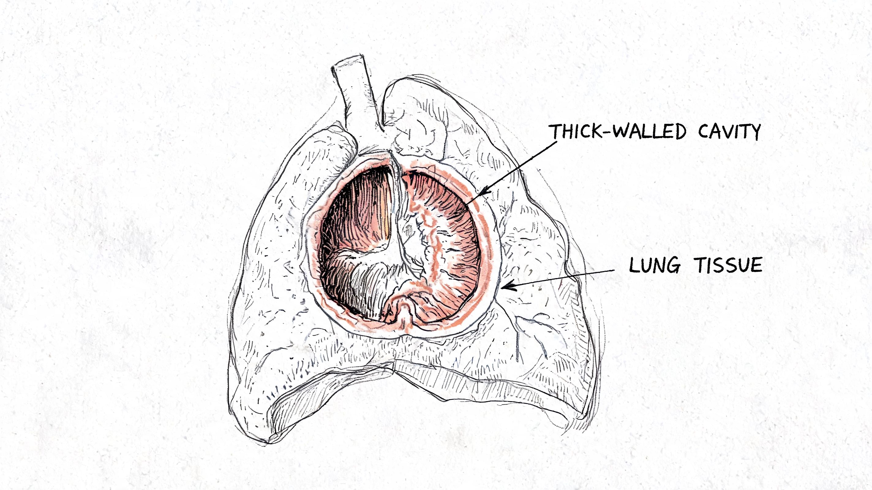
At autopsy, I do not stop at the phrase “cavitating mass.” I examine the lungs directly. That includes the lesion's location, size, contents, wall character, relation to the airways, and whether there are other nodules or signs of spread elsewhere in the body.
What I look for during the gross examination
The gross exam is the direct visual and hands-on examination of the organs. For a lung cavity, that usually includes:
- Location in the lung: Upper lobe, lower lobe, central, or peripheral.
- Wall features: Thin, thick, smooth, shaggy, nodular, or irregular.
- Cavity contents: Air, blood, pus, necrotic material, or mixed debris.
- Nearby findings: Consolidation, scarring, enlarged lymph nodes, pleural involvement, or additional lesions.
These observations matter because a cavity does not exist in isolation. A single thick-walled cavity with a destructive nearby mass raises different concerns than scattered cavities in both lungs.
Why the wall gets sampled first
One of the most useful parallels between clinical biopsy and autopsy pathology is where we aim the sample. In a biopsy of a cavitating mass, the needle tip is deliberately positioned in the thick peripheral wall, avoiding the central void, because that is where viable diagnostic tissue is most likely to be found, as described in an AJR discussion of CT-guided biopsy technique.
The same principle applies at autopsy. If I want to determine whether a cavity came from infection, tumor necrosis, or another process, I sample the wall, not just the empty center.
The cavity wall is the evidence-rich part of the lesion. The center may only show the aftermath.
Microscopy, cultures, and documentation
After gross sampling, the tissue is processed into slides for microscopic review. That is how I identify malignant cells, tissue necrosis, inflammatory patterns, microorganisms, or a healing response. If infection is a serious possibility, additional microbiology may be appropriate.
For attorneys, accurate records matter too. Good forensic work depends on careful documentation, clear specimen labeling, and chain of custody. In cases involving recorded interviews, dictated observations, or clinical audio, secure handling of health information also matters. Some firms and medical teams use tools for safely managing sensitive medical voice recordings so the record remains organized and compliant.
This is also where an independent autopsy can matter. Services such as Private Autopsies allow families or attorneys to obtain a separate postmortem review when questions remain about cause of death, diagnosis, or timing.
Distinguishing Disease from Postmortem Changes
This is the issue that clinical articles often leave out, and it is one of the most important points in forensic pathology. A cavity seen at autopsy does not automatically mean that the person had a true cavitating lung lesion during life.
After death, bodies undergo predictable changes. Gas can form. Tissue can soften. Structures can separate. In some circumstances, those changes can create an appearance that resembles a preexisting cavity.
What makes a true ante-mortem cavity different
The key distinction is tissue reaction. True pathological cavities show evidence of cellular reaction and inflammation at the margins, while postmortem changes do not, as discussed in a forensic pathology review on postmortem imaging and interpretation.
That means I am looking for signs that the body responded to the lesion during life. Those signs may include inflammation, tumor cells lining the wall, granulation tissue, fibrosis, or other microscopic features of a living disease process.
Why this matters so much
Families may fear that a cavity proves an undiagnosed cancer. Attorneys may wonder whether the finding supports a negligence claim. Those are serious questions, and they cannot be answered from appearance alone.
A decomposition artifact does not carry the same meaning as a lesion that formed while the person was alive. If that distinction is missed, the final interpretation can be wrong in a way that affects grief, legal analysis, and even family medical history.
A postmortem cavity-like change can look alarming. Histology tells us whether the body reacted to it in life.
The forensic approach
When timing is in question, I correlate:
- Body condition and decomposition stage
- Gross appearance of the lung tissue
- Microscopic appearance of the cavity margin
- Clinical history, imaging, and records if available
That combination is often what separates a true disease process from a postmortem artifact.
What a Cavitating Mass Means for a Cause of Death Investigation
A family may receive an autopsy report that mentions a cavitating mass in the lung and immediately fear an undiagnosed cancer. An attorney may read the same line and wonder whether it points to missed treatment, a delayed diagnosis, or a disease that had already spread. In a cause of death investigation, that single finding can matter a great deal, but only after we answer a more basic question. Did this cavity represent a disease process that developed during life, or a change that appeared after death?
That distinction shapes everything that follows.
A cavitating mass can be the main finding that explains death, or it can be a secondary finding that helps complete the medical story without being the event that caused death. In forensic practice, I treat it like a damaged room in a house after a fire. The damage may show where the fire started, or it may be one area affected after the fire was already spreading. The cavity matters, but its role has to be placed in sequence.
When the cavity is part of the fatal chain
If the lesion reflects advanced lung cancer, metastatic cancer, a destructive infection, or a complication such as abscess formation, bleeding, or severe pneumonia, it may belong directly in the cause of death statement. Cause of death refers to the disease or injury that started the chain of events ending in death.
Manner of death is a different question. That is the medicolegal classification, such as natural or accident. A cavitating cancer or infection usually supports a natural manner of death unless the surrounding facts show some separate injury, exposure, or neglect issue that changed the outcome.
Why the source of the mass changes the interpretation
One of the most important questions is whether the cavity began in the lung or arrived there from somewhere else. Some cavitating masses are primary lung cancers. Others are metastases from cancers that started in the head and neck, skin, gastrointestinal tract, or another organ. Radiology references describe cavitation as a recognized pattern in pulmonary metastatic disease, especially with some squamous malignancies.
That difference is not academic. If the lung lesion was metastatic, the death investigation may need to focus on the original cancer, the stage of disease, and whether the lung finding was one chapter in a larger process. If it was a primary lung lesion, the analysis may center more on local destruction of lung tissue, infection in the cavity, airway compromise, or spread within the chest.
For families, this can answer a painful question. A cavity in the lung does not automatically mean the lung was where the disease started.
The questions that help determine significance
When I review a cavitating mass in an autopsy, I want the report to answer a few practical questions in plain terms:
- Did the lesion contribute directly to death, indirectly to death, or was it incidental?
- Was there evidence of cancer, infection, or another defined disease process?
- Was the cavity linked to a primary lung lesion or to metastatic disease from another site?
- Did the person have complications such as sepsis, hemorrhage, respiratory failure, or aspiration?
- Do the autopsy, microscopy, and available medical records support a lesion that formed during life?
Those answers are what connect an abnormal finding to a legally and medically sound cause of death opinion.
Families and attorneys who want a plain-language overview of that process may find it helpful to read how a coroner determines cause of death. The final interpretation in a case like this still depends on autopsy examination, histology, and the full medical history, but the framework is useful.
A cavitating mass is a clue. In some deaths, it is the clue that explains everything. In others, it is only one piece of the timeline, and the most important forensic task is proving whether that piece belonged to life, to death, or to the changes that followed death.
Frequently Asked Questions About Lung Findings
A family receives an autopsy report, sees the words cavitating mass in the lung, and immediately wonders whether that finding explains the death. That is a fair question. In forensic practice, the answer depends on two separate issues. First, what the lesion was during life. Second, whether any part of what we see after death could be change that happened after circulation stopped.
FAQ on Cavitating Lung Masses
| Question | Answer |
|---|---|
| Can a cavitating mass in lung tissue be the direct cause of death? | Yes. A cavitating mass can be the disease process that led to death, or it can produce complications such as major infection, bleeding, or respiratory failure. It can also be an incidental finding that was present but did not cause death. The autopsy question is not simply whether the lesion existed, but whether it played a real role in the final sequence of events. |
| Does a cavity always mean cancer? | No. Cancer is one cause, but infection, inflammatory disease, and tissue breakdown from other illnesses can also produce a cavity. After death, decomposition and related artifact can sometimes create a hollowed appearance that mimics a true cavity formed during life. |
| How is a cavity different from a cyst? | The simplest way to separate them is by the wall. A cavity usually has a thicker, more irregular wall and often forms within an area of disease, while a cyst usually has a thinner, more uniform wall. That difference helps guide the diagnosis, much like the shape and edges of a hole in drywall can suggest whether it came from slow water damage or a single sharp impact. |
| Can imaging alone give the final answer? | Usually no. Imaging can suggest whether a lesion looks more like infection, cancer, or another process, but the final answer often requires tissue examination. At autopsy, microscopy helps determine whether the lining of the cavity, the surrounding inflammation, and any necrosis were present during life or are better explained by postmortem change. |
| Why seek a second opinion autopsy or independent review? | A second review can help when the timeline is disputed, when the report leaves the lesion poorly characterized, or when a family or attorney needs a clearer explanation of whether the finding represents true disease, postmortem artifact, or both. |
A few final points families often need
Cause of death is the disease or injury that started the fatal chain of events.
Manner of death is the medicolegal classification, such as natural, accident, suicide, homicide, or undetermined.
Chain of custody is the documented handling of tissue, slides, photographs, and other evidence so the medical and legal record remains reliable.
One point often causes the most confusion. A cavity seen at autopsy is not automatically proof of a disease process that formed before death. The pathologist has to compare the gross examination, microscope slides, radiology if available, toxicology, and the clinical timeline. That process works much like checking whether a crack in a wall happened during construction or after the building settled. The appearance matters, but context matters just as much.
If you need help understanding a lung finding in an autopsy report, or if you need an independent review for a family member, legal matter, or county case, you can contact Texas Autopsy Services. I approach these cases with the same priorities every time: clear explanations, careful tissue-based analysis, and respect for the person who died and the people now seeking answers.