September 22, 2026 · Texas Autopsy Services
Lung Fibrosis Death Explained Causes and Autopsy Insights
Learn how lung fibrosis death occurs, from respiratory failure to acute exacerbation, and how autopsy clarifies cause of death in Texas.
On this page
- Introduction When Lung Fibrosis Raises Questions After Death
- How Lung Fibrosis Leads to Death
- Common Complications That Cause Death in Fibrotic Lung Disease
- What Autopsy Reveals About Lung Fibrosis Death
- Understanding Cause of Death Versus Contributing Factors
- When a Private Forensic Autopsy Brings Clarity in Texas
- Finding Answers and Next Steps After a Lung Fibrosis Death
A family may receive a death certificate that lists pulmonary fibrosis, yet still be left with difficult questions. Was the disease itself the immediate reason for death? Did an acute exacerbation, infection, blood clot, heart complication, medication effect, or another condition change the outcome? When the hospital course was rapid or records are incomplete, the phrase “lung fibrosis death” may describe only part of what happened.
- Lung fibrosis can cause death through respiratory failure, but sudden complications may be the final event.
- Acute exacerbation can place new, widespread lung injury on top of established scarring.
- A forensic autopsy uses medical history, gross examination, histology, imaging, and ancillary testing to identify the sequence of events.
- Cause of death and contributing cause are different entries in a death investigation.
- An independent examination or second-opinion autopsy review can help families, attorneys, and agencies assess whether the death certificate accurately reflects the medical findings.
Introduction When Lung Fibrosis Raises Questions After Death
A loved one may have lived with pulmonary fibrosis for years, used supplemental oxygen, and managed a gradual loss of stamina. Then the person may have become suddenly short of breath during a hospitalization or died unexpectedly at home. The medical records may mention pulmonary fibrosis, respiratory failure, pneumonia, or cardiac arrest, but those terms don't always explain how the conditions connected.
Families often want a clear answer rather than a list of diagnoses. They may need to know whether fibrosis started the fatal chain, whether another condition caused the final decline, or whether the disease was present but only contributed to death. Attorneys and medical professionals may face a related question, especially when the hospital course was complex or the official investigation didn't address every concern.
Idiopathic pulmonary fibrosis, or IPF, is a specific disease. “Lung fibrosis” is a broader description of scarring in lung tissue and may result from different diseases, exposures, medications, or connective tissue disorders. That distinction matters because the underlying disease affects how a forensic pathologist interprets tissue, imaging, and the death certificate.
Our board-certified forensic pathologists approach these cases by examining the complete sequence. We review the clinical history, oxygen and respiratory findings, imaging, treatment records, laboratory studies, and circumstances surrounding death. We then correlate those records with the postmortem examination and any indicated testing.
This process doesn't assume that fibrosis was the sole cause. It asks a more precise question: what medical event caused death, what disease began that process, and what other conditions made the outcome worse? The answer can bring medical clarity, support a legal review, and help families understand what happened without reducing the person's death to a single vague label.
How Lung Fibrosis Leads to Death
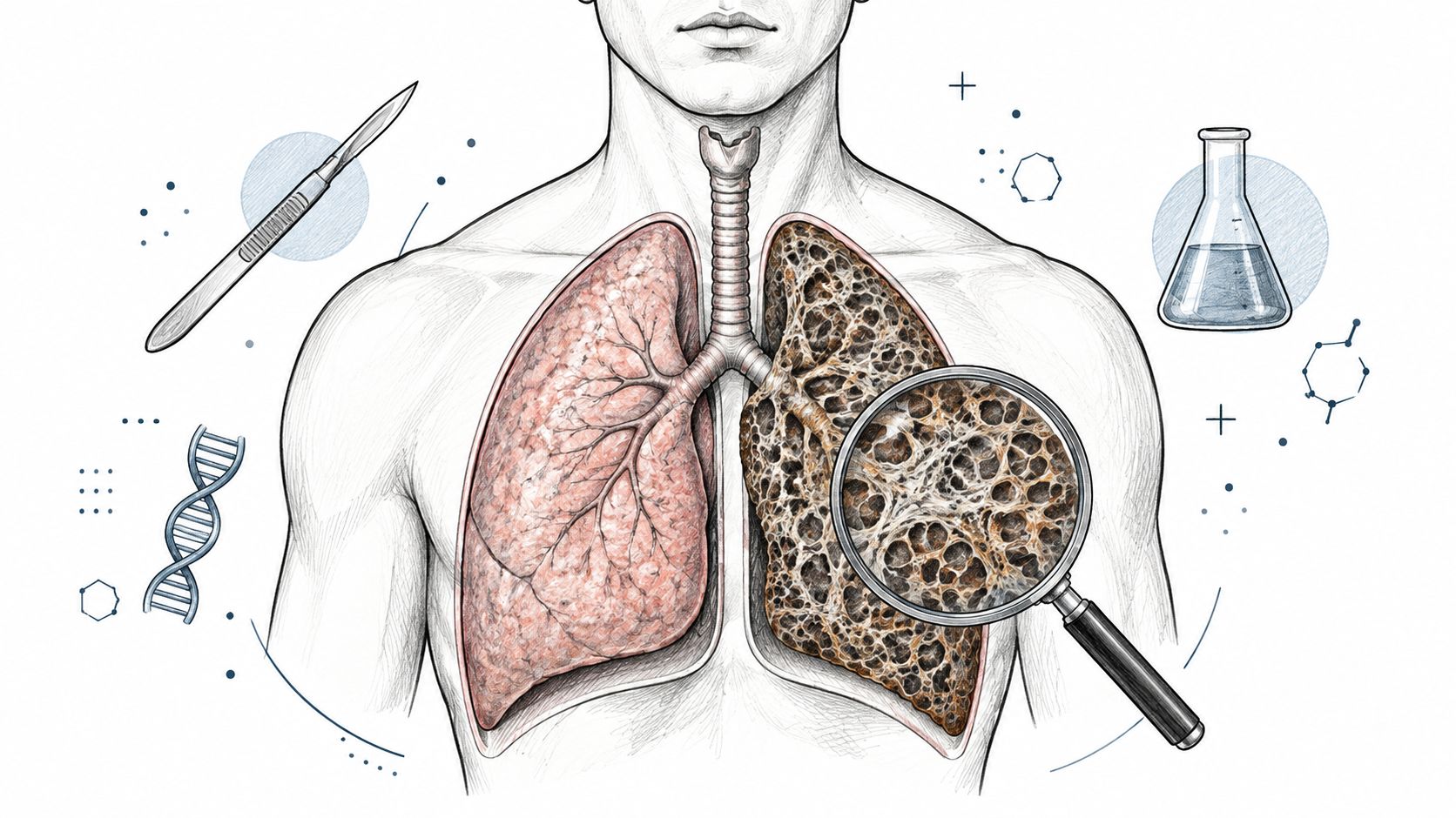
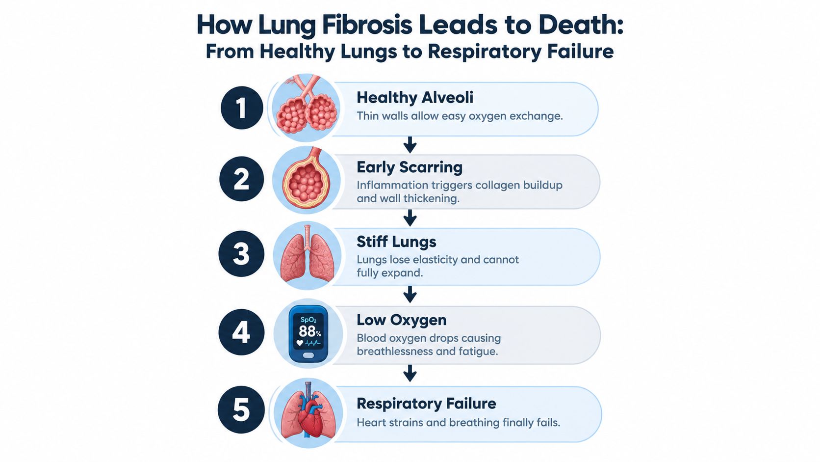
A person may appear to be breathing, yet scarred lungs may no longer move enough oxygen into the blood. Healthy lungs contain millions of tiny air sacs called alveoli. Their thin walls let oxygen enter the bloodstream and carbon dioxide leave it. Each breath also depends on lung tissue expanding and recoiling.
Fibrosis replaces flexible tissue with stiff scar tissue. The change is similar to a soft sponge becoming thick and rigid. The lungs cannot expand as easily, and oxygen must cross a more difficult barrier. The body may compensate by breathing faster or working harder, even while the chest continues to rise and fall.
Chronic decline and sudden deterioration
Idiopathic pulmonary fibrosis can cause a prolonged loss of breathing capacity. Reviews describe median survival of about 3 to 5 years after an IPF diagnosis. One systematic review reported 31% mortality at 5 years without antifibrotic therapy, with mean overall survival of about 4 years (systematic review of IPF prognosis).
The terminal process does not always follow a slow, predictable course. Some people deteriorate gradually. Others worsen over days or weeks after an acute exacerbation, infection, pulmonary embolism, or heart complication. These events can use up the remaining ability to exchange oxygen, leading to severe hypoxemia, or dangerously low blood oxygen, and respiratory failure.
An educational resource on how allergies worsen asthma symptoms may clarify why respiratory illnesses can share symptoms. Asthma and pulmonary fibrosis remain separate diseases, so similar shortness of breath does not establish the same cause.
At autopsy, the key question is whether established fibrosis alone explains the death or whether a new event caused the final collapse. Histology may show chronic interstitial scarring, an acute injury pattern, infection, or another process layered onto the fibrosis. Medical records, imaging, toxicology, microbiology, and other ancillary testing can help connect those findings to the clinical course.
Reading the death certificate
Cause of death identifies the disease, injury, or medical event that caused death. It may appear as a sequence, such as respiratory failure caused by an acute lung injury, with chronic fibrotic disease underlying that injury.
Manner of death is the broad classification, such as natural, accident, suicide, homicide, or undetermined. Several medical conditions may be involved in a natural death.
A contributing cause worsens the outcome without necessarily starting the fatal sequence. Pulmonary fibrosis may reduce respiratory reserve while infection triggers the final respiratory collapse. The death certificate may therefore list more than one relevant condition.
Common Complications That Cause Death in Fibrotic Lung Disease
Fibrotic lung disease leaves little respiratory reserve. The lungs become stiff and have less working surface for oxygen exchange. A new injury that a person with healthy lungs might survive can become fatal when extensive scarring is already present. The final collapse may happen quickly, even though the underlying disease developed over years.
Acute exacerbation is a major mechanism in deaths from IPF. Reviews report that roughly 35% to 46% of IPF deaths are associated with acute exacerbation. In-hospital mortality is commonly above 50%. Reported one-month mortality is about 37% to 53%, and three-month mortality is about 63.8% to 73.7% (review of acute exacerbation in IPF). After an exacerbation, median survival is typically only 1 to 4 months, according to the same review.
An exacerbation adds diffuse alveolar damage to established fibrosis. This acute injury further disrupts the thin air sacs where oxygen enters the blood, and respiratory failure may follow. Infection can be present, suspected, or excluded. That distinction may require review of medical records, postmortem tissue, and microbiology results.
Comparing terminal complications
| Complication | Typical Course | Clue for Cause of Death |
|---|---|---|
| Acute exacerbation | Rapid worsening over days to weeks, often with severe hypoxemia | New diffuse lung injury superimposed on established fibrosis |
| Respiratory failure | Progressive or sudden inability to maintain adequate oxygen exchange | Clinical records, oxygen findings, imaging, and lung examination support a fatal respiratory mechanism |
| Pulmonary hypertension and right-heart strain | Chronic pressure burden may worsen breathing tolerance and circulation | Cardiac enlargement, right-heart changes, and clinical history may show a meaningful contribution |
| Infection or pneumonia | Fever, inflammation, or respiratory decline may occur, but symptoms can overlap with exacerbation | Histology, cultures, molecular testing, and records help assess whether infection caused or contributed to death |
| Thromboembolic disease | A pulmonary embolus may cause sudden collapse or worsen pre-existing respiratory disease | Identification of clot in the pulmonary arteries and supporting medical findings can change the cause-of-death sequence |
Invasive mechanical ventilation requires careful interpretation. Among patients hospitalized with acute exacerbation of interstitial lung disease, reported mortality with invasive ventilation has exceeded 90% in some summaries (review of IPF prognosis and treatment). This figure applies to a particularly severe clinical setting, not to every person living with pulmonary fibrosis.
The forensic question is which process ended the person's life. Chronic fibrosis may be the primary disease, while an acute exacerbation, infection, clot, or respiratory failure supplies the final mechanism. Autopsy findings, histology, clinical records, imaging, toxicology, cultures, and other ancillary tests may be needed to separate the primary cause from contributing conditions.
What Autopsy Reveals About Lung Fibrosis Death
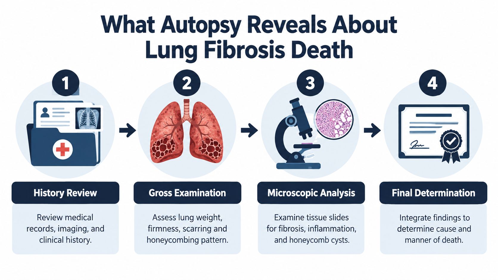
A forensic autopsy begins before the first incision. Our board-certified forensic pathologists review the circumstances of death, prior diagnoses, imaging, hospital notes, respiratory support, laboratory results, medication history, and relevant exposure information. This background helps the examination answer a specific question rather than only document that scarring was present.
The gross examination evaluates the lungs directly. The pathologist assesses their appearance, firmness, distribution of scarring, and patterns such as honeycombing. The examination also considers whether the findings are diffuse or localized and whether other organs show evidence of heart strain, infection, clotting, or another disease process.
Histology and ancillary testing
Microscopic examination, or histology, allows the pathologist to assess the architecture of lung tissue. Depending on the case, slides may show patterns associated with usual interstitial pneumonia, areas of established fibrosis, fibroblastic foci, honeycomb cysts, inflammation, or diffuse alveolar damage associated with acute exacerbation.
A guide to understanding a histology report can help families and attorneys understand why microscopic findings are reported separately from the final cause and manner of death. Histology is interpreted with the clinical history and gross findings, not in isolation.
Ancillary testing is selected according to the circumstances. Toxicology may address medication or substance exposure. Microbiology may evaluate bacterial, viral, or fungal infection. Molecular studies may be considered when a specific diagnostic question remains after the initial examination.
Connecting findings to the fatal event
Autopsy findings don't automatically prove that fibrosis caused death. They must be compared with antemortem imaging, oxygenation data, treatment response, and the documented sequence of deterioration.
A postmortem series of 42 IPF patients found immediate causes of death were respiratory in 64%, cardiovascular in 21%, and noncardiopulmonary in 14%. Acute exacerbation of IPF was the single most common immediate cause at 29% (postmortem study of IPF deaths).
Chain of custody means documenting who handled the body, specimens, records, and evidence, when each transfer occurred, and how materials were secured. Our team maintains documentation and uses HIPAA-compliant reporting practices so that findings can be reviewed by families, attorneys, agencies, and courts when appropriate.
Understanding Cause of Death Versus Contributing Factors
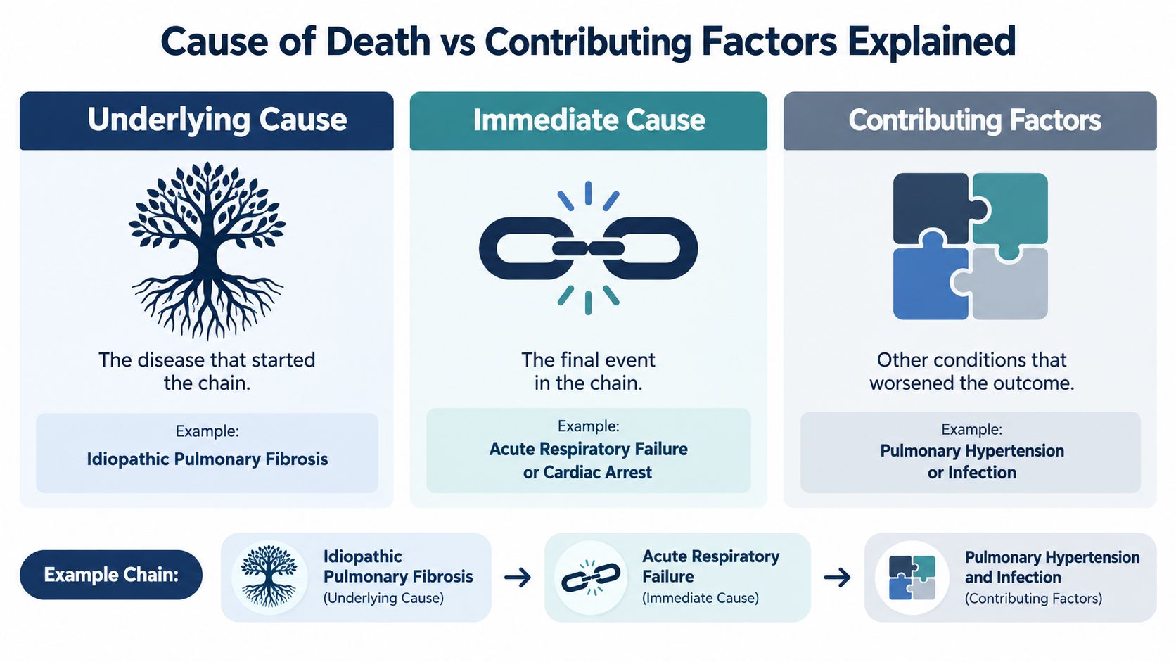
A death certificate can contain several medically accurate terms without answering the family's central question. The underlying cause is the disease that started the fatal chain. The immediate cause is the final disease or event that directly produced death. A contributing factor is a separate condition that made survival less likely or worsened the course.
For example, idiopathic pulmonary fibrosis may be the underlying disease, acute respiratory failure may be the immediate cause, and pneumonia or pulmonary hypertension may be contributing factors. Another case may show fibrosis as a chronic background condition while a different disease, injury, or cardiac event caused death.
Why the distinction matters
Recent U.S. mortality data identified 67,843 deaths during 2020–2022 in which IPF appeared as either the underlying or contributing cause. Of those deaths, 42.7% were contributing-cause deaths, showing that IPF often forms part of a broader cause-of-death picture rather than being the sole explanation (CDC mortality report).
IPF also needs to be distinguished from other fibrotic lung diseases. Scarring may be associated with occupational exposure, medication effects, connective tissue disease, chronic hypersensitivity, or another interstitial lung disorder. Those possibilities can change the medical interpretation and may affect whether the disease is described as idiopathic.
Age also provides context without determining an individual cause of death. A global-trends study reported that 87.5% of IPF deaths occurred in people aged 65 or older, while crude mortality increased from 2.10 per 100,000 in 2001 to 3.14 per 100,000 in 2022 (international mortality review). These population findings don't replace an individual investigation.
Families reviewing a certificate may want to ask:
- What condition started the fatal sequence?
- What event caused the final collapse?
- Was fibrosis primary, contributing, or incidental?
- Were infection, clot, medication effects, or heart disease evaluated?
- Were imaging, histology, and laboratory findings interpreted together?
A plain-language explanation of cause and manner of death may help organize those questions before requesting a review.
When a Private Forensic Autopsy Brings Clarity in Texas
A private autopsy may be appropriate when the medical examiner or justice of the peace declines a case, when the hospital course is unclear, or when the official findings don't address an important concern. Families and attorneys may also seek an independent examination when they need to distinguish acute exacerbation from infection, evaluate possible medication or exposure effects, or preserve evidence for a legal proceeding.
Texas Autopsy Services is an independent private autopsy and forensic pathology practice based in Elgin, Texas. Every examination is performed by a forensic pathologist certified by the American Board of Pathology. The practice serves all 254 Texas counties, with in-house licensed transport, secure storage, digital imaging, and direct communication with the team.
What families should prepare
Records help the pathologist establish the sequence before death. Useful materials may include hospital records, imaging reports and images, medication lists, prior pulmonary diagnoses, death certificate information, and details about occupational or environmental exposure. When records are transmitted, families should use an authorized release or an authorized fax of medical records so protected health information is handled appropriately.
The examination may include:
- Record review, including imaging and the documented clinical deterioration.
- External and internal examination, with focused evaluation of the lungs, heart, pulmonary vessels, and other organs.
- Histology and ancillary testing, such as toxicology, microbiology, or molecular studies when indicated.
- Written reporting, with findings, interpretation, cause of death, and manner of death.
Texas requirements and professional standards remain important throughout the process. Our team works within the relevant provisions of Texas Health & Safety Code Chapters 711, 716, and 651, along with applicable Texas Funeral Service Commission standards. The practice uses flat-rate, all-inclusive pricing and explains logistics directly, without promising a particular result or outcome. Most cases are completed within 24 to 48 hours, although the final report may depend on histology, testing, records, and case complexity.
For attorneys or agencies needing broader forensic support, forensic consultant services can provide a structured path for reviewing records, pathology, and disputed findings.
Finding Answers and Next Steps After a Lung Fibrosis Death
A lung fibrosis death isn't always the result of slow scarring alone. The fatal mechanism may be respiratory failure, acute exacerbation, infection, thromboembolic disease, heart strain, or another event occurring on a background of chronic fibrosis. Autopsy, histology, imaging review, and selected ancillary testing can help determine whether fibrosis was the underlying cause, an important contributor, or an incidental finding.
Accurate certification matters to families, clinicians, attorneys, and public health officials. If questions remain, gather the death certificate, medical records, imaging, medication history, and information about the final illness before seeking an independent review. Families can learn about private autopsies, while county officials and agencies can review county forensic autopsies.
Seeking a careful explanation isn't an accusation. It's a respectful effort to understand the deceased person's final medical course and preserve an accurate record for those who remain.
Texas Autopsy Services offers independent autopsy examinations and second-opinion reviews for suspected lung fibrosis deaths, with every examination performed by a forensic pathologist certified by the American Board of Pathology. For confidential guidance about records, transport, or the appropriate next step, please visit Texas Autopsy Services and contact our team.