March 18, 2026 · Texas Autopsy Services
What Autopsy Smoker Lungs Reveal About Cause of Death
A forensic pathologist explains what autopsy smoker lungs reveal, from physical damage to direct links with the cause of death. Get clear answers.
On this page
- A Firsthand View of a Smoker's Lungs
- What We See With The Naked Eye
- The Unseen Damage Revealed Under The Microscope
- Connecting Smoking Directly to the Cause of Death
- How Toxicology Testing Confirms Smoking History
- Why A Private Autopsy Provides Clear Answers For Families
- Frequently Asked Questions About Smoker Autopsies
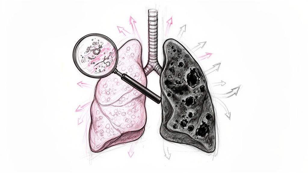
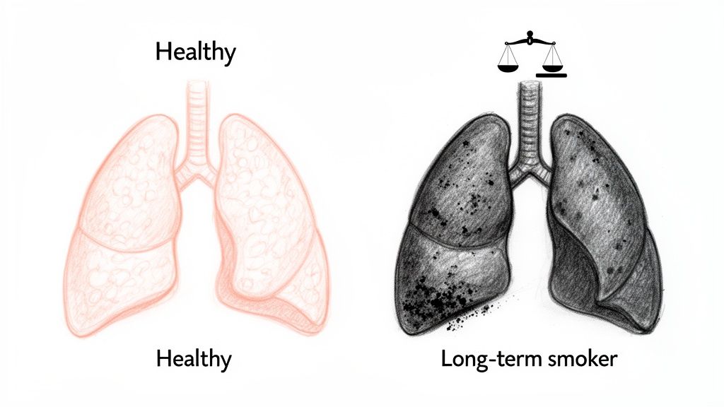
In my work as a forensic pathologist, I have conducted over a thousand autopsies. The difference between a smoker's lungs and a non-smoker's is one of the most striking things I see. Healthy lungs are light, soft, and have a healthy pink color. The lungs of a heavy smoker, however, are often heavy, blackened, and stiff from years of inhaling carbon, a condition called anthracosis. My goal isn't to be graphic but to explain the reality of what I observe.
- A smoker's lungs are visibly different in color, texture, and weight due to inhaled tar and carbon.
- The damage isn't just cosmetic; it includes destroyed air sacs (emphysema) and thickened airways (chronic bronchitis).
- A "pack-year" history helps me quantify the lifetime toxic exposure and link it directly to the cause of death.
- Toxicology testing for cotinine, a byproduct of nicotine, provides objective chemical proof of recent tobacco use.
- A Private Autopsy provides families and attorneys with objective, clear answers when a cause of death is uncertain or needs further clarification.
A Firsthand View of a Smoker's Lungs
When I perform a private autopsy, my duty is to find objective, undeniable answers. For a long-term smoker, the evidence is usually visible before I even examine a slide under the microscope. The way the lungs look and feel tells a clear story about their exposure. This goes far beyond color; it’s about the very texture, weight, and function of the organ.
A non-smoker’s lungs get their healthy, pink look from a rich network of blood vessels and delicate, air-filled tissue. When I handle them, they feel light and springy, almost like a sponge.
The Stark Contrast in Smoker Lungs
A smoker’s lungs are a world apart. The tissue is often a dark gray or even black, stained by the tar and carbon particles inhaled daily. This pigment doesn’t just sit on the surface; it gets locked into the lung tissue and the surrounding lymph nodes. It's permanent.
But it’s not just the color. The entire structure of the lung changes. Years of smoking destroys the tiny, delicate air sacs (alveoli) that are essential for getting oxygen into the blood. This is emphysema, and it makes the lungs stiff and far less elastic. They often feel heavy in my hands, weighed down by the combination of inflammation, scar tissue, and trapped fluid.
As a forensic pathologist, my examination goes beyond simply noting these changes. It's about connecting the physical evidence of damaged lungs to the specific sequence of events that led to death, providing families with a complete and factual medical narrative.
For families and attorneys here in Texas, understanding this physical evidence is a critical step. Many people request Private Autopsies to get concrete answers that connect what's written on a death certificate to the reality inside the body. An independent autopsy can clarify how a loved one's illness progressed and confirm if smoking-related damage was the primary cause of death or a major contributing factor. My role is to provide those scientific facts with both medical precision and compassion.
What We See With The Naked Eye
The moment I begin the internal examination, the story of a person's smoking history starts to unfold. You don't need a microscope to see the damage. The evidence is right there, written in the color, feel, and overall structure of the lungs. We call this the gross examination—what's visible to the naked eye.
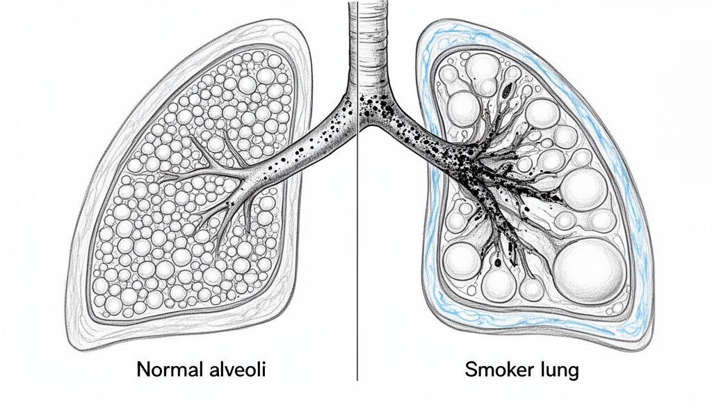
The first thing that stands out is the color. A healthy lung has a uniform pinkish-gray appearance. In contrast, a smoker’s lungs are often covered in a dark, mottled black staining. This is a condition called anthracosis, caused by years of inhaling and trapping carbon particles from tobacco smoke. This isn't a light dusting from city pollution; it’s a deep, permanent discoloration that permeates the tissue.
Macroscopic Findings In Smoker Lungs vs Healthy Lungs
The table below gives you a clear, side-by-side look at the stark differences I observe on the examination table. These are the changes that tell the story of a lifetime of smoking.
| Characteristic | Healthy Lungs (Non-Smoker) | Smoker Lungs (Advanced Changes) |
|---|---|---|
| Color | Uniform pink or light gray. | Mottled black and gray (anthracosis). |
| Texture | Soft, spongy, and highly elastic. | Firm, less elastic, often feels "heavy." |
| Air Sacs (Alveoli) | Intact and uniform. Lungs feel full. | Destroyed sacs, forming large holes (bullae). |
| Airways (Bronchi) | Thin, delicate walls. | Thickened, inflamed walls. |
| Surface | Smooth and clean. | May have bumpy, irregular texture from bullae. |
| Overall Size | Normal size, easily fits within the chest cavity. | Often over-inflated and enlarged due to trapped air. |
Looking at these differences, you can start to build a physical picture of why a smoker struggles to breathe. The very architecture of the organ has been compromised.
The Loss of Sponginess And Elasticity
Beyond just the color, the feel of the lungs tells a huge part of the story. Healthy lungs are incredibly light and springy. Think of a new sponge. But in a long-term smoker, that sponginess is gone. The lungs often feel heavier and have lost much of their natural elasticity. This is the physical evidence of emphysema.
Emphysema is the disease process that breaks down the walls of the tiny, delicate air sacs (alveoli) where oxygen passes into the blood. Instead of millions of tiny, efficient sacs, the lung tissue becomes riddled with large, empty pockets called bullae. These are essentially dead zones in the lungs—holes that do nothing but take up space. I can see and feel them during the exam, and they are the direct cause of the severe shortness of breath so many smokers endure.
On the autopsy table, I'm not just noting if these changes are present. I’m gauging their severity. How extensive is the blackening? How large and numerous are the bullae? This gives us a direct physical measurement of the damage accumulated over a lifetime.
Thickened Airways From Chronic Irritation
Another hallmark I look for is evidence of chronic bronchitis, which is almost a given in heavy smokers. The constant irritation from smoke causes the walls of the main airways (the bronchi) to become inflamed and noticeably thickened.
When I make cross-sections of the lungs, it's not unusual to see these airways plugged with thick, tenacious mucus. This blockage is precisely what causes the chronic, rattling "smoker's cough." Seeing this firsthand—the inflamed tissue and the mucus buildup—provides a clear anatomical reason for the symptoms the person experienced in life.
For families and attorneys, connecting these visible findings from an autopsy of smoker lungs to a loved one's daily struggles can be incredibly powerful. It closes the loop between the symptoms they witnessed and the physical reality inside the body. This is often a key reason people seek definitive answers through Private Autopsies—to get that complete and factual medical story.
The Unseen Damage Revealed Under The Microscope
While looking at a smoker's lungs on the autopsy table gives a stark picture of the damage, the real story unfolds when I get the tissue samples under a microscope. This is where histology comes in. It’s like zooming in on a battlefield, allowing me to see exactly how the body’s cells fought—and ultimately lost—a long war against the poisons in tobacco smoke. What I find here provides the concrete evidence linking a person’s smoking history to a specific disease.
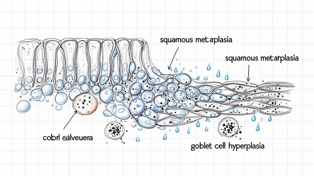
Almost immediately, I can see signs of intense, chronic inflammation. The airways are packed with immune cells that have rushed to the scene. Among them, I always find huge numbers of macrophages, which are the immune system’s cleanup crew. In a smoker, these cells are literally engorged with black carbon pigment. We even call them "smoker's macrophages"—a dead giveaway for long-term inhalation of cigarette smoke.
Cellular Changes That Impair Function
Beyond the general inflammation, the very cells lining the airways begin to change in a desperate attempt to protect themselves. There are two key transformations I specifically look for: goblet cell hyperplasia and squamous metaplasia.
Goblet Cell Hyperplasia: Goblet cells are the little factories that produce mucus. In a smoker's lungs, constant irritation puts these cells into overdrive, causing them to multiply again and again. This is what's behind the chronic cough and phlegm that define chronic bronchitis. Under the microscope, it’s clear that these mucus-producing cells have completely taken over the normal airway lining.
Squamous Metaplasia: This change is much more alarming. The airway is normally lined with delicate, hair-like cells (cilia) that sweep out mucus and debris. In a smoker, the body replaces them with tougher, flatter cells called squamous cells. Think of it like swapping out a soft carpet for durable tile. The tile is tougher, but it can’t do the original job—in this case, cleaning the lungs.
Squamous metaplasia is not yet cancer, but it is considered a precancerous condition. It’s a major step on the road to squamous cell carcinoma, a very common type of lung cancer. Finding this during an autopsy draws a direct line from the person's smoking habit to their heightened risk of developing a malignancy.
Correlating Microscopic Findings To Disease
These microscopic findings aren’t just interesting observations; they are the puzzle pieces that form the larger picture of disease. When I see the tiny walls of the air sacs (alveoli) destroyed, that confirms emphysema and explains why the person was so short of breath. The combination of inflammation, mucus-clogged airways, and damaged cilia is the textbook definition of Chronic Obstructive Pulmonary Disease (COPD).
Turning small tissue sections into a final diagnosis is a meticulous process. If you’re interested in the details, you can read our guide on how a histology report is created. This microscopic work is what transforms what we see with the naked eye into a solid medical narrative. For families and legal teams, this level of detail provides the irrefutable scientific proof needed to connect a lifetime of smoking to a clear and precise cause of death.
Connecting Smoking Directly to the Cause of Death
After I’ve examined the lungs and seen the damage firsthand, my job is to connect those physical findings to the actual chain of events that led to a person’s death. It’s one thing to see diseased lungs; it's another to prove they were the reason someone died. This is how we move from observation to a firm, medically sound conclusion, giving families and legal teams the clear answers they need.
The evidence I find on the autopsy table lets me determine if a smoking-related disease was the primary killer or if it was a major accomplice in the death.
The Dose-Dependent Relationship: Measuring the Damage
A fundamental principle I rely on is the dose-dependent relationship between smoking and disease. Put simply, the more someone smoked and the longer they did it, the higher their risk of dying from it. We have a specific way to measure this: "pack-years."
A pack-year is a straightforward calculation: multiply the number of cigarette packs smoked per day by the number of years the person smoked. For instance, if someone smoked two packs a day for 30 years, they have a 60 pack-year history. This isn't just an abstract number; it's a powerful tool that quantifies the total toxic load the lungs have been forced to handle over a lifetime.
The higher that pack-year number, the stronger the causal link I can draw between the smoking history and the diseases I find. This objective data helps turn a likely cause of death into a medical certainty.
This becomes especially important when a cause of death is in dispute. A high pack-year history, paired with the classic visual evidence of severe emphysema or a lung tumor, builds an undeniable case. It allows a pathologist like me to state with a high degree of medical certainty that smoking was the driving force behind the fatal illness.
When Smoking is a Contributing Cause
Not everyone with a history of smoking dies from a lung-specific disease. Often, the final event is a heart attack or a stroke. But even then, the autopsy can show exactly how smoking set the stage for that catastrophe.
Heart Disease: Smoking is a primary driver of atherosclerosis, which is the hardening and clogging of your arteries. During an autopsy, I can literally see and feel the plaque buildup inside the heart’s own blood vessels. When I find severe atherosclerosis in a heavy smoker who died from a heart attack, I can confidently list smoking as a major contributing cause of their death.
Stroke: It's the same story with the brain. Smoking wreaks havoc on the blood vessels leading to the brain, dramatically increasing the odds of a stroke. The autopsy provides direct proof of that vascular damage, showing why a fatal stroke was not just possible, but probable.
In cases like these, the death certificate will name the heart attack or stroke as the immediate cause of death. But just as importantly, it will list smoking-related cardiovascular disease as the underlying condition that made it happen. Clarifying this distinction is one of the most valuable services a private autopsy can provide.
The Unmistakable Link to Lung Cancer
The line between smoking and lung cancer isn't just a statistic; it's something I see physically manifested in my work. The fact is, cigarette smoking is tied to about 80% to 90% of all lung cancer deaths in the United States, making it the single biggest risk factor by a huge margin.
Smokers are 15 to 30 times more likely to develop or die from lung cancer than non-smokers. If you want to dig deeper into the numbers, the CDC offers extensive research on these risk factors.
The Lingering Risk for Former Smokers
What about people who quit? Quitting is the absolute best step anyone can take for their health, but unfortunately, the damage doesn't just disappear. The risk of dying from smoking-related diseases like COPD and lung cancer stays elevated for years, sometimes even decades, after a person's last cigarette.
When I perform an autopsy on a former smoker, I often find the permanent scars of their past habit, like emphysema and the dark staining of anthracosis. This is why a complete medical history, including past smoking habits, is so critical. It helps me assemble a full and accurate picture of every factor that contributed to their death, even those from long ago. This is where a private autopsy in Texas can be so crucial, providing the objective proof needed to understand the entire story.
How Toxicology Testing Confirms Smoking History
The physical damage I see in a smoker's lungs tells a powerful story on its own. But to get the full picture, I always turn to toxicology. This chemical analysis of blood and tissue gives me objective, scientific proof to back up what my eyes are seeing, helping me confirm recent tobacco use and build a complete medical narrative.
This is the straightforward, tragic path I often document: from a history of smoking to the irreversible lung damage that ultimately leads to death.
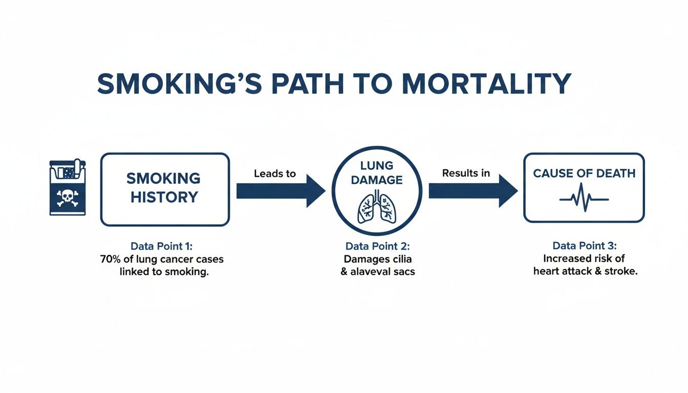
It’s a direct line connecting a specific behavior to a fatal outcome, and toxicology helps me prove that connection.
Identifying The Chemical Fingerprint of Smoking
When we test for tobacco use, we’re not actually looking for nicotine. Nicotine leaves the body fairly quickly. Instead, we’re searching for its primary byproduct, cotinine.
Cotinine is the key. It hangs around in the body for much longer—often for several days—giving us a much wider window to detect it. Finding cotinine is definitive proof of recent tobacco or nicotine product use. This becomes incredibly important in cases where the person’s smoking history was unknown, unclear, or even hidden from their family.
Think of cotinine as an unbiased chemical witness. It can confirm what we’ve been told, but just as often, it reveals a hidden habit that was a major, but unknown, contributor to the person’s final illness.
For families, this kind of confirmation provides answers and helps connect a loved one’s health decline to a clear cause. For legal matters, it provides objective evidence that can be crucial for insurance or occupational health claims.
Building a Complete Medical and Toxicological Profile
Toxicology does more than just confirm smoking; it helps me piece together the entire medical puzzle. Most people with a long history of smoking are also on medications for the conditions it causes.
During the toxicology screen, I’m also looking for other substances that fill in the details of their health story, such as common drugs used to treat:
- COPD: Bronchodilators and steroids are common findings.
- Heart Disease: Medications to manage high blood pressure or cholesterol.
- Blood Clots: Anticoagulants, since smoking significantly raises the risk of clotting.
We may also find chronically elevated levels of carbon monoxide in the blood, a classic sign of a heavy smoker. Finding these substances helps me connect the physical organ damage with the specific medical treatments the person was receiving before they died.
Of course, getting accurate toxicology results depends entirely on having precise, reliable instruments. Forensic labs go to great lengths to find the right lab equipment and furniture because that precision is what allows me to deliver a definitive report.
By combining these toxicology results with what I find during the physical and microscopic examination, I can build a complete, detailed narrative. To learn more about how all this information comes together, our guide on understanding a toxicology report from an autopsy is a great resource. This approach ensures every factor contributing to the cause of death is properly identified and documented.
Why A Private Autopsy Provides Clear Answers For Families
When a loved one with a long history of smoking passes away, families are often left with lingering, painful questions. Was it the smoking that directly caused their death, or was something else going on? A private autopsy is often the only way to find the definitive answers you need.
Hospital autopsies are quite rare today. And a county medical examiner typically only steps in for specific legal reasons, like an accident or suspected crime. This leaves a real gap for families who simply want to understand the truth behind a natural death. That’s where a private autopsy, requested by the next of kin, can make all the difference.
An Independent Path to Clarity
An independent autopsy is an objective investigation into the cause of death. My focus as a forensic pathologist isn't limited by hospital policies or legal jurisdictions; it's centered entirely on giving your family clear, unbiased answers. This is especially critical when the person who passed had a complicated medical history that included smoking.
A death certificate might just say "cardiopulmonary arrest," but that doesn't explain why the heart and lungs stopped. A proper postmortem examination provides that missing context.
My job is to look past the surface-level diagnosis and piece together the complete medical story. It's about confirming what we think we know, finding any other contributing factors, and documenting the full extent of smoking-related damage to give your family the certainty it deserves.
For instance, someone might have died from a heart attack. The autopsy can show us that the underlying coronary artery disease was made much, much worse by years of smoking. Or, we might find that a simple respiratory infection turned fatal because the lungs were already badly damaged by emphysema.
The Value of an Unbiased Examination
As a board-certified forensic pathologist at Texas Autopsy Services, my sole commitment is to your family. I am not tied to any hospital or insurance company, which guarantees that my findings are completely impartial. The entire process is handled with the highest level of dignity and respect for your loved one.
A private autopsy is particularly helpful for a few key reasons:
- Confirming a Diagnosis: We can definitively determine if a condition like COPD or lung cancer was present and understand just how severe it was.
- Uncovering Hidden Factors: I can identify other diseases that may have contributed to the death, painting a much fuller medical picture for you.
- Providing Legal and Insurance Clarity: The final, detailed report is a factual, medical-legal document that can be crucial for settling insurance claims or other legal questions.
- Offering Peace of Mind: For many families, just having a clear, scientific explanation of what happened is an essential part of grieving and finding closure.
I perform every autopsy in full compliance with all Texas laws, including regulations from the Texas Funeral Service Commission. The final report brings together all the gross, microscopic, and toxicology findings into a clear story you can understand. If you are considering this for your family, you can learn more about how a private autopsy service works. My ultimate goal is to deliver the truth with medical precision and give you the answers you need to move forward.
Frequently Asked Questions About Smoker Autopsies
Losing a loved one is incredibly difficult, and the autopsy process can bring up a lot of questions. As a forensic pathologist, part of my role is to help families and their attorneys understand the process. Here are answers to some of the most common concerns I hear from people right here in Texas.
Can We Still Have an Open Casket Viewing After an Autopsy?
Yes, absolutely. This is usually the first and most important question families ask, and I want to be very clear: an autopsy will not prevent you from having an open-casket service.
I treat every examination with the same respect and technical skill a surgeon would for a living patient. All incisions are made in a way that they won't be seen once your loved one is dressed. My team and I, along with our partners at the Waterloo Mortuary Partnership, are committed to preserving the dignity of your loved one above all else.
How Long Does It Take To Get Autopsy Results?
The timeline for autopsy results has two parts. The physical autopsy itself is usually done within a few hours. This means you can move forward with funeral home arrangements right away. I can typically give the family preliminary verbal findings within 24 to 48 hours.
The final, comprehensive report takes more time. That’s because we’re waiting on detailed lab work, like toxicology reports and microscopic tissue analysis. Getting those definitive answers right takes time, so you can generally expect the final written report in about 60 to 90 days.
Is It Too Late to Request a Private Autopsy if the Death Certificate Is Signed?
No, it's usually not too late. The legal next-of-kin can request a Private Autopsy even after a doctor has signed the death certificate. Many families do this because the official cause of death seems vague or doesn't tell the whole story.
As long as your loved one has not yet been buried or cremated, we can perform the examination. If our findings differ from the initial death certificate, we can file an official amendment to make sure the permanent record is accurate.
If you have other questions or want to determine if an independent autopsy is the right step for your family, please don't hesitate to call. My team at Texas Autopsy Services is here to offer clear, compassionate guidance. You can contact our office for a confidential consultation at https://www.texasautopsyservices.com.