June 1, 2026 · Texas Autopsy Services
A Guide to Fetal Autopsy Measurements
We explain key fetal autopsy measurements, their meaning, and how they provide answers for families and legal professionals. An expert guide.
On this page
- Introduction Why These Measurements Matter
- The Primary Goals of Fetal Measurements
- A Catalogue of Standard Fetal Autopsy Measurements
- Protocols for Ensuring Measurement Accuracy
- How Measurements Help Estimate Gestational Age
- Interpreting the Data Using Normative Ranges
- Understanding Discrepancies and Limitations
- How Measurements Appear in the Final Report
- Frequently Asked Questions About Fetal Measurements
TL;DR
- Fetal autopsy measurements do specific work. They help assess gestational age, growth, body proportion, and whether the physical findings fit the reported history.
- A number is only as useful as the method behind it. Weight, crown-rump length, foot length, head circumference, and organ weights must be taken in a consistent way and interpreted in context.
- Different measurements answer different questions. Foot length can be especially helpful in early gestation. Organ weights and organ-weight ratios can add support when growth restriction or internal disease is a concern.
- Condition of the body affects confidence. Maceration, fragmentation, edema, and postmortem change can distort some values and leave others more dependable.
- The report should state both findings and limits. Families, clinicians, and attorneys need to know what was measured, what those measurements support, and where the uncertainty remains.
A family may ask whether the baby was developing as expected. An attorney may ask whether the physical findings match the stated gestational age. Both questions often start with the same place on the autopsy table. Careful measurement.
I approach fetal autopsy measurements as a board-certified forensic pathologist who has to answer both human and legal questions at once. These measurements are part of the evidentiary record. They help establish what can be said with confidence, what remains uncertain, and why.
Each measurement has a practical purpose. Body weight helps assess growth, but only if fluid loss, edema, and tissue condition are considered. Foot length often holds up better than soft-tissue measurements in early gestation and can be useful when the body is fragile or partially disrupted. Head, chest, and abdominal circumferences can show whether growth was proportionate. Organ weights can support concerns about growth restriction, hydrops, infection, or other disease processes, but they are never interpreted in isolation.
That last point matters to families and lawyers alike. A single discordant number does not settle the case. A macerated fetus may measure smaller than expected in some ways and remain closer to expected in others. A fetus with fragmentation may not permit every standard measurement at all. In practice, the work is not just collecting numbers. It is deciding which numbers are dependable, which are qualified, and how plainly that limitation should be stated in the final report.
Many articles stop at listing the standard measurements. The harder and more useful question is what those measurements mean when real-world conditions interfere with precision. That is where the pathologist's judgment matters most, and where clear wording matters just as much as technical skill.
Introduction Why These Measurements Matter
Fetal autopsy measurements are one of the first ways we turn loss into documented medical evidence. They create a physical record of the body at the time of examination. For a grieving family, that record may help answer whether the fetus was growing appropriately, whether development matched the expected stage of pregnancy, and whether there are clues that support or challenge what was believed before delivery.
For attorneys, measurements matter for a different reason. They are objective. They can be reviewed, compared, and interpreted against accepted reference standards. They don't replace clinical history, placental examination, imaging, or microscopy, but they give the investigation a measurable foundation.
Why numbers matter in a sensitive case
A fetal autopsy is not a collection of random dimensions. It is a structured attempt to answer several basic questions:
- How developed was the fetus: Does the body match the expected gestational age?
- Was growth appropriate: Are the weight and proportions in line with expected development?
- Is there disproportion: Do the measurements suggest dysmorphology, altered growth patterns, or organ-specific disease?
- Do the findings fit the reported history: Are the physical findings consistent with the timeline and clinical records?
Measurements are often the most durable part of the gross examination because they can be reviewed later, compared with standards, and discussed in plain terms.
In my practice, one of the most important parts of this process is explaining that measurements are not there to reduce a child to data. They are there to preserve facts carefully and respectfully. In fetal and perinatal cases, details can fade quickly if they aren't documented early and precisely.
Why a complete record matters
The more complete the examination, the more useful the measurements become. A weight without a gestational estimate is limited. A head circumference without body proportions may be misleading. An organ weight without context may raise concern when the underlying issue is developmental stage.
That is why I tell families and referring professionals the same thing. A meaningful interpretation depends on the full picture. Measurements are one part of that picture, but they are a foundational part.
The Primary Goals of Fetal Measurements
The most practical way to understand fetal autopsy measurements is to think of them as answers to a set of structured medical questions. Each measurement contributes to one or more of those questions. Some confirm what was already suspected. Others expose a mismatch that needs further explanation.
Establishing gestational age
The first task is often to estimate or confirm gestational age. In plain language, gestational age is the developmental age of the fetus, usually expressed in weeks of pregnancy. That estimate becomes the baseline for everything else. Without it, a weight or organ size has little meaning.
In earlier gestations, external measurements are especially helpful because body size and organ development change quickly over short periods. A fetus that appears small may be appropriately developed for an earlier gestation, or growth restricted for a later one. Those are very different conclusions.
Assessing growth and proportional development
Once gestational age is estimated, the next question is whether growth was appropriate. To determine this, body weight, crown-rump length, crown-heel length, head circumference, chest circumference, abdominal circumference, and organ weights begin to work together.
A pathologist is looking for patterns such as:
- Global small size: many measurements are reduced together
- Relative head sparing: the head appears better preserved than other growth measures
- Asymmetry: one body region or organ system doesn't match the rest
- Disproportion: the relationship among measurements suggests a developmental anomaly rather than simple smallness
Identifying dysmorphology and developmental clues
Measurements also help identify dysmorphology, meaning unusual body proportions or physical features that may suggest a congenital anomaly or syndrome. A single measurement rarely settles that issue. A pattern across several measurements may raise concern and guide additional review, photography, radiology, or genetic testing when available.
Practical rule: The number itself is never the diagnosis. The pattern is what matters.
Supporting cause of death analysis
Families often ask whether measurements can tell us exactly why the death occurred. Usually, the answer is no. They support the interpretation. They rarely stand alone.
They can show that growth was restricted, that gestational dating is inconsistent with the history, or that organ proportions are abnormal. Those findings may strengthen a conclusion when combined with placental pathology, microscopy, and clinical records. They may also narrow the list of likely explanations. That's useful in both family counseling and medical-legal review.
A Catalogue of Standard Fetal Autopsy Measurements
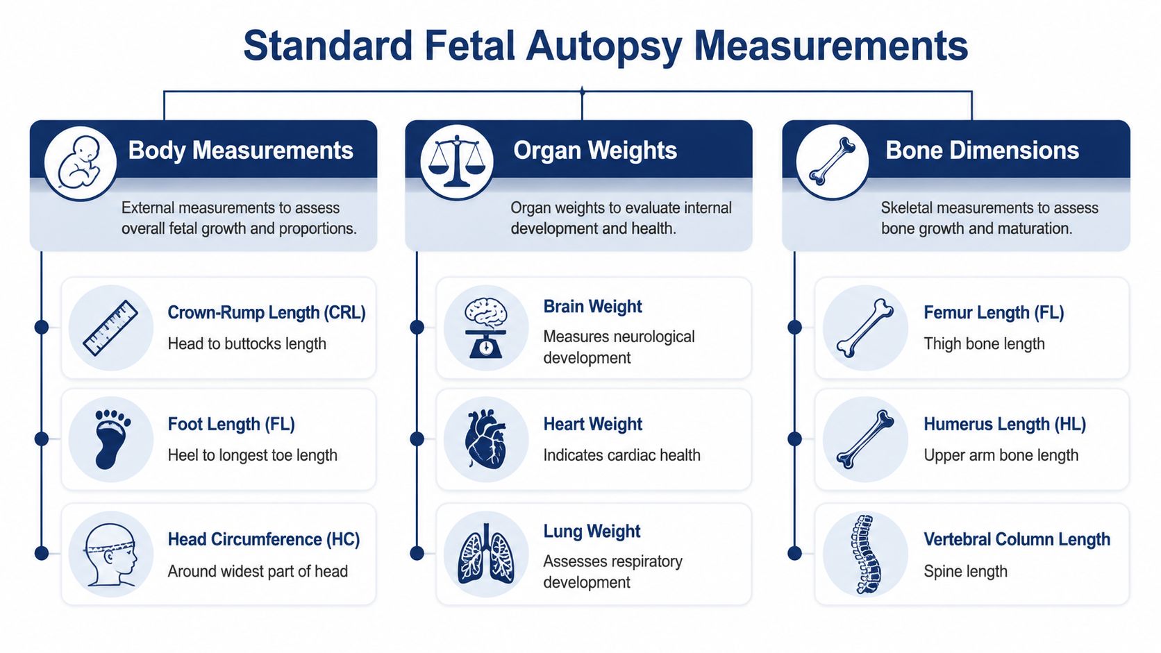
A complete fetal examination starts with the external body. According to ACOG guidance on management of stillbirth, those measurements should be recorded before dissection and should include body weight, crown-rump length, crown-heel length, head circumference, chest circumference, abdominal circumference, and foot length. The same guidance also recommends photographs of the whole body and any abnormalities so that the measurements remain tied to visible anatomy.
For families who want a clearer picture of that first step, this overview of the external examination in an autopsy shows how documentation fits into the broader process.
External body measurements
These are the standard measurements typically referenced in a fetal autopsy report:
- Body weight helps assess overall growth and compare the fetus with expected values for gestational age.
- Crown-rump length measures from the top of the head to the buttocks and is especially helpful in earlier gestations.
- Crown-heel length gives total body length and helps assess overall body development.
- Head circumference helps evaluate cranial growth and proportionality.
- Chest circumference provides context for thoracic development.
- Abdominal circumference may reflect body habitus, organ size, and growth pattern.
- Foot length is often one of the most practical external dating tools in earlier fetal cases.
Internal organ weights
After the external exam, internal dissection allows organ weights to be recorded. In a thorough fetal autopsy, that often includes the brain, lungs, heart, liver, and kidneys, along with review of the placenta when available. Organ weights help answer whether internal development matches the external appearance and the estimated gestational age.
An examination transcends a visual survey. A fetus may appear externally small, yet show organ patterns that suggest selective growth restriction. Another may look proportionate externally but have internal findings that shift the interpretation.
Paired organs and asymmetry
Certain organs should be considered in relation to each other. Right and left kidneys, for example, may be weighed and compared if asymmetry is a concern. The same principle applies to other paired structures when internal findings raise that question.
A practical point matters here. A small difference can be ordinary. A meaningful difference has to be interpreted with the rest of the case, not isolated from it.
Bone and skeletal dimensions
In some cases, long bone measurements and skeletal proportions are also relevant. These can be useful when there is concern for a developmental disorder, skeletal dysplasia, or a mismatch between body size and stated gestational age. They are not required in every case, but they can become important when external proportions are unusual.
A complete examination records what can be measured reliably, not just what is easy to measure.
That distinction matters. The best fetal autopsy measurements are the ones that are documented carefully, tied to anatomy, and interpreted in context rather than treated as stand-alone facts.
Protocols for Ensuring Measurement Accuracy
The value of a fetal autopsy measurement depends on how it was taken. Poor technique creates false precision. A number may look exact on paper and still be misleading if the method was inconsistent, rushed, or anatomically off target.
Technique before interpretation
Technical standards summarized in The Autopsy Book on measurement and weights stress comparability. Head circumference should be taken at the widest point. Chest circumference is measured at the nipple line. Abdominal circumference is measured at the umbilicus. Foot length is measured from the heel to the tip of the longest toe. For paired organs such as kidneys and adrenal glands, weighing together and separately can help detect asymmetry.
Those details may sound small. They aren't. If one examiner measures head circumference too high and another too low, the number changes even when the fetus doesn't.
What consistency looks like in practice
In my own work, accuracy starts before any incision. The body is examined, positioned, and documented first. Weight should be measured with equipment sensitive enough for very small fetuses. Circumference measurements should be taken with the tape or string snug against the body but not pressing into the tissue.
A consistent approach usually includes:
- Recording before dissection so the body hasn't been altered by the examination itself
- Using the same landmarks each time so numbers can be compared with published references
- Photographing the body and notable findings so later review has visual context
- Separating measurement from interpretation so the raw data stay clear even if the final opinion evolves after microscopy or records review
If records or reports may need review across languages, precise wording matters as much as precise measuring. In those situations, Translators USA's medical translation is the type of resource that can help preserve technical meaning when clinical or legal documents are shared internationally.
Why process matters to families and attorneys
A careful method protects everyone involved. Families deserve an honest record. Attorneys need documentation that can withstand scrutiny. Physicians need findings that can be correlated with prenatal imaging, obstetric history, and placental pathology.
For readers who want a broader overview of the sequence, the procedure of an autopsy places these measurements within the larger postmortem workflow.
If the method isn't sound, the interpretation won't be sound either.
How Measurements Help Estimate Gestational Age
A common question arrives early in the case. Do the body measurements match the pregnancy dates in the chart, or do they point to a different developmental stage?
That question matters to families trying to understand what happened, and to attorneys and clinicians who need a defensible opinion. Sometimes the dating from prenatal care is clear and well supported. Sometimes the records are sparse, late, or inconsistent. In those cases, postmortem measurements help estimate gestational age, but they do not replace clinical history or ultrasound findings.
The measurements that help most
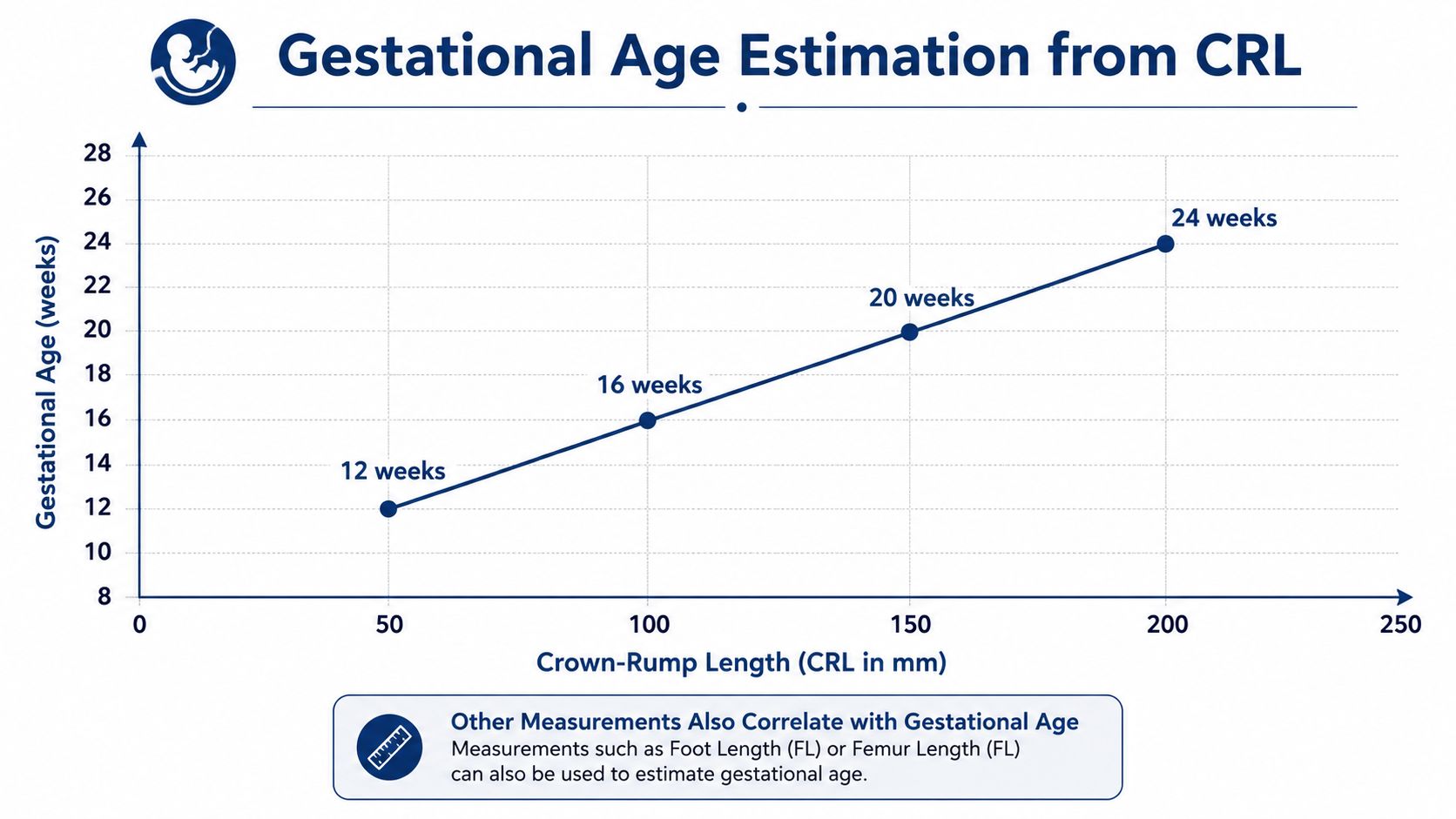
Several external measurements contribute to age estimation, but a few are used more often because they remain relatively reproducible across observers. In earlier gestations, crown-rump length and foot length are usually the most informative starting points. As noted earlier in the article, standard references and practice guidance support using physical measurements and external scoring as part of gestational age assessment, especially in younger fetuses.
Foot length deserves special mention. In daily practice, it is often easier to obtain consistently than some other measurements, particularly in small fetuses or cases with fragile tissue. That does not make it infallible. It makes it useful.
Why a measurement can point in the wrong direction
No experienced pathologist dates a fetus from one number alone if the rest of the examination raises concern. A crown-rump length may suggest one age range, while foot length, head size, body weight, and organ development suggest another. When those findings line up, the estimate becomes more reliable. When they do not, the job shifts from simple measurement to careful interpretation.
The most common reasons for disagreement are practical and familiar:
- Growth restriction, which can make size-based dating appear younger than the actual gestational age
- Maceration, which can soften tissue, alter contour, and reduce confidence in some external measurements
- Congenital anomalies or skeletal dysplasia, which can make one body part a poor proxy for overall development
- Uncertain clinical dating, including late prenatal care or conflicting record documentation
Experience matters in these situations. The pathologist has to decide whether a discrepancy reflects normal variation, pathologic growth disturbance, postmortem change, or an error in the recorded dating.
How uncertainty is handled in a real case
Families often want a single answer. Sometimes we can give one with reasonable confidence. Sometimes the honest answer is a range.
If a fetus is significantly macerated, for example, I may place more weight on measurements that are less distorted by tissue breakdown and less weight on those that depend on body posture or surface integrity. If the feet are preserved but the trunk is markedly altered, foot length may carry more interpretive value than crown-rump length. If skeletal proportions are abnormal, neither number may reflect true gestational age well enough to stand alone.
That judgment should appear plainly in the final report. The report may state that the measurements are consistent with an estimated gestational age, or that the estimate is approximate because postmortem change, growth restriction, or anomaly limits precision. For families, that wording can feel frustrating. For medical-legal review, it is the correct way to communicate uncertainty without overstating what the examination can prove.
Interpreting the Data Using Normative Ranges
A measurement means very little until it's compared with what is expected for that stage of development. A body weight, head circumference, or liver weight has to be read against gestational age. Otherwise, the number is just a raw observation without clinical meaning.
Comparison is the whole point
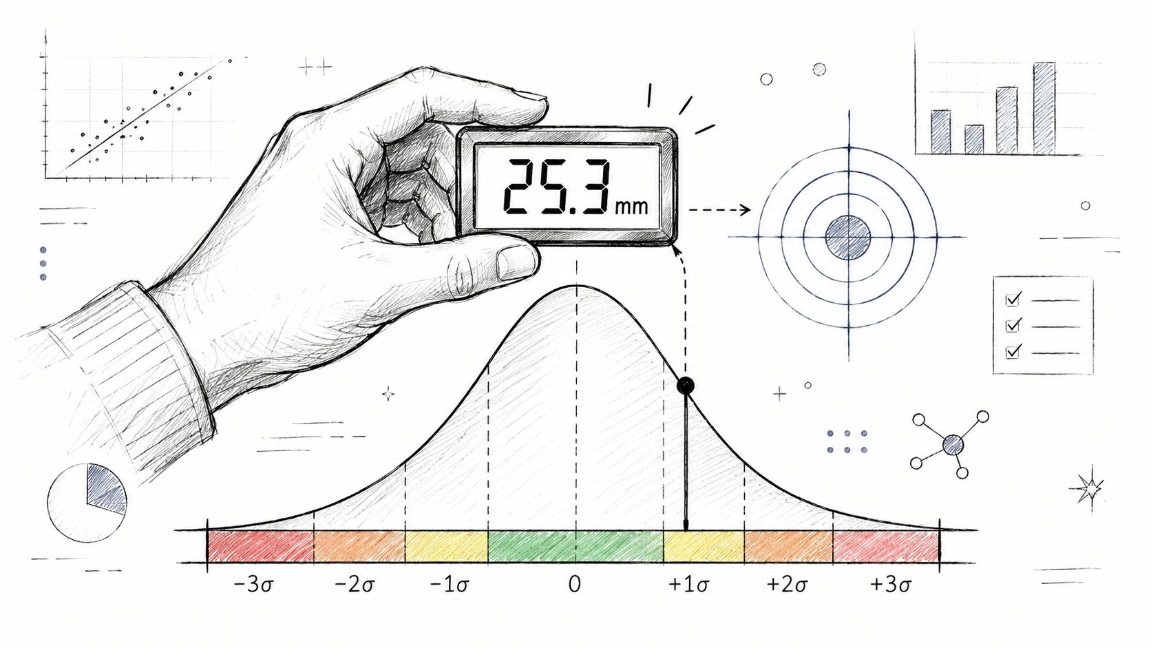
The fetal autopsy literature uses week-specific means and standard deviations for many body and organ measurements. In practice, that means the pathologist asks whether a given value is within an expected range for that gestation, clearly below it, clearly above it, or proportionally unusual compared with the rest of the body.
A practical interpretation often looks at several relationships at once:
- Body size versus estimated age
- Head size versus body size
- Abdominal size versus overall growth
- Organ weights versus developmental stage
- One organ versus another organ
Why ratios can matter
One of the best-known examples is the brain:liver weight ratio. In a UCL study on postmortem fetal organ-weight ratios, a ratio of 6 was reported as a clinically useful cut-off. It identified pathological fetal growth restriction with 92% specificity and 55% sensitivity when separating it from other causes of death. In the same work, it showed 80% specificity and 53% sensitivity when distinguishing growth-restricted fetuses with placental abnormalities from small fetuses without those findings.
That matters because it shows how fetal autopsy measurements can become quantitative markers rather than simple descriptions. It also matters because the same study emphasized the limit. These ratios can support interpretation of intrauterine growth status, but they do not by themselves prove the cause of death.
A ratio may strengthen an argument for growth restriction. It does not, by itself, answer why the fetus died.
That distinction is critical in legal review. A strong pattern is evidence. It is not permission to overstate the conclusion.
Turning numbers into findings
This short video gives a useful visual frame for thinking about how statistical comparison works in pathology and measurement-based interpretation.
In real practice, interpretation is rarely built on one threshold alone. A pathologist combines normative comparison with placental findings, body proportions, organ examination, and the known clinical course. The best reports show the logic clearly enough that another physician, lawyer, or family member can follow it.
Understanding Discrepancies and Limitations
The hardest part of fetal autopsy measurements is not taking them. It's deciding how much confidence to place in them when the body has been altered by time, environment, or condition after death.
Maceration changes the playing field
Maceration is the tissue breakdown that can occur when a fetus remains in utero after death. It can affect skin integrity, fluid balance, tissue firmness, and the ease with which landmarks can be identified. That means some measurements become less reliable even if they can still be obtained.
Research summarized in this study on fetal foot length and maceration found that foot length correlated strongly with ultrasound-based gestational age overall, with r = 0.85, but the estimate became much less reliable with maceration. The gestational-age difference increased from about 4 days in non-macerated fetuses to 19 days with mild maceration and 34 days with severe maceration.

Those numbers capture a reality families often don't hear plainly enough. A measurement can still be useful while no longer being precise.
Fragmentation and nonintact remains
Some fetuses are not intact enough for every standard measurement. In those cases, the examination doesn't become worthless, but the strategy changes. The pathologist documents what can be measured reliably, what can only be estimated, and what should not be over-interpreted.
That may include situations where:
- Crown-heel length can't be trusted because the body position or integrity is altered
- Circumferences are distorted by tissue loss or slippage
- Weight is misleading because of fluid shifts or decomposition
- An isolated foot length remains available but must be treated as approximate
How uncertainty should be communicated
Experience is key. The right answer is not to force certainty out of poor data. It's to explain the limitation in plain language. I want the report to tell the reader whether a value is dependable, approximate, or significantly limited by condition.
When tissue condition reduces reliability, the report should say so directly. That isn't weakness. That's good pathology.
In my view, honest uncertainty is one of the clearest signs that a pathologist is being objective. Families may not like hearing that a number is approximate. Attorneys may wish the answer were cleaner. But overconfidence helps no one, especially in a sensitive fetal case.
How Measurements Appear in the Final Report
A good final report doesn't bury measurements in technical clutter. It presents them in a way that lets the reader see what was found and how those findings support the final opinion.
What is usually documented
Most reports list the major external measurements and key organ weights in a dedicated gross examination section. The report may also include the estimated gestational age, whether the body appears proportionately developed, and whether any measurements fall outside expected ranges for that stage.
The report should also define key terms clearly. Cause of death means the disease, injury, or condition that led to death. Manner of death is the classification of how the death occurred, such as natural or accident, though in fetal cases the framework may differ from standard adult forensic usage. Chain of custody means the documented handling of the body and any evidence from transfer through examination and storage.
How interpretation is written
When measurements are meaningful, the final report should say what they support. When they are limited, the report should explain why. The strongest reports separate direct observation from interpretive opinion.
Examples of report language often sound like this in substance:
- Observed finding: body weight, lengths, circumferences, and organ weights are recorded
- Comparative interpretation: the measurements are consistent with, smaller than expected for, or limited in interpretation for the estimated gestational age
- Qualification: tissue maceration or fragmentation reduces confidence in exact dating or growth assessment
- Integrated opinion: the measurements support, but do not alone establish, a broader conclusion
For families reviewing unfamiliar terminology, this guide on how to read a pathology report can help translate the structure into plain language.
Why this matters in medical-legal settings
Attorneys often focus on whether the report explains its reasoning. It should. If growth restriction is suggested, the measurements and comparison logic should be visible. If gestational age is approximate, the basis for that estimate should be stated. If the body condition limited interpretation, that limitation should not be hidden.
That is what turns fetal autopsy measurements into usable evidence rather than a list of disconnected numbers.
Frequently Asked Questions About Fetal Measurements
Families and attorneys usually ask practical questions, not textbook questions. The table below addresses the ones I hear most often.
Common questions and direct answers
| Question | Answer |
|---|---|
| Do fetal autopsy measurements tell you the exact cause of death? | No. They can support important conclusions about gestational age, growth, and proportional development, but they are interpreted alongside history, placental findings, microscopy, and other evidence. |
| Which measurement is usually most helpful for dating an early fetus? | Foot length is often very useful in earlier gestations, especially when other history is limited. It still has to be interpreted in context. |
| What if the measurements don't all agree with each other? | That happens. A mismatch may reflect growth restriction, anomaly, inaccurate clinical dating, or postmortem change. The pathologist should explain the discrepancy rather than force a single simplistic answer. |
| Can maceration make the measurements unreliable? | Yes. Some measurements remain helpful, but confidence drops as tissue degradation increases. A careful report should say which findings are dependable and which are only approximate. |
| When are these examinations appropriate outside a hospital setting? | Families, attorneys, and agencies may seek an independent autopsy or second-opinion review when they need an objective assessment of growth, development, and cause of death issues. In Texas, options can include Private Autopsies, County Forensic Autopsies, or coordination through partners such as the Waterloo Mortuary Partnership when logistics require it. |
A final practical note
Fetal autopsy measurements are at their best when they are taken carefully, interpreted conservatively, and explained clearly. That combination gives families something real to hold onto. It also gives attorneys and clinicians a record they can evaluate without guessing what the pathologist meant.
Texas law and postmortem handling requirements can affect transportation, authorization, release, and disposition. Those issues may involve the Texas Funeral Service Commission and provisions of the Texas Health & Safety Code, including Chapters 711, 716, and 651, depending on the circumstances. For that reason, case-specific procedural questions should be addressed directly with the responsible professionals rather than assumed from general online information.
If you need help understanding whether a fetal or perinatal autopsy may provide useful answers, contact Texas Autopsy Services. I approach these cases with the same standard I would want for my own family: a careful examination, clear communication, and honest limits where the evidence doesn't support more.