June 26, 2026 · Texas Autopsy Services
A Guide to Forensic Pathology Pics for Texas Families
What are forensic pathology pics and why are they taken? Our guide explains the types, purpose, and legal process for families and attorneys in Texas.
On this page
- Answering Difficult Questions After a Loss
- The Purpose of Images in a Forensic Investigation
- The Different Types of Forensic Images We Produce
- How and When We Create Photographic Records
- Upholding Legal and Evidentiary Standards in Texas
- How Families Can Access and Review Images
- Your Questions Answered A Guide to Our Reports
- Frequently Asked Questions About Forensic Images
What Families Should Know About Forensic Pathology Pics
- Forensic pathology pics are evidence, not spectacle. They create a permanent visual record that supports the medical findings after death.
- Cause of death means the disease or injury that led to death. Manner of death is the classification, such as natural, accident, suicide, homicide, or undetermined.
- In Texas, a medical examiner is legally required only to perform an inquest, and a full autopsy is left to the pathologist's discretion, which is one reason some families seek an independent autopsy for fuller documentation through Private Autopsies.
- Forensic images must meet strict legal standards. A critical requirement is that they be non-editable and captured with the camera's date and time set precisely so the evidence can be defended if challenged in court.
- Families do not always need to view graphic images directly. In many cases, the written report and a guided explanation provide the clearest and most respectful path to understanding.
A family often reaches this point after the first urgent decisions are over. The death certificate may feel too brief. A hospital explanation may feel incomplete. An attorney may be trying to understand whether the physical findings support the legal narrative.
At that moment, people often ask about photographs. They may call them autopsy photos, forensic images, or forensic pathology pics. What they usually mean is simpler. They want to know what was documented, who can review it, and whether those images can help answer hard questions with honesty and dignity.
Those are fair questions. The images created during a forensic examination are not taken for shock value. They are made to preserve facts carefully, in a way that can be reviewed later by family, counsel, or another qualified expert when appropriate.
Answering Difficult Questions After a Loss
A daughter calls after a sudden death. She has already heard one explanation from the hospital, another from the county, and now she learns that photographs were taken during a private examination. Her first question is usually simple and painful at the same time. Why were pictures necessary?
That question deserves a careful answer.
In forensic pathology, images are created to document findings faithfully, the same way a chart preserves a patient's history or an X-ray preserves a view that can be reviewed later. Families often worry that photographs exist for shock value. Attorneys often ask whether they are only illustrative, or whether they are part of the proof. Under an ethical medical-legal practice, the answer starts in the same place for both groups. The images exist to record what was present, in a form that can be reviewed, explained, and tested.
Forensic photography exists to preserve facts that words alone may not fully convey.
That point often becomes clearer after reading a report. A sentence can state that there was an injury, a disease process, or evidence of medical treatment. A photograph can show where that finding was located, how large it was, how it was oriented, and how it related to nearby structures. The sequence is important because meaning depends on context. A single image rarely stands alone. It works like one frame in a longer medical record.
Families are often surprised to learn that a death investigation does not always include every procedure they expected. In Texas, the legal process may answer some questions while leaving others open for the family or counsel. That is one reason independent review is sometimes requested. It can help clarify whether the existing record is sufficient, whether more documentation is needed, and whether the conclusions match the observed findings.
For that reason, questions about forensic pathology pics are usually questions about truth, proof, and understanding. In a respectful practice, the deceased is never treated as an object of display. The photographs are handled as evidence and as part of a careful effort to give families and attorneys clear, defensible answers after a loss.
The Purpose of Images in a Forensic Investigation
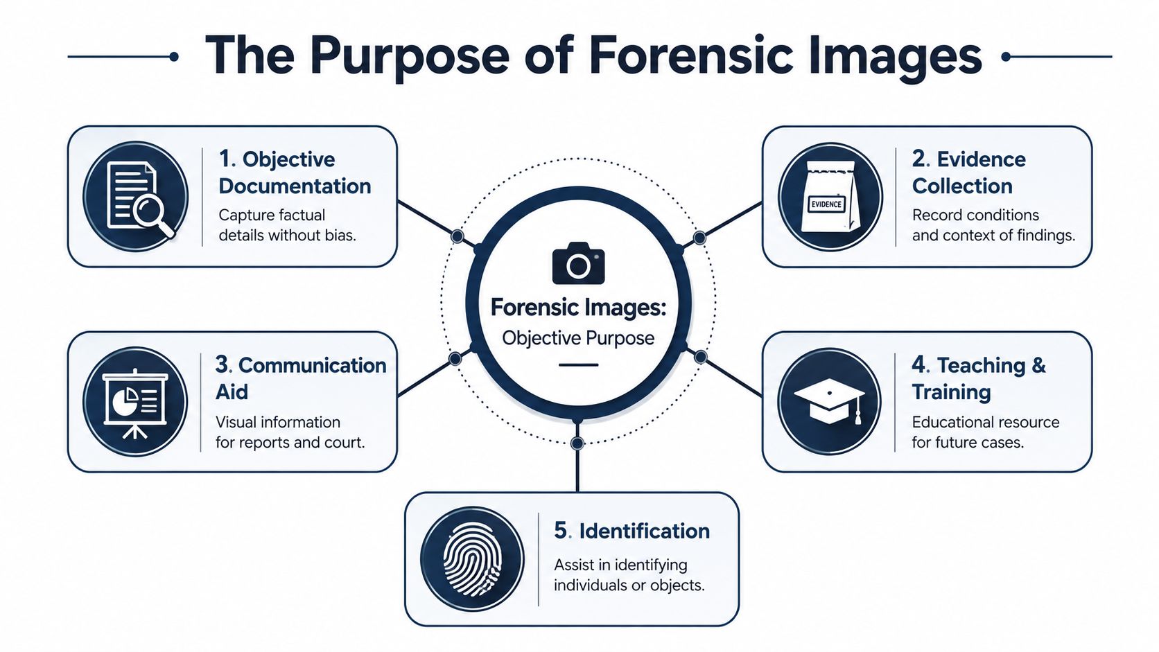
A family calls after receiving an autopsy report and asks a hard, understandable question: “Why were photographs taken at all?” In our work, the answer is straightforward. The images are made to preserve medical and legal facts with care, so the findings can be reviewed later by the pathologist, the family's attorney, or another qualified expert.
A permanent factual record
A forensic photograph records what was present at a specific point in the examination. That may include injuries, medical devices, natural disease, identifying features, or the appearance of organs and tissues. In the same way a chart preserves a doctor's observations from a hospital visit, a photograph preserves a visual finding so it does not depend on memory alone.
That record serves both compassion and legal rigor. Families deserve clear answers. Attorneys need documentation that can be examined, questioned, and compared with the written report. An ethical forensic practice treats photographs as part of the evidentiary file, with the same seriousness given to notes, specimens, and final opinions.
Images also help explain two terms that are often confused. Cause of death is the disease or injury that led to death. Manner of death is the classification, such as natural, accident, suicide, homicide, or undetermined. A photograph does not make those decisions by itself, but it can support or challenge an interpretation when the finding is reviewed in context.
A tool for explanation and professional review
Photographs are often associated with court, but their role is broader and more practical. They help a pathologist explain what was seen, where it was located, and how it relates to the final medical opinion. That can be important for a consulting physician, a reviewing attorney, or a family member who needs more than a technical sentence in a report.
A useful image may clarify questions such as:
- Where an injury was located and what body structures were involved
- Which marks came from medical treatment such as lines, tubes, incisions, or resuscitation efforts
- How a visual finding relates to the written opinion in the report
A report and a photo work together. One gives the medical interpretation. The other preserves the visual reference point.
That pairing is especially important when people encounter forensic pathology pics without explanation. An isolated image can mislead even well-meaning viewers. Sequence, labeling, and chain of custody give the picture meaning. Without that framework, a photograph is only a fragment.
Respectful use, not exposure
Some images are appropriate for direct review with families. Others are better explained through selected photographs, diagrams, or a guided conference. We choose that approach carefully, because the goal is understanding, not shock.
In a Texas forensic setting, that restraint is part of good practice. The deceased is never treated as a subject for display. The photographs exist to document findings in a way that is medically accurate, legally defensible, and handled with respect for the person who died and the people left behind.
The Different Types of Forensic Images We Produce
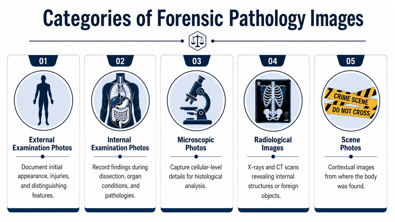
Forensic pathology pics are not one category. They include several types of records, and each serves a different purpose in a complete examination.
External Examination Photos
These are usually the first photographs created during an autopsy examination. They document the body as received, including identifying marks, scars, tattoos, medical intervention, and any visible injuries or natural changes.
External photographs often answer questions that seem basic but matter greatly later. Was there bruising? Where exactly was it located? Did the person have surgical incisions, hospital lines, or defibrillator pad marks that might be confused with injury by a nonmedical viewer? A clear set of external images helps separate treatment effects from disease or trauma.
They also preserve orientation. A written note may say there was an injury on the left side of the neck or the back of the scalp. A photograph shows that placement directly.
Internal Examination Photos
Once the internal examination begins, photographs document the condition of organs, body cavities, hemorrhage, disease, and injuries that cannot be seen from the outside. These images are often the most medically important, even though they are not always the most suitable for direct family review.
Internal images can help explain why a final opinion was reached. For example, they may show the pattern of bleeding, the location of a rupture, the appearance of the heart, or the relationship between injury and surrounding anatomy.
A short comparison is helpful here:
| Image type | What it commonly shows | Why it matters |
|---|---|---|
| External | Surface findings, identifying features, treatment marks | Establishes visible condition and injury location |
| Internal | Organ findings, hemorrhage, disease, trauma | Supports the medical basis of the final opinion |
| Microscopic | Tissue-level changes | Confirms disease or injury at the cellular level |
| Radiographic | Internal structures before or alongside dissection | Reveals findings that may be hard to see otherwise |
Microscopic Images
Some findings can only be confirmed under the microscope. Histology slides may show disease, inflammation, tissue reaction, or subtle injury that is not obvious on gross examination.
Microscopic images are not always included in the same way as standard autopsy photographs, but they can be vital when a case turns on timing, tissue response, or the presence of underlying natural disease. These images are usually most useful to medical professionals and attorneys working with retained experts.
Radiographic Images
Modern forensic work often includes imaging before or alongside dissection. Postmortem computed tomography, or PMCT, serves as the mandatory baseline forensic imaging examination and requires a whole-body scan to ensure thorough data capture, according to the American Journal of Roentgenology forensic imaging protocol. For enhanced resolution of specific pathologies, a second high-resolution scan is performed. That protocol helps pathologists visualize issues that traditional autopsy might miss.
PMCT is especially useful because it documents the body before dissection changes the anatomy. It can preserve the position of fractures, foreign objects, gas collections, and other internal relationships.
Postmortem magnetic resonance imaging, or PMMRI, is used more selectively. It is not a routine standalone exam. It is used when external inspection and baseline PMCT suggest a need for better assessment of soft-tissue injuries or hemorrhagic sites, as described in this review of postmortem neuroimaging and forensic imaging practice. That matters because relying on CT alone limits detection of some soft-tissue injury and some hemorrhagic brain sites.
Some findings are easiest to understand with a camera. Others are easier to understand with a microscope or scanner. A careful forensic examination may use all three.
How and When We Create Photographic Records
A family member may ask us, "When were these pictures taken, and why this one instead of another?" That is a fair question. In forensic pathology, photographs are created as the examination unfolds so each image can be tied to a specific step, observation, and specimen. That order protects both understanding and fairness.
The record follows the examination
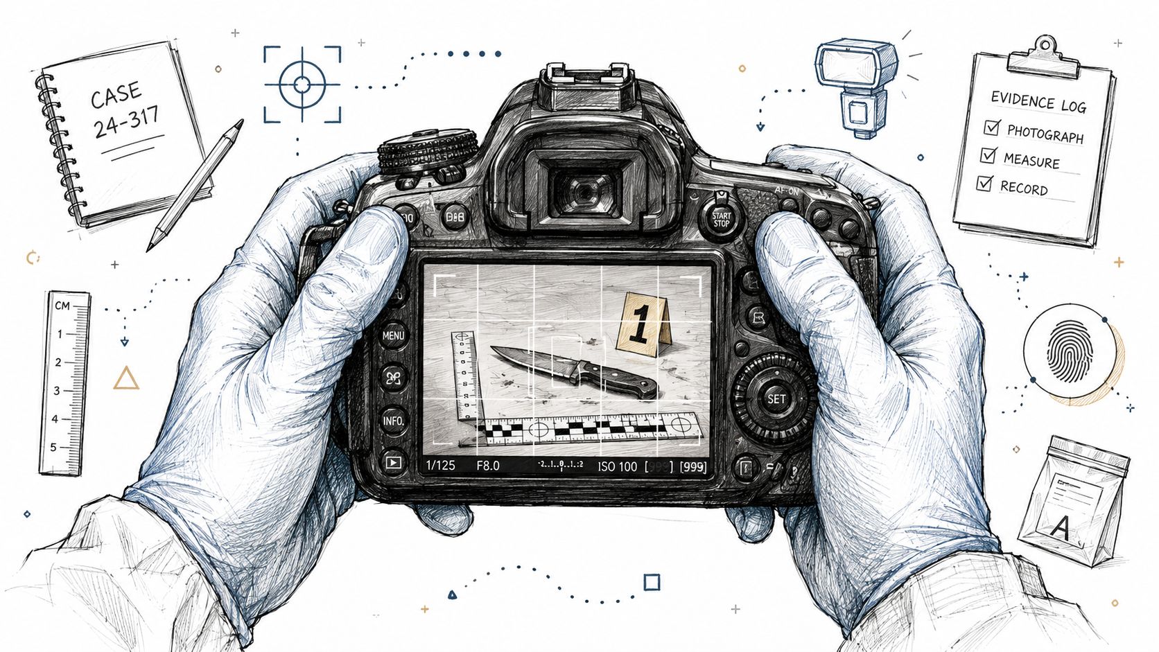
We usually photograph in sequence. That may begin when the body is received, continue through the external examination, and extend into the internal examination when a finding needs to be documented clearly. The goal is a medical record in images, arranged so a reviewer can tell what was seen, when it was seen, and how one finding relates to another.
Context is what gives a photograph meaning. A close-up of an injury, by itself, can be misleading. A wider orientation image shows where that injury sits on the body, and a later internal image may show whether there is related damage beneath the surface. Those images work together the way pages of a chart work together. One page may be useful. The sequence is what makes the record understandable.
Timing matters for another reason. Some findings are best documented before cleaning, before dissection, or before organs are removed and examined separately. Once anatomy changes, the original relationship between structures may no longer be visible in the same way.
Why timing and scope matter in Texas cases
In Texas practice, the amount of photography depends on the scope of the examination that is performed. As noted earlier, an official death investigation does not always mean a full autopsy with full photographic documentation. Families and attorneys are often surprised by that. They may receive a conclusion, yet still have limited visual documentation of the findings that led to it.
That is one reason an independent examination can be helpful. At Texas Autopsy Services, we approach imaging with two duties in mind. One duty is compassion for the people trying to understand a death. The other is legal rigor, because these records may later be reviewed by counsel, insurers, or the court.
A private examination may include:
- External photographs that document identifying features, visible injuries, medical treatment effects, and the body as received
- Internal photographs that show organ findings, hemorrhage patterns, fractures, or the relationship between injuries and underlying anatomy
- Specimen and evidence documentation tied to labeling and handling procedures, which should match the case file and chain-of-custody procedures for forensic evidence and images
- Case review photographs selected for explanation during meetings with families or attorneys, so the images answer a medical question rather than create confusion
We also handle storage and release with privacy in mind. That includes careful file organization, controlled access, and practices similar to those discussed in optimizing privacy in document handling.
In a large state, local practices can differ. A consistent photographic method helps reduce confusion. It gives families and attorneys a clearer record of what was observed, in what order, and under what conditions.
Upholding Legal and Evidentiary Standards in Texas
A forensic image is not just a picture file. If it may become part of litigation, an insurance dispute, or a formal review, it has to be handled as evidence.
Why image integrity matters
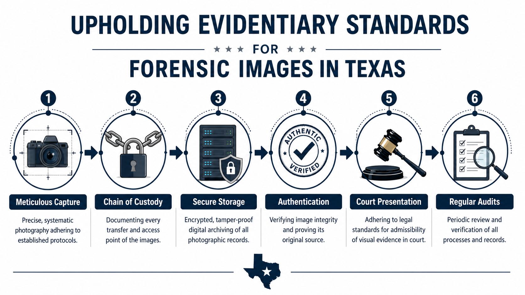
A critical standard in forensic photography is that the image must be non-editable, and the camera's date and time must be set precisely. That requirement helps prevent evidence from being contested in court and is part of proper chain-of-custody verification, as discussed in this forensic photography review in the medical literature.
That may sound technical, but the principle is simple. If no one can show when an image was made, whether it was altered, and who handled it afterward, the image becomes easier to challenge.
Chain of custody in plain language
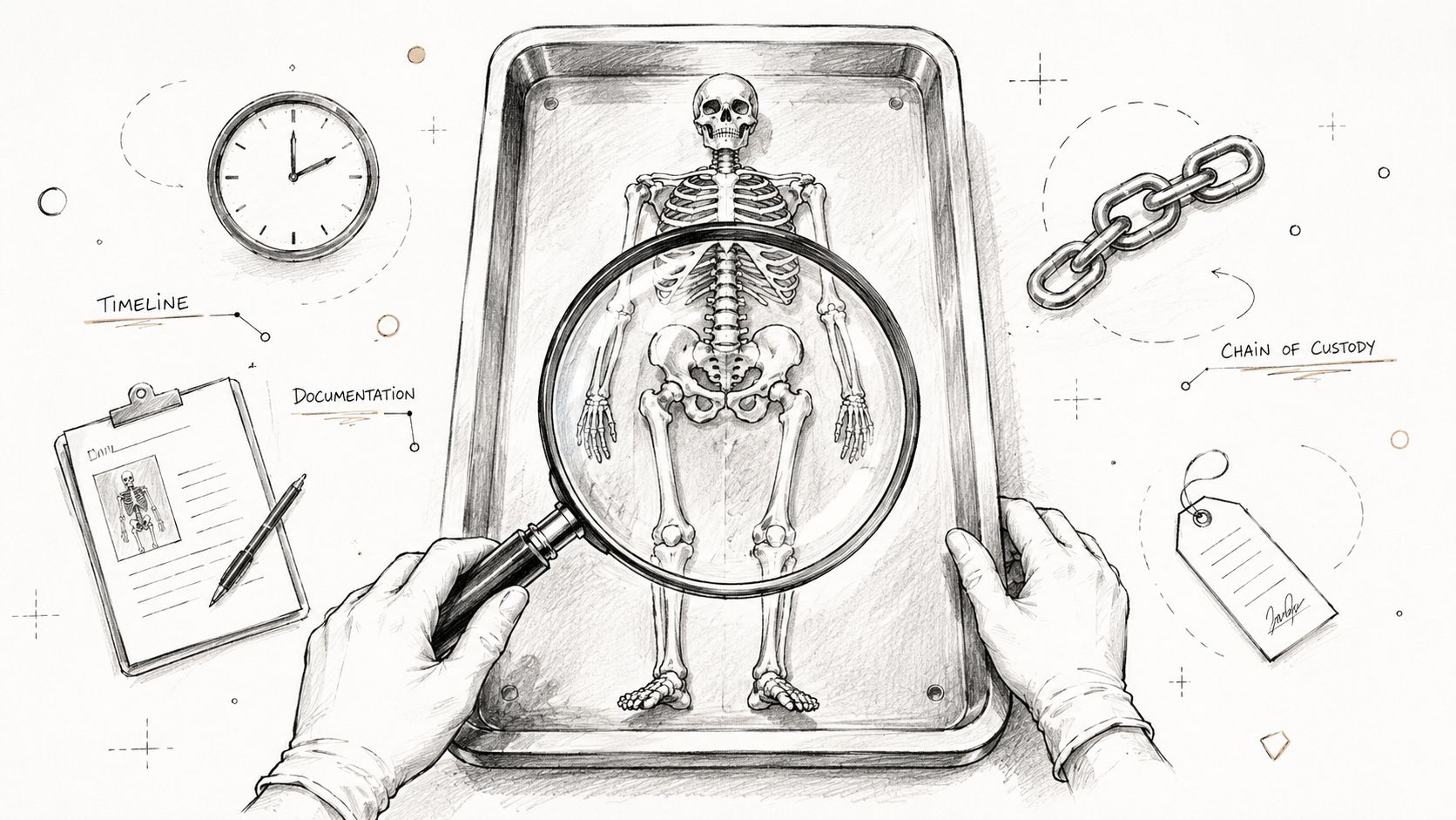
Chain of custody means the documented history of evidence from creation through storage, access, and release. For digital photographs, that includes who captured the image, how it was stored, who was permitted to review it, and how it was transmitted if it was later released.
Evidence is strongest when each handoff is documented and each file remains traceable to its original source.
For attorneys, that documentation helps establish authenticity. For families, it protects privacy. The same safeguards that make evidence defensible in court also reduce the risk of misuse or unauthorized circulation.
A simple checklist often includes:
- Controlled capture with correct camera settings and case association
- Secure storage so files remain protected from alteration
- Documented access showing who reviewed or received the material
- Verified release procedures when records are provided to an authorized party
People who handle sensitive case files often benefit from broader guidance on secure workflows outside the autopsy setting as well. Practical reading on optimizing privacy in document handling can help families and legal teams think carefully about how records are stored and transmitted after release.
A deeper discussion of these procedures is available in our article on chain of custody procedures.
This short overview helps illustrate why those steps matter in practice:
Texas legal context
Any discussion of records in Texas should be understood within the larger legal framework that also touches funeral, transport, and postmortem handling issues, including the Texas Funeral Service Commission and Texas Health & Safety Code Chapters 711, 716, and 651. Those rules do not turn every photograph into public material. They reinforce that these records are sensitive, controlled, and tied to lawful authority.
How Families Can Access and Review Images
For many families, the hardest part is not whether photographs exist. It is deciding whether they want to see them and, if so, how to do that safely.
Requesting access
Access depends on legal authority and the holder of the records. In practical terms, that usually means the next of kin or an authorized representative, such as an attorney acting for the proper party. The process should be formal and documented, not casual.
When families ask for images, it helps to be specific about the need. Some want confirmation of an injury location. Some want to understand medical treatment. Others need records for a second opinion autopsy review. A narrower request is often easier to manage and easier to explain.
Preparing for review
Not every person should review a full set of autopsy photographs alone. Some people do better with a selected subset. Others should have a trusted support person present, such as counsel, clergy, or a family member who can help them process what they are seeing.
A good alternative is a guided review. In that setting, one of our board-certified forensic pathologists explains what the image shows, why it matters, and what it does not prove on its own. That clinical explanation often reduces confusion because nonmedical viewers can easily mistake treatment effects or postmortem changes for something else.
You are allowed to ask for explanation without asking to see every image.
That approach is often gentler and more useful than unrestricted release.
Privacy after release
Once records are released lawfully, families should still treat them as highly private materials. They are not keepsakes and they are not meant for broad sharing. If records need to be shared within a family, the goal should be controlled access and clear understanding of who is viewing them.
People understand this more easily when they compare it to ordinary photo sharing. A casual tool built for an online wedding photo gallery is designed for convenience and broad access. Forensic images are the opposite. They call for restraint, secure handling, and a very limited audience.
For readers who want more background on the difference between curiosity-driven searches and proper medical-legal review, our discussion of real autopsy pics addresses that concern directly.
Your Questions Answered A Guide to Our Reports
The written report remains the primary document in an autopsy case. It contains the formal findings, the interpretation of those findings, and the final opinion on cause of death. The photographs support that report. They do not replace it.
That distinction often brings relief to families. Many people can understand the important conclusions through a report conference alone. They do not need to view graphic material to learn what happened. If selected images would help clarify one point, those can sometimes be reviewed in a limited and clinically explained way.
For attorneys, the report also provides the structure that makes the images useful. Without that framework, photographs can be misread. With it, they become supporting documentation that can be evaluated in context.
Readers who are concerned about image misuse after release may also find a practical professional guide for unpermitted photos useful as a general privacy resource. Questions about forensic image interpretation and litigation support are also discussed in our article on photography expert witness.
Our work also extends beyond family-requested examinations. County officials may need County Forensic Autopsies, and postmortem logistics may involve partners such as the Waterloo Mortuary Partnership. In each setting, the same principle applies. The report is central, and the images exist to document and support the findings with care.
Frequently Asked Questions About Forensic Images
| Question | Answer |
|---|---|
| Can any relative obtain forensic pathology pics? | Usually, no. Access generally depends on legal authority, such as next-of-kin status or documented authority to act for the family or estate. |
| Are forensic images the same as the autopsy report? | No. The report is the primary written explanation of findings and conclusions. Images are separate supporting records. |
| Do families have to look at the photographs to understand the findings? | No. Many families prefer a report conference or a guided explanation instead of direct image review. |
| Why are the camera date and time settings so important? | They help verify when the image was created and support authenticity. In forensic work, that is part of preserving evidentiary value. |
| Can these images be used in court? | They can be, if they were captured, stored, and released under proper evidentiary procedures and are relevant to the issue being litigated. |
If you need careful guidance after a death, Texas Autopsy Services is available to speak directly with families, attorneys, medical professionals, and county officials. Every examination is performed by a forensic pathologist certified by the American Board of Pathology. We serve all 254 Texas counties, handle logistics with in-house licensed transport, and provide a transparent process with full chain of custody. If you're trying to understand whether an independent autopsy, a second opinion, or a guided review of findings would help, contact our team. We'll explain the options clearly and respectfully.