October 15, 2026 · Texas Autopsy Services
Autopsy Lungs of a Smoker: What Forensic Findings Show
Autopsy lungs of a smoker can reveal emphysema, pigment, and airway damage, but lung appearance alone does not prove cause of death. Learn what pathologists
On this page
- Why Lung Appearance Alone Cannot Answer the Question
- What a Pathologist Looks for on Gross Examination
- How Microscopic and Laboratory Analysis Add a Second Layer of Evidence
- The Dose Response Between Smoking and Structural Lung Damage
- Why Pigment Alone Is Not a Smoking Meter
- How Lung Findings Connect to Medical History, Toxicology, and Cause of Death
- Why Smoking Is Rarely the Official Cause of Death on the Certificate
- Questions Families and Attorneys Should Ask Before an Autopsy
Families often hear that a black lung proves smoking and explains a death. That shortcut sounds decisive, but it misses the forensic question. In an autopsy of smoker lungs, the pathologist has to answer three separate questions: what was inhaled, what structural disease is present, and what caused death.
Why Lung Appearance Alone Cannot Answer the Question
A dark or heavy lung can be a clue, but it is not a verdict. Pigment can show exposure, emphysema can show destructive disease, and neither one by itself proves the immediate cause of death. We separate those issues because a person may have smoked, may have smoking-related lung injury, and still have died from something else.
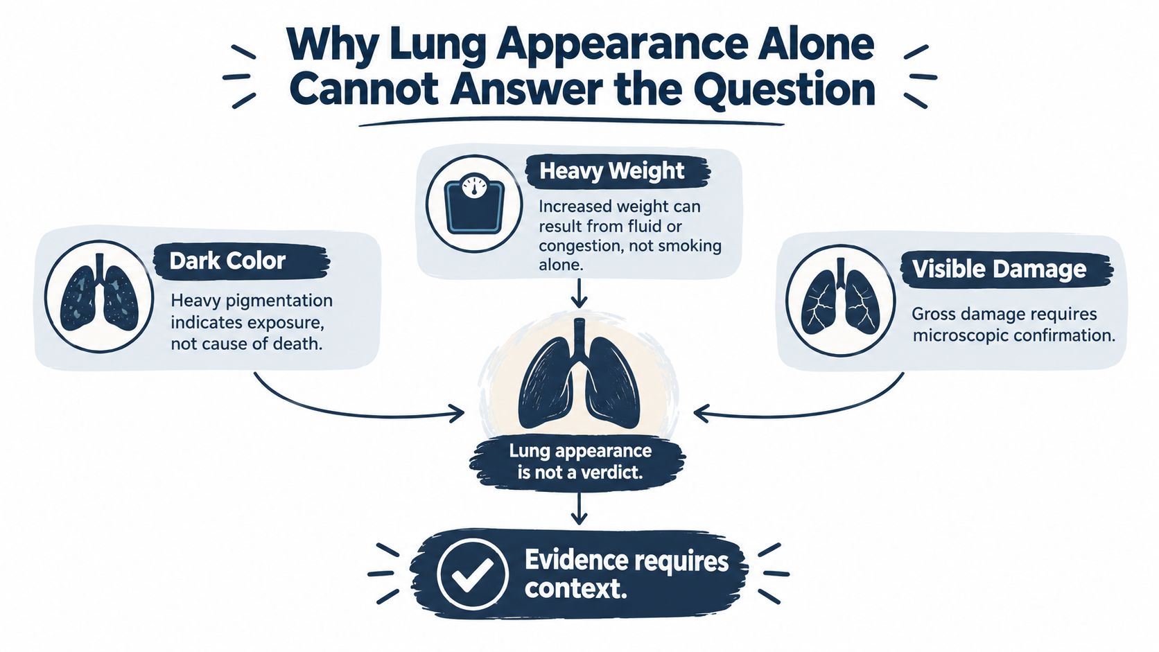
Practical rule: black color, heavy weight, and visible damage each raise questions, but none of them answers the case alone.
A study of 131 autopsy cases found bullous lung disease in 75.3% of smokers compared with 38% of nonsmokers, with bullae present in 40.7% versus 6% respectively, which shows association, not proof of cause of death (source). That distinction matters because a forensic pathologist isn't judging the lungs by appearance alone. We're separating exposure, structural injury, immediate mechanism, and documentation on the death certificate.
The four questions we ask
First, did the person have a smoking or other inhalational exposure history. Second, is there structural lung disease such as emphysema, chronic bronchitis, or scarring. Third, was the fatal event something like pneumonia, embolism, heart disease, cancer, or respiratory failure. Fourth, does the medical record support what the final report says.
Those questions keep a family, an attorney, or a court from confusing a striking photograph with a medical conclusion. A lung can look dramatic and still not explain the death. It can also look less dramatic than the disease burden is, which is why gross appearance is only the starting point.
What a Pathologist Looks for on Gross Examination
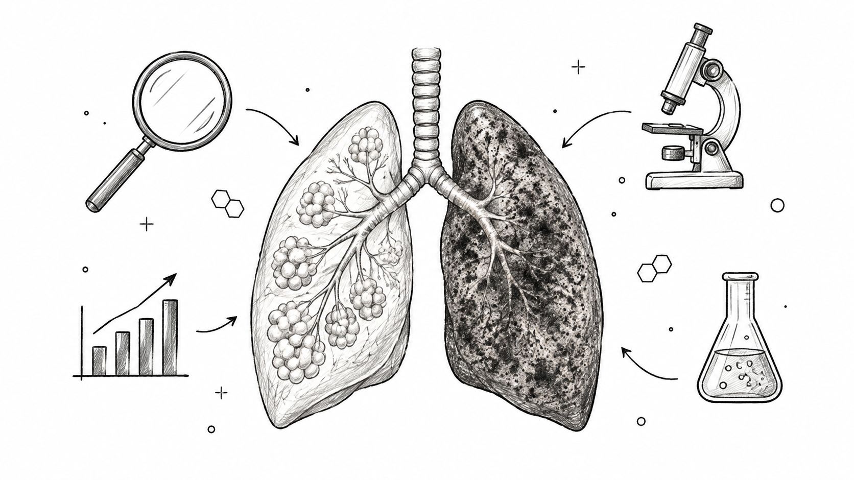
The gross exam is the first look with the naked eye, and it answers only part of the question. We note lung color, weight, inflation, firmness, scarring, airway mucus, bullae, collapse, and any mass that could point to another disease. Emphysema matters because it changes structure and function, not just appearance.
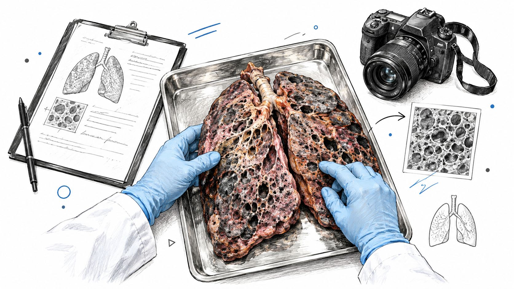
Emphysema is defined by destruction of alveolar walls, with adjacent air sacs merging into larger air spaces, reducing the surface area available for oxygen and carbon-dioxide exchange (source). In plain language, the tiny gas-exchange units are breaking apart. That is why a smoker's lung can lose elasticity and trap air even when the first visible clue is darkness or overinflation.
What the naked eye can and can't tell us
Gross inspection can show bullae, which are larger air spaces that form when lung tissue is destroyed. It can show anthracotic pigment, which is retained carbon and soot-like material. It can also show overinflation, scarring, congestion, and tumor nodules. None of those findings, by itself, says why the person died.
A pathologist also examines the airways, because smoking-related injury often affects the bronchi and bronchioles, not just the air sacs. Thick mucus, wall thickening, and irregular narrowing can point toward chronic airway disease. If a lung is heavy, we also ask whether fluid, congestion, or edema added to the weight, because those can occur for reasons unrelated to smoking.
A gross exam should describe what is seen, not rush to a conclusion. The conclusion comes only after the tissue, records, and circumstances agree.
Findings recorded at the table
| Gross finding | What it may suggest | Why it still needs context |
|---|---|---|
| Dark pigment | Exposure to carbonaceous material | Pigment alone doesn't prove smoking caused death |
| Overinflation | Air trapping or emphysema | Other lung disease can also change inflation |
| Bullae | Structural destruction | Bullae can coexist with unrelated fatal events |
| Scarring or mass | Old injury or tumor | Microscopy and records decide significance |
Later, the lung is sampled in several places, because a single visible lesion can miss the broader pattern. That helps connect the surface exam to the disease process that is present.
A well-documented case summary includes this kind of gross description along with tissue sampling and chain-of-custody documentation.
The following walkthrough shows how a gross lung examination is documented at the table.
How Microscopic and Laboratory Analysis Add a Second Layer of Evidence
A lung can look one way at the table and tell a different story under the microscope. Surface pigment, airway injury, emphysema, and the cause of death are separate questions, and pathology answers them one layer at a time. The naked eye can suggest a direction, but tissue sections show whether the lining is inflamed, whether the airways have changed shape, and whether emphysema follows a pattern that fits smoke exposure or something else.
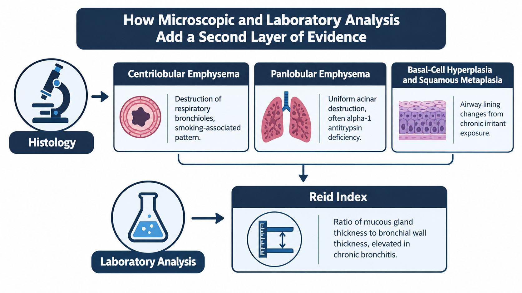
In a study of 400 adults who died of nonrespiratory, noncancer causes, current smokers showed higher rates of basal-cell hyperplasia, squamous metaplasia, cellular atypia, mucous-cell metaplasia, and a higher Reid Index than nonsmokers, while former smokers fell between the two groups (source). Those changes fit long-term irritant injury. They support exposure history, but they do not identify the final cause of death on their own.
What the microscope reveals
Microscopy also helps sort out the type of emphysema. Smoking-related injury is often centrilobular, which means the damage centers on the small airways and nearby air spaces. That pattern fits smoke exposure better than a surface glance does. Other patterns can point toward different causes, so no one should treat every emphysematous lung as the same disease.
Sampling matters just as much as the slide itself. A pathologist does not rely on one piece of tissue from one spot. Sections from central bronchi, segmental bronchi, membranous bronchioles, and suspicious peripheral lesions help build a more accurate view of the whole lung. For a plain-language overview of what microscopic examination adds in autopsy work, see what is microscopic examination.
What laboratory measurement adds
Laboratory testing can support what the microscope shows. In one quantitative autopsy study, mean elemental-carbon concentrations were 56.68 µg/g in never-smokers, 449.56 µg/g in cigarette smokers, and 6,678.2 µg/g in coal miners, with the smoker average about eight times the never-smoker average and linked with pack-years (source). That does not establish cause of death, but it gives objective support for exposure.
| Group | Histologic emphysema | Source |
|---|---|---|
| Male nonsmokers | Approximately 26% | NLM archive |
| Male smokers with more than 500,000 lifetime cigarettes | About 70% | NLM archive |
For families and attorneys, the key point is that microscopic evidence turns a visual impression into documented findings. It helps the autopsy report separate pigment from structural damage, and structural damage from the final medical cause of death.
The Dose Response Between Smoking and Structural Lung Damage
Smoking injury is graded, not all-or-nothing. More exposure makes structural change more likely, but there is no single line where damaged tissue suddenly appears. That is why a pathologist asks for pack-year history, not just a yes-or-no smoking label.
Exposure history also has to be translated into usable medical language. A smoker with a short history of light use and a long-term heavy smoker may both say “yes” to smoking, yet their lungs do not need the same sampling approach. That is the kind of distinction clinicians document in how to write a COPD care plan, and the same habit of precise recording helps an autopsy report avoid vague conclusions.
The pattern matters more than a dramatic photo. In one autopsy series, histologic emphysema appeared far more often in male smokers with very heavy lifetime exposure than in nonsmokers, which supports a dose-response relationship without turning it into a simple cutoff. A pathologist uses that kind of history to decide how much tissue to examine and where to look for injury.
Why visible findings can mislead
Visible bullous disease has been reported more often in smokers than nonsmokers, yet surface color alone does not measure smoking exposure. A 72-specimen autopsy study found that smoking was not a major driver of lung-surface carbon in that group (source). Those two observations point in the same direction. A black or dark lung surface is only one clue.
Air pollution, occupational dust, prior infection, age, and chronic lung disease can all change how tissue looks. A coal worker, a city resident, and a heavy smoker may each show dark discoloration, but the reason for that color can differ. The forensic task is to separate exposure from injury and injury from cause of death.
Reading the pattern, not the photo
The gross view and the tissue sections have to agree. If the lungs look heavily pigmented but the sampled areas do not show structural injury, the report should say so plainly. If the history suggests heavy smoking, the sampling plan should reach deeper and wider, not stop at the most obvious surface.
For families and attorneys, the key question is whether the findings fit the history and whether they explain death. That distinction is the difference between a visual impression and a defensible medical opinion.
Why Pigment Alone Is Not a Smoking Meter
Black pigment is one of the easiest findings to see and one of the easiest to misread. It shows that particulate material reached the lungs, but it does not tell us where that material came from or whether it caused death. A pathologist has to answer those questions separately.
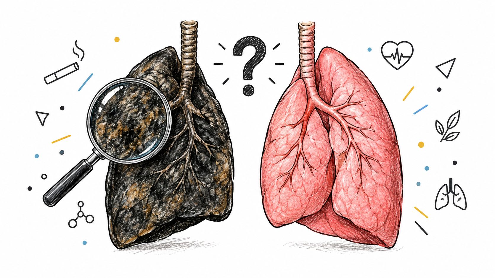
Dark coloration can come from several kinds of inhaled particles. City air, workplace dust, and old healed injury can all leave pigment behind. A coal worker's lung, for example, may look dark because of anthracotic dust, while a smoker's lung may show pigment along with airway injury and emphysema. The surface color alone cannot separate those possibilities.
Microscopy does that work. Under the microscope, anthracosis usually appears as fine carbon in macrophages, the scavenger cells that collect debris in the air spaces and around the small airways. Smoking-related injury often adds other changes, such as enlarged air spaces, scarring around bronchioles, or inflammation that fits chronic smoke exposure. That is the difference between seeing soot in a chimney and seeing heat damage in the bricks.
Laboratory and record review add another layer. If the tissue is dark but the microscopic pattern does not match smoking injury, the report should say so. If the pattern does match, the findings still need to be read with the medical history and toxicology, because more than one exposure can act on the same lung. A single dark photograph cannot do that job.
The practical lesson is simple. Pigment is a clue, not a conclusion. An autopsy has to show whether the darkness reflects anthracosis, smoking-related change, or another source of inhaled material before anyone ties it to cause of death.
How Lung Findings Connect to Medical History, Toxicology, and Cause of Death
A lung finding matters only when it is tied to the person's medical story. Emphysema, chronic bronchitis, vascular change, or even lung cancer can be present without being the event that caused death. The autopsy must separate the underlying cause, the immediate mechanism, and any contributing condition.
The old public health lesson is settled. Cigarette smoking has long been linked to serious lung disease at the population level, and that is not the same question as what killed one person in one case. In an autopsy, the issue is individual attribution, not general causation. The pathologist still has to connect the findings to the timeline, the symptoms, and the final event.
A simple example shows why toxicology matters. A patient may have heavy smoking-related lung damage, but blood testing can also show another drug or exposure that helps explain the death. In that setting, the lungs may show chronic injury while toxicology points to a separate acute problem, such as a poison, sedative effect, or heart stress from another substance. A careful forensic toxicology exam helps separate those possibilities before the opinion is finalized.
The same principle applies to the medical record. Prior imaging, symptoms, smoking history, and the circumstances of death all have to be read together. A dark or damaged lung does not answer the whole question by itself.
The final opinion should state what the lung findings mean, and what they do not mean. Sometimes they explain the death. Sometimes they helped weaken the person over time but did not cause the last event. Sometimes they are important background findings that sit beside a different cause of death.
That distinction is the heart of autopsy interpretation. A pathologist is not deciding whether the lungs looked abnormal. The task is to decide whether those abnormalities were part of the chain that ended life, or whether they were findings that belonged to the medical history without being the fatal process.
Why Smoking Is Rarely the Official Cause of Death on the Certificate
Families are often surprised when the death certificate does not say smoking, even when the lungs clearly show smoking-related injury. The reason is simple. A death certificate records the chain of events that led to death, not a habit or exposure by itself. Smoking may be the background driver, while the official cause names the fatal event that ended life.
As noted above, smoking can shape the disease process without appearing as the listed cause. That pattern is one reason the certificate has to be read as a medical summary, not a visual label for damaged lungs.
Why modern exposure history matters
The exposure history is more complicated than cigarettes alone. WHO reported in 2025 that more than 100 million people worldwide were vaping, so a postmortem exam has to consider cigarettes, e-cigarettes, dual use, and other inhalational exposures. That does not replace the smoking history, it broadens it.
A family asking about a cause-of-death entry should also ask whether the pathologist considered chronic lung injury, recent nicotine use, and competing disease. For background on how that paperwork is framed, the cause-of-death certificate guide from Texas Autopsy Services separates medical findings from certification language.
Questions that help clarify the report
- What was the immediate cause? Ask whether the final event was respiratory failure, infection, embolism, heart disease, or something else.
- What was the underlying disease? Ask whether smoking-related emphysema, chronic bronchitis, or cancer was the long-term driver.
- Were other exposures checked? Ask whether vaping, dual use, or occupational inhalation were considered.
- Was the microscopic exam done? Ask whether representative tissue sections were reviewed.
- Did toxicology add anything? Ask whether nicotine-related or other relevant findings were tested.
Those questions keep the report focused on causation, not assumptions.
Questions Families and Attorneys Should Ask Before an Autopsy
A useful private or second-opinion autopsy starts with the right questions. The aim is not a dramatic answer. It is an answer that can stand up to review. Families and attorneys need to know what history was reviewed, what tissue was sampled, what tests were ordered, and how the final opinion separates exposure from cause.
The microscopic changes described earlier are exactly why the report should not rest on appearance alone. Pigment can be present, emphysema can be severe, and neither one automatically explains how the person died.
Printable checklist to bring to the appointment
- Smoking and vaping history: List every known tobacco or nicotine exposure, including dual use and any recent use.
- Prior lung disease: Bring any history of COPD, asthma, pneumonia, lung cancer, or unexplained shortness of breath.
- Sampling plan: Ask which bronchi, bronchioles, and peripheral areas will be examined.
- Laboratory testing: Ask whether toxicology, elemental-carbon analysis, or special stains are appropriate.
- Competing causes: Ask how heart disease, embolism, infection, or malignancy will be weighed.
- Report language: Ask how the final report will distinguish exposure, disease, and cause of death.
If the case involves litigation, an outside review, or a disputed certificate, a medical-legal team such as the one described on the medical malpractice page may also benefit from a clear autopsy record.
Families deserve an explanation, not a photograph without context. A careful review can show what the lungs do reveal and what they do not. If you need a second opinion on a smoker's lungs, or you want help understanding whether lung findings changed the cause of death, visit Texas Autopsy Services to speak with our team and get clear guidance on the next step.