October 24, 2026 · Texas Autopsy Services
Digital Pathology in Forensic Cases: A Practical Guide
Learn how digital pathology supports forensic cases through digital imaging, telepathology, faster review, and secure, HIPAA-compliant reporting in Texas.
On this page
- What Digital Pathology Means in Practice
- How Digital Imaging and Telepathology Work Together
- Faster Review and Remote Consultation for Forensic Cases
- Secure Reporting and Chain of Custody in Digital Workflows
- Limits of Digital Review and Common Misconceptions
- How Texas Autopsy Services Uses Digital Tools
- Next Steps for Families, Attorneys, and Agencies
A family receives an autopsy report with photographs and microscopic findings, and an attorney asks whether those slides can be reviewed by another pathologist without shipping fragile glass across Texas. A county official faces a similar question when a specialist opinion is needed quickly. Digital pathology can help in both situations, but only when the image, the original specimen, and the written record stay clearly connected.
- Digital images support review: A scanned slide can be viewed and shared without moving the original glass.
- Validation remains local: Each laboratory must test its own scanner, software, display, and staff competency.
- Documentation creates credibility: Metadata, access records, and chain of custody matter as much as image quality.
- A pathologist stays responsible: Digital tools and artificial intelligence assist interpretation. They don't replace the forensic pathologist.
What Digital Pathology Means in Practice
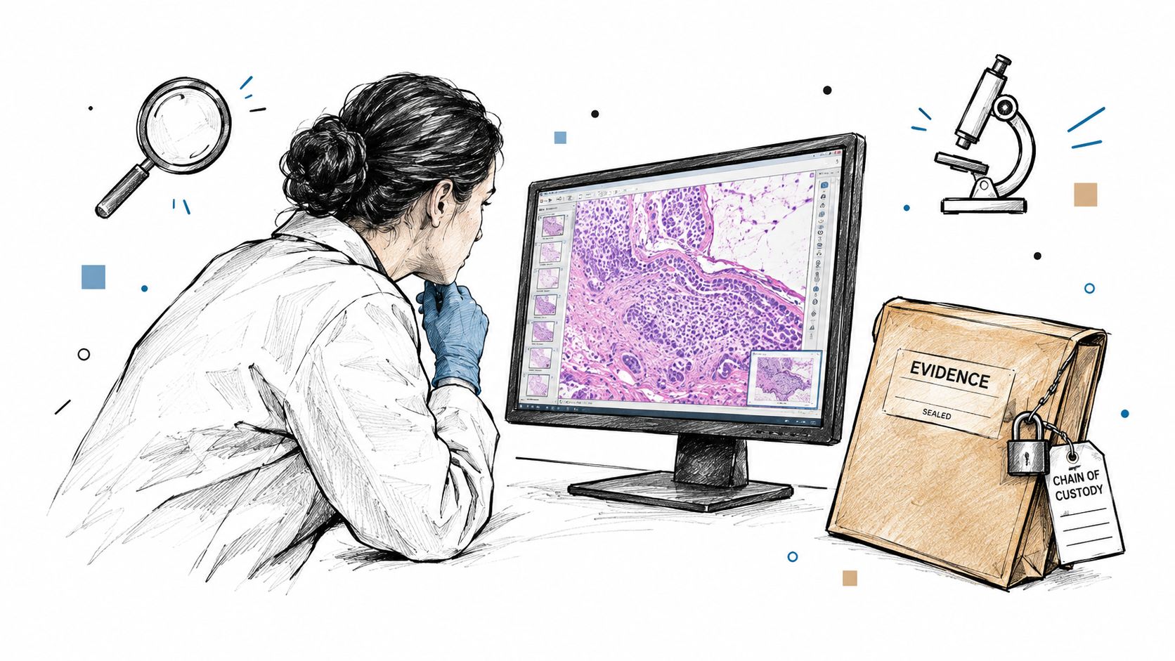
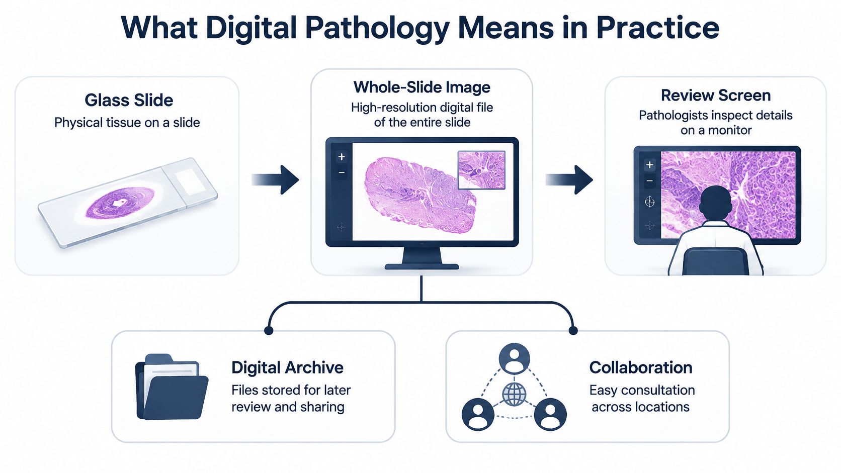
Digital pathology begins with a familiar object, a glass slide holding a thin section of stained tissue. In a conventional workflow, a pathologist places that slide under a microscope and moves it by hand. In a digital workflow, a scanner photographs the entire slide at high resolution and creates a whole-slide image, often called a WSI. That file can then be viewed, stored, shared, and analyzed on a computer screen rather than only through microscope eyepieces.
Think of the change as moving from a single paper map to a detailed digital map of the same place. The territory, the tissue, hasn't changed. The way a pathologist explores it, marks areas of interest, and shares the view with a colleague has changed. That distinction matters because the quality of the digital map depends on the scanner, the preparation of the slide, and the screen used for review.
The Parts of a Working System
A working system needs more than a scanner. It requires an image management system to organize cases, secure storage for large files, a calibrated display, and procedures that link each image to the correct person and specimen. Early digitization led to clinically usable whole-slide workflows that later supported remote review, subspecialty consultation, image archives, and computer-aided diagnosis, as described in the cross-continent survey of anatomic pathology laboratories.
Adoption has been gradual rather than universal. In that survey of 127 laboratories, 72 laboratories, or 56.7%, had fully or partially implemented a digital pathology workflow, while 55 had no in-house digital pathology. Among users, 36 of 72, or 50%, digitized less than 5% of their total caseload, and 16 laboratories, or 22.2%, scanned more than 75% of cases. Fourteen laboratories digitized all routine histology, including immunohistochemistry.
Those figures show a practical pattern. Many laboratories start with selected cases, consultations, or research, then expand toward routine practice. Digital pathology is therefore both an imaging change and a workflow change. Scanners, software, viewing environments, storage, and quality checks must work together. If one part is weak, the image may be hard to trust, even when the underlying tissue preparation was sound.
How Digital Imaging and Telepathology Work Together
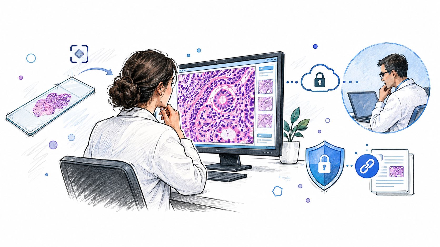
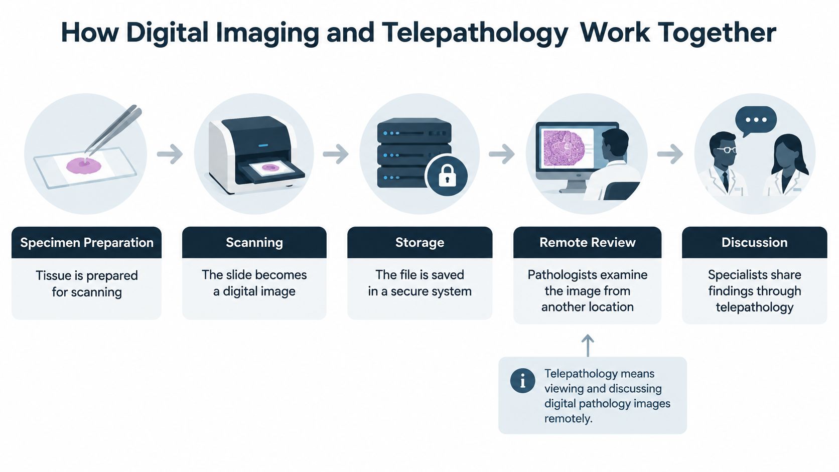
A digital case follows a sequence that begins before scanning. Tissue is fixed, processed, sectioned, placed on glass, and stained so cellular structures become visible. Staff verify identification, check slide quality, and scan the slide. The resulting file enters secure storage with case information. A pathologist opens the image on a validated display, examines it at different magnifications, records findings, and correlates those findings with the gross examination and other test results.
Telepathology is the viewing and discussion of those digital images between people in different locations. It doesn't create a new specimen. It allows another qualified pathologist to see the same scanned field, review annotations, and discuss whether additional sections, stains, or examination of the original glass are needed. Our related overview of digital imaging examples in forensic review shows how that sequence fits into case documentation.
Why Local Validation Matters
A scanner that performs well in one laboratory can't be assumed to perform the same way in another. Staining, tissue preparation, scanner settings, software versions, monitors, and reader training all vary. A defensible workflow documents scanner qualification, staining consistency, display calibration, image-quality checks, secure case identification, and pathologist competency using the actual hardware and software configuration.
Practical rule: Validate the exact system that will be used for the case, not a similar system described in a brochure or a published study.
Evidence supports careful use, not blind confidence. In a large verification and validation study, 2,745 of 3,001 cases, or 91%, showed complete concordance between digital pathology and glass-slide reports, with sensitivity of 98.07% for digital pathology versus 99.07% for glass microscopy, as reported in the verification and validation study of whole-slide imaging. The small difference shows that digital review can be highly concordant without being error-free.
Folds, tissue loss, out-of-focus areas, compression artifacts, and subtle color differences can affect interpretation. When a region looks unclear on screen, the correct response is to rescan, review the glass, or prepare additional material. Telepathology works best when remote access is paired with that discipline, so distance doesn't become an excuse to accept an image that isn't adequate for the question being asked.
Faster Review and Remote Consultation for Forensic Cases
Remote consultation offers a clear practical benefit. A digital file can be made available to a consulting pathologist while the original slide and tissue block remain in controlled storage. That reduces handling, avoids repeated shipping of fragile glass, and helps families, attorneys, and agencies obtain a second opinion autopsy review when distance would otherwise slow communication. It also allows two pathologists to discuss the same annotated region rather than describe separate microscope fields from memory.
Speed still depends on factors outside the scanner. Staff must confirm authorization, locate prior reports and medical records, verify specimen identity, assess image quality, and determine whether toxicology or additional testing affects the interpretation. For that reason, we don't treat digital access as a promise of instant answers. We explain timing by case factors, and when discussing our general process we refer to most cases within 24 to 48 hours for the examination phase rather than offering an unconditional guarantee.
What Concordance Data Shows
Digital review has strong support for many routine diagnostic tasks. A systematic review in the Journal of Clinical Pathology covering 25 studies and 10,410 histology samples reported overall clinical concordance of 98.3% (95% confidence interval, 97.4% to 98.9%) between digital pathology and conventional microscopy, as summarized in the Journal of Clinical Pathology systematic review and meta-analysis. That finding explains why digital files are useful for consultation, documentation, and review of prior findings.
The same review recorded 546 major discordances, and more than half, 57%, were associated with nuclear atypia, dysplasia grading, or malignancy assessment. Those are areas where subtle cellular detail matters. Focus, staining, display conditions, and image quality can change how that detail appears. High overall concordance therefore doesn't remove the need for expert judgment, especially when a finding may affect a cause of death investigation or legal proceeding.
Attorneys and families also need reports they can read, search, and share appropriately. Secure digital reporting pairs the pathologist's written opinion with the relevant images and records. For general document handling outside our laboratory systems, teams that manage large case files may find reviewing PDFs with browser-based tools useful for organizing their own copies. That convenience never replaces controlled access to the official forensic record.
For a family searching for an autopsy near me, the central benefit isn't technology for its own sake. It's access to a qualified forensic pathologist, a clear explanation of limits, and a record that another qualified reviewer can follow. Remote consultation strengthens that access when the image is adequate and the original material remains available for correlation.
Secure Reporting and Chain of Custody in Digital Workflows
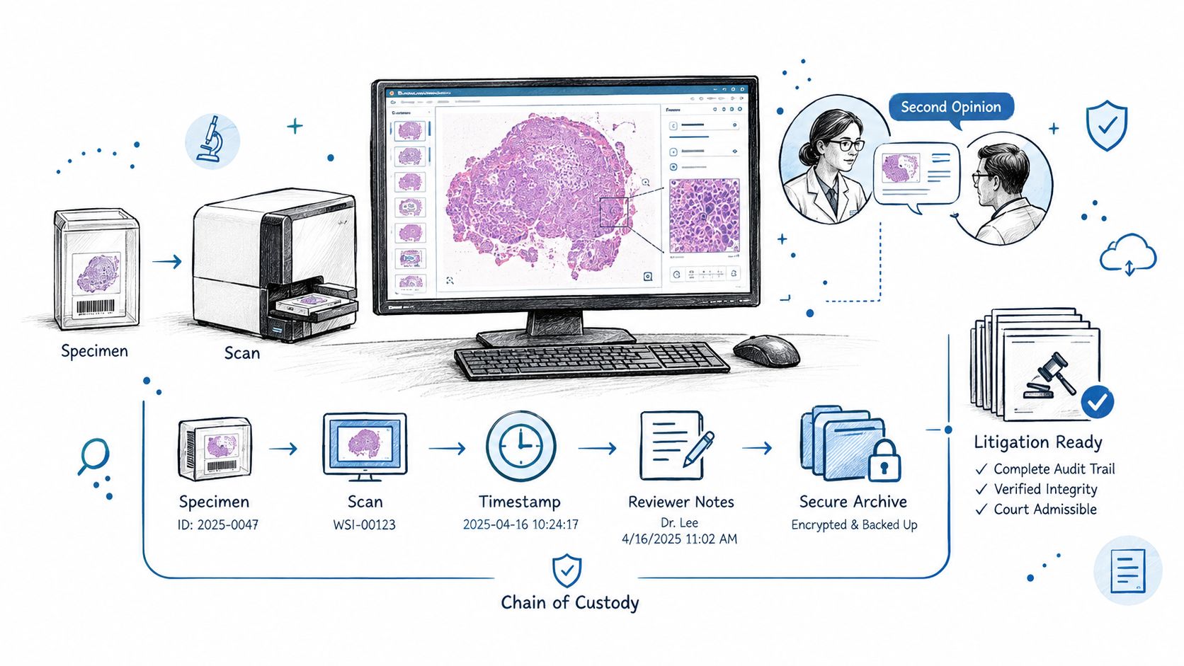
A digital image becomes credible through documentation. Chain of custody means the written record showing who received, handled, stored, transferred, and accessed a specimen or record, and when each step occurred. In digital work, that record must connect the body or specimen, the tissue block, the glass slide, the scanned image, the reviewing pathologist, and the final report. If that connection breaks, another reviewer can't be certain that the image represents the correct case.
A defensible file should preserve the original glass slide when retention rules require it, along with image metadata, scanner and software information, rescans, annotations, and access logs. Access should be limited to authorized people, and transmission should use secure systems that protect health information. Our discussion of chain of custody and cyber security controls addresses how access control supports that protection.
Records That Support a Digital Finding
- Original material: Retained slides, blocks, and specimens allow correlation when an image is incomplete or disputed.
- Image metadata: File identifiers, scan date, scanner information, and software version help establish provenance.
- Change history: Rescans, annotations, and amended reports should be recorded rather than overwritten.
- Access record: Logs show who viewed or received a file and under what authorization.
Families and attorneys sometimes need to send records from hospitals, funeral homes, or other offices. Those transmissions deserve the same care as laboratory transfers, and general guidance on faxing sensitive paperwork can help senders think through confirmation and privacy before documents move. Within our practice, HIPAA-compliant reporting and direct communication reduce the chance that records pass through unnecessary intermediaries.
Digitization doesn't automatically improve evidentiary quality. Without validated workflows and documented provenance, it can add another layer of uncertainty. Barriers involving proprietary formats, legacy systems, vendor lock-in, cost, reimbursement, and compliance are discussed in the review of barriers to defensible digital pathology use. The answer for a specific case is local validation, concordance testing when appropriate, monitoring after deployment, and documentation that can be explained under scrutiny.
Limits of Digital Review and Common Misconceptions
One common misconception is that a scanned image is always more accurate than a microscope. A scan is a representation of a slide, and its value depends on tissue preparation, focus, color rendering, and display quality. Areas can be folded, torn, missing, or out of focus. Compression can alter fine detail. Our overview of digital image quality in pathology review explains why a pathologist may request the glass slide even after receiving a digital file.
A second misconception is that artificial intelligence can issue an independent diagnosis. AI tools can highlight regions, assist with quantification, support triage, and help a pathologist review a large image. Those functions are decision support. They don't transfer medical responsibility to software, and they don't replace examination of the body, review of circumstances, toxicology, or correlation with medical history.
Reading AI Performance Carefully
Pooled figures can look reassuring while hiding variation. A 2024 systematic review and meta-analysis reported mean sensitivity of 96.3% and mean specificity of 93.3% for AI tools analyzing whole-slide images across diseases and tasks, as reported in the 2024 systematic review and meta-analysis of AI in whole-slide imaging. Performance differed by domain. Gastrointestinal pathology averaged 93% sensitivity and 94% specificity, uropathology 95% and 96%, and breast pathology 83% and 88%, respectively.
A headline performance number doesn't prove that a tool is reliable for a different tissue type, scanner, stain, population, or diagnostic question.
Deployment requires task-specific validation, calibrated thresholds, and review of false negatives and false positives. Models can degrade when staining protocols, scanners, tissue quality, prevalence, or patient populations differ from development data. For postmortem work, the safer role for AI is a reproducible aid for highlighting suspicious regions or measuring features while our board-certified forensic pathologists retain interpretive responsibility.
Reports should distinguish algorithm output from the final medical opinion. The relevant image, model version, threshold, and audit trail should be preserved when an algorithm contributes to review. That transparency helps families and attorneys understand what was automated, what was personally examined, and why the final opinion does or doesn't follow a highlighted finding. Digital review also can't determine every issue in a death investigation. Physical findings, scene information, records, laboratory testing, and specimen handling remain essential parts of the autopsy process.
How Texas Autopsy Services Uses Digital Tools
We use digital imaging as one part of an independent autopsy and consultation process, not as a substitute for examination. At Texas Autopsy Services, every examination is performed by a forensic pathologist certified by the American Board of Pathology. Our team serves all 254 Texas counties from Elgin, with in-house licensed transport, secure temperature-controlled storage, toxicology and molecular testing when indicated, and HIPAA-compliant reporting with full chain-of-custody documentation.
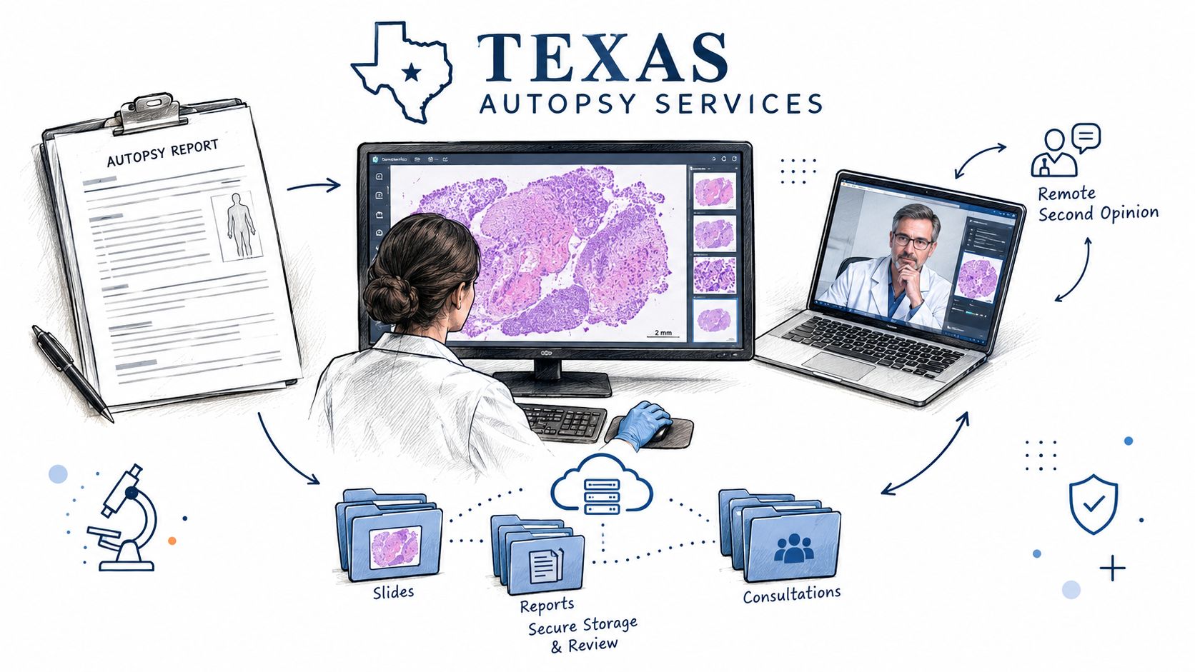
In practice, digital tools help us document microscopic findings, organize case images, consult when a focused question arises, and review prior material for second opinions. They also support clear communication with families, attorneys, healthcare professionals, and county officials. A cause of death is the disease or injury that starts the sequence leading to death. Manner of death is the classification of circumstances, such as natural, accident, homicide, suicide, or undetermined. Digital microscopy may inform cause, but manner requires correlation with circumstances and records beyond the slide.
Regulatory Context for Digital Systems
The U.S. Food and Drug Administration has cleared whole-slide imaging systems through the 510(k) pathway to aid pathologists who review and interpret digital images. In June 2024, Roche announced FDA 510(k) clearance for its Digital Pathology Dx system, including the VENTANA DP 200 scanner, workflow software, and display, in the Roche announcement of FDA clearance for its digital pathology solution. The clearance language describes assistance to a pathologist rather than replacement of a pathologist.
That distinction guides our approach. Clearance of one product doesn't validate every scanner, monitor, software configuration, staining protocol, or intended use. Documented local procedures for image quality, access control, retention, cybersecurity, and correlation with original material still matter. Those controls are especially important when images may support independent review, litigation, or expert testimony.
The short video below offers additional visual context for how digital review fits into a broader forensic workflow.
Cost is another practical concern. Scanners, storage, software, integration, cybersecurity, training, maintenance, and validation create ongoing expenses, and hardware cost and integration burden are leading adoption barriers in the U.S. clinical diagnostic laboratory survey on digital pathology adoption. Our flat-rate, all-inclusive pricing for private autopsy services Texas families request is designed to keep technology-related costs from appearing as unexpected charges. We also work within applicable Texas requirements, including oversight involving the Texas Funeral Service Commission and the Texas Health & Safety Code Chapters 711, 716, and 651 where those provisions apply to disposition, cremation authorization, and funeral service practice.
Next Steps for Families, Attorneys, and Agencies
Before sending digital images or prior reports for review, it helps to confirm a few practical points. Ask which original slides and tissue blocks are retained, how image metadata is preserved, who can access the files, and how each image is linked to the chain of custody. Ask whether the reviewing forensic pathologist may need the glass slides, additional sections, medical records, toxicology results, or scene information before offering an opinion. Clear answers at the start prevent delays and misunderstandings later.
Different requesters need different records. Families often need a plain-language explanation of cause and manner of death and guidance on authorization. Attorneys may need a complete, court-admissible report, preserved images, and a record suitable for disclosure or testimony. County officials and justices of the peace may need documentation that supports medicolegal decision-making in counties without a medical examiner. Law offices managing record requests and deadlines sometimes use virtual legal assistants to organize administrative follow-up, while medical interpretation remains with the forensic pathology expert.
Common Questions From Families and Attorneys
Can a digital slide replace an autopsy? No. A slide shows sampled tissue under magnification. It can't replace external and internal examination, specimen collection, toxicology, records review, or correlation with circumstances.
Can prior slides be reviewed for a second opinion? Often, yes, when identity, authorization, image quality, and chain of custody can be established. Original glass or additional material may still be necessary if the scan is incomplete or a finding is disputed.
Who interprets the image? A qualified pathologist interprets the image and signs the medical opinion. Software may organize, display, measure, or highlight areas, but it doesn't assume responsibility for the conclusion.
Readers who want service details can review our information on Private Autopsies and County Forensic Autopsies. Funeral professionals coordinating logistics may also review the Waterloo Mortuary Partnership. We know these decisions often arise during grief, conflict, or uncertainty. Our team is available 24/7 at (806) 230-1889 for direct guidance, without call centers or middlemen.
If you're considering a private autopsy, second-opinion slide review, or county forensic consultation, we can explain which records and original materials are needed and how digital pathology may support the review. Every examination is performed by a board-certified forensic pathologist, with statewide transport, full chain-of-custody documentation, and flat-rate pricing. Visit Texas Autopsy Services or call our team for calm, direct guidance.